- Featured Product
- KD/KO Validated
Ki-67 Polyklonaler Antikörper
Ki-67 Polyklonal Antikörper für IHC, IF/ICC, IF-P, FC (Intra), ELISA
Wirt / Isotyp
Kaninchen / IgG
Getestete Reaktivität
human und mehr (5)
Anwendung
IHC, IF/ICC, IF-P, FC (Intra), ELISA
Konjugation
Unkonjugiert
Kat-Nr. : 27309-1-AP
Synonyme
 at dilution of 1:8000 (under 20x lens). Heat mediated antigen retrieval with Tris-EDTA buffer (pH 9.0).")
 at dilution of 1:8000 (under 20x lens). Heat mediated antigen retrieval with Tris-EDTA buffer (pH 9.0).")
 at dilution of 1:10000 (under 10x lens). Heat mediated antigen retrieval with Tris-EDTA buffer (pH 9.0).")
 at dilution of 1:400 (under 10x lens). Heat mediated antigen retrieval with Tris-EDTA buffer (pH 9.0).")
 at dilution of 1:400 (under 40x lens). Heat mediated antigen retrieval with Tris-EDTA buffer (pH 9.0).")
 at dilution of 1:400 (under 10x lens). Heat mediated antigen retrieval with Tris-EDTA buffer (pH 9.0).")
 at dilution of 1:400 (under 40x lens). Heat mediated antigen retrieval with Tris-EDTA buffer (pH 9.0).")
 at dilution of 1:200 (under 10x lens. Heat mediated antigen retrieval with Tris-EDTA buffer (pH 9.0).")
 at dilution of 1:200 (under 40x lens. Heat mediated antigen retrieval with Tris-EDTA buffer (pH 9.0).")
 at dilution of 1:16000 (under 10x lens. Heat mediated antigen retrieval with Tris-EDTA buffer (pH 9.0).")
 at dilution of 1:16000 (under 40x lens. Heat mediated antigen retrieval with Tris-EDTA buffer (pH 9.0).")
 at dilution of 1:1000 (under 10x lens. Heat mediated antigen retrieval with Tris-EDTA buffer (pH 9.0).")
 at dilution of 1:1000 (under 40x lens. Heat mediated antigen retrieval with Tris-EDTA buffer (pH 9.0).")
 at dilution of 1:1000 (under 10x lens. Heat mediated antigen retrieval with Tris-EDTA buffer (pH 9.0).")
 at dilution of 1:1000 (under 40x lens. Heat mediated antigen retrieval with Tris-EDTA buffer (pH 9.0).")
 at dilution of 1:16000 (under 10x lens. Heat mediated antigen retrieval with Tris-EDTA buffer (pH 9.0).")
 at dilution of 1:16000 (under 40x lens. Heat mediated antigen retrieval with Tris-EDTA buffer (pH 9.0).")
 at dilution of 1:16000 (under 10x lens. Heat mediated antigen retrieval with Tris-EDTA buffer (pH 9.0).")
 at dilution of 1:16000 (under 40x lens. Heat mediated antigen retrieval with Tris-EDTA buffer (pH 9.0).")
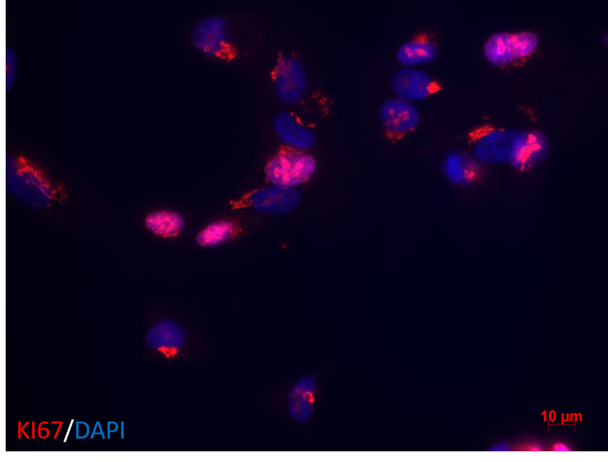
 fixed paraffin-embedded human lung cancer tissue using Ki-67 antibody (27309-1-AP) at dilution of 1:200 and CoraLite®488-Conjugated Goat Anti-Rabbit IgG(H+L) (SA00013-2). Heat mediated antigen retrieval with Tris-EDTA buffer (pH 9.0).")
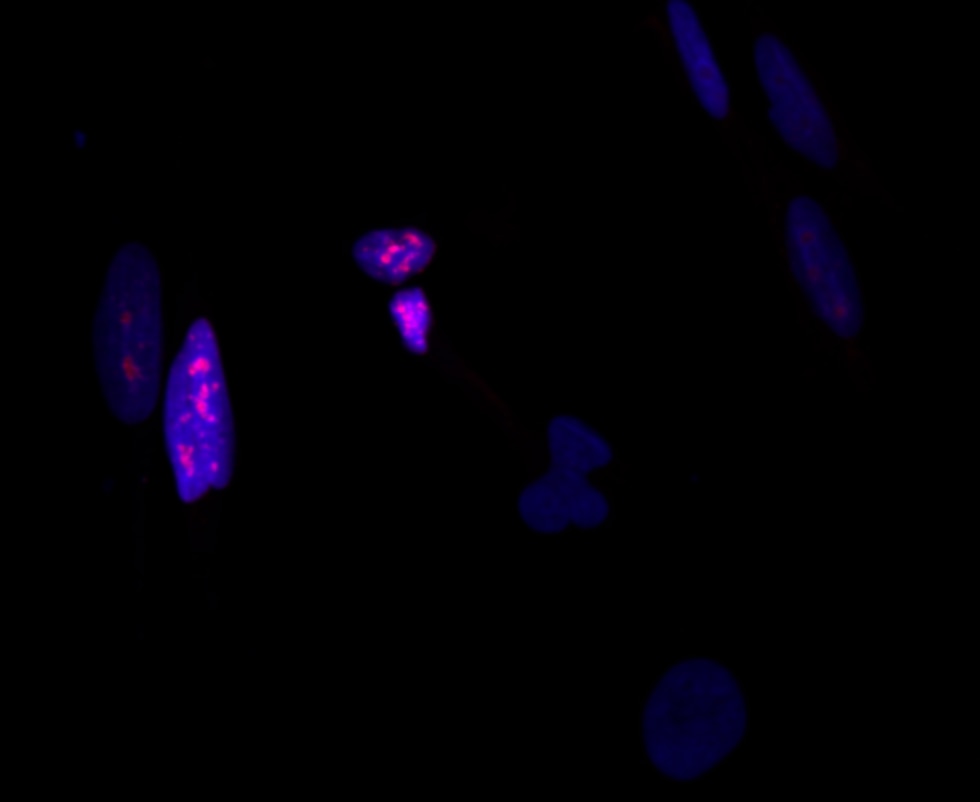
 fixed HeLa cells using KI67 antibody (27309-1-AP) at dilution of 1:200 and CoraLite®488-Conjugated AffiniPure Goat Anti-Rabbit IgG(H+L).")
 fixed HeLa cells using 27309-1-AP (KI67 antibody) at dilution of 1:100 and CoraLite488-Conjugated AffiniPure Goat Anti-Rabbit IgG(H+L). F-actin is stained using CL555-phalloidin (red).")
 fixed HEK-293 cells using KI67 antibody (27309-1-AP) at dilution of 1:400 and CoraLite®488-Conjugated AffiniPure Goat Anti-Rabbit IgG(H+L).")
 fixed HeLa cells using KI67 antibody (27309-1-AP) at dilution of 1:200 and CoraLite®488-Conjugated AffiniPure Goat Anti-Rabbit IgG(H+L), CL594-Phalloidin (red).")
 and CoraLite®488-Conjugated AffiniPure Goat Anti-Rabbit IgG(H+L) at dilution 1:1000 (red), or 0.4 ug x. Cells were fixed and permeabilized with Transcription Factor Staining Buffer Kit (PF00011).")
Geprüfte Anwendungen
| Erfolgreiche Detektion in IHC | humanes Tonsillitisgewebe, humanes Mammakarzinomgewebe, humanes Kolonkarzinomgewebe, humanes Gliomgewebe, humanes Lungenkarzinomgewebe, humanes Lymphomgewebe, humanes Hautkrebsgewebe, Insulinomgewebe, K-562-Zellen Hinweis: Antigendemaskierung mit TE-Puffer pH 9,0 empfohlen. (*) Wahlweise kann die Antigendemaskierung auch mit Citratpuffer pH 6,0 erfolgen. |
| Erfolgreiche Detektion in IF-P | humanes Lungenkarzinomgewebe |
| Erfolgreiche Detektion in IF/ICC | HeLa-Zellen, HEK-293-Zellen |
| Erfolgreiche Detektion in FC (Intra) | Jurkat-Zellen |
Empfohlene Verdünnung
| Anwendung | Verdünnung |
|---|---|
| Immunhistochemie (IHC) | IHC : 1:4000-1:16000 |
| Immunfluoreszenz (IF)-P | IF-P : 1:50-1:500 |
| Immunfluoreszenz (IF)/ICC | IF/ICC : 1:50-1:500 |
| Durchflusszytometrie (FC) (INTRA) | FC (INTRA) : 0.40 ug per 10^6 cells in a 100 µl suspension |
| It is recommended that this reagent should be titrated in each testing system to obtain optimal results. | |
| Sample-dependent, check data in validation data gallery | |
Veröffentlichte Anwendungen
| KD/KO | See 1 publications below |
| IHC | See 996 publications below |
| IF | See 253 publications below |
Produktinformation
27309-1-AP bindet in IHC, IF/ICC, IF-P, FC (Intra), ELISA Ki-67 und zeigt Reaktivität mit human
| Getestete Reaktivität | human |
| In Publikationen genannte Reaktivität | human, hamster, Hausschwein, Hund, Zebrafisch, Ziege |
| Wirt / Isotyp | Kaninchen / IgG |
| Klonalität | Polyklonal |
| Typ | Antikörper |
| Immunogen | Ki-67 fusion protein Ag26266 |
| Vollständiger Name | antigen identified by monoclonal antibody Ki-67 |
| Berechnetes Molekulargewicht | 359 kDa |
| GenBank-Zugangsnummer | NM_002417 |
| Gene symbol | KI67 |
| Gene ID (NCBI) | 4288 |
| Konjugation | Unkonjugiert |
| Form | Liquid |
| Reinigungsmethode | Antigen-Affinitätsreinigung |
| Lagerungspuffer | PBS with 0.02% sodium azide and 50% glycerol |
| Lagerungsbedingungen | Bei -20°C lagern. Nach dem Versand ein Jahr lang stabil Aliquotieren ist bei -20oC Lagerung nicht notwendig. 20ul Größen enthalten 0,1% BSA. |
Hintergrundinformationen
The Ki-67 protein (also known as MKI67) is a cellular marker for proliferation. Ki67 is present during all active phases of the cell cycle (G1, S, G2 and M), but is absent in resting cells (G0). Cellular content of Ki-67 protein markedly increases during cell progression through S phase of the cell cycle. Therefore, the nuclear expression of Ki67 can be evaluated to assess tumor proliferation by immunohistochemistry. It has been demonstrated to be of prognostic value in breast cancer. In head and neck cancer, several studies have reported an association between high proliferative activity and poorer prognosis.
Protokolle
| PRODUKTSPEZIFISCHE PROTOKOLLE | |
|---|---|
| IHC protocol for Ki-67 antibody 27309-1-AP | Protokoll herunterladenl |
| IF protocol for Ki-67 antibody 27309-1-AP | Protokoll herunterladen |
| STANDARD-PROTOKOLLE | |
|---|---|
| Klicken Sie hier, um unsere Standardprotokolle anzuzeigen |
Publikationen
| Species | Application | Title |
|---|---|---|
Signal Transduct Target Ther FBXW7β loss-of-function enhances FASN-mediated lipogenesis and promotes colorectal cancer growth | ||
Mol Cancer Cell surface CD55 traffics to the nucleus leading to cisplatin resistance and stemness by inducing PRC2 and H3K27 trimethylation on chromatin in ovarian cancer | ||
Mol Cancer lncRNA ZNRD1-AS1 promotes malignant lung cell proliferation, migration, and angiogenesis via the miR-942/TNS1 axis and is positively regulated by the m6A reader YTHDC2 | ||
Cancer Discov Stress Granules determine the Development of Obesity-associated Pancreatic Cancer. | ||
Adv Mater Supramolecular Hydrogel with Ultra-Rapid Cell-Mediated Network Adaptation for Enhancing Cellular Metabolic Energetics and Tissue Regeneration | ||
Protein Cell NDFIP1 limits cellular TAZ accumulation via exosomal sorting to inhibit NSCLC proliferation |
Rezensionen
The reviews below have been submitted by verified Proteintech customers who received an incentive for providing their feedback.
FH Udesh (Verified Customer) (08-12-2025) | Worked well in Western, also detected a non specific band at 70 kDa
|
FH Celine (Verified Customer) (07-09-2025) | Works well in immunoflurescence with fixed cells
 |
FH Guo-rong (Verified Customer) (08-30-2022) | Clear staining
 |
FH Alessandro (Verified Customer) (07-27-2022) | No aspecific staining, great outcome
|
FH Ana (Verified Customer) (02-15-2022) | Both, 1:50 and 1:100 dilutions worked well
|
FH kes (Verified Customer) (01-17-2022) | Works at 1:200 dilution for IHC
|
FH Yan (Verified Customer) (02-18-2020) | 1:100 work for cell.
|

