- Featured Product
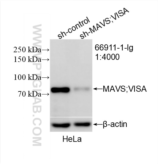
- KD/KO Validated
MAVS; VISA Monoklonaler Antikörper
MAVS; VISA Monoklonal Antikörper für WB, IHC, ELISA
Wirt / Isotyp
Maus / IgG1
Getestete Reaktivität
human und mehr (3)
Anwendung
WB, IHC, IF, IP, CoIP, ELISA
Konjugation
Unkonjugiert
CloneNo.
1A8E9
Kat-Nr. : 66911-1-Ig
Synonyme
Geprüfte Anwendungen
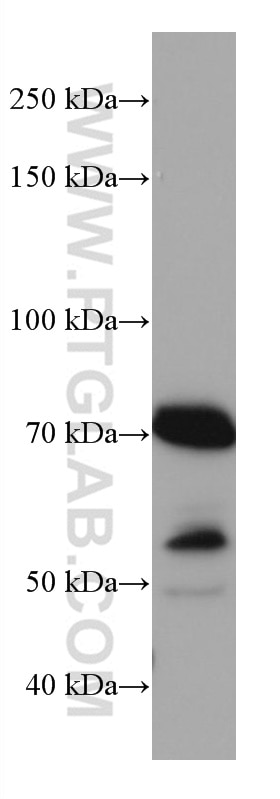
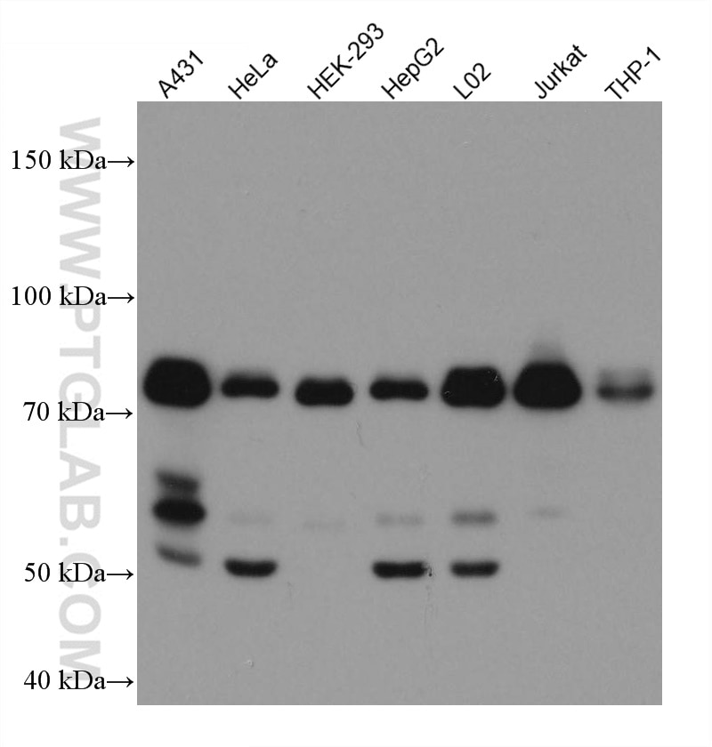
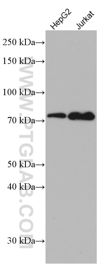
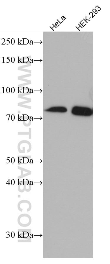
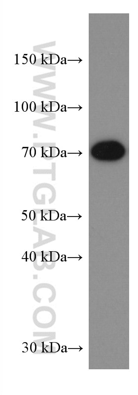
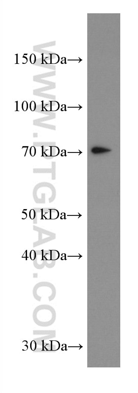
| Erfolgreiche Detektion in WB | A431-Zellen, HEK-293-Zellen, HeLa-Zellen, HepG2-Zellen, Jurkat-Zellen, L02-Zellen |
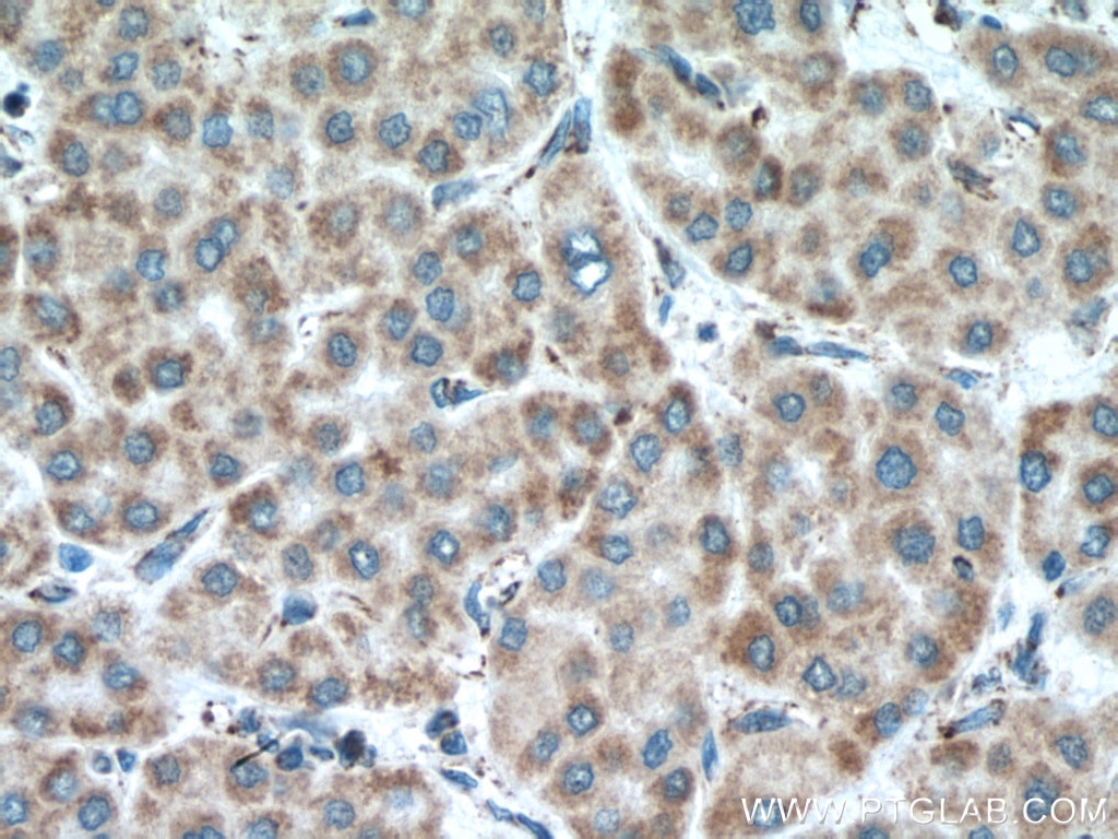
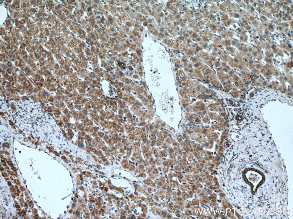
| Erfolgreiche Detektion in IHC | humanes Leberkarzinomgewebe Hinweis: Antigendemaskierung mit TE-Puffer pH 9,0 empfohlen. (*) Wahlweise kann die Antigendemaskierung auch mit Citratpuffer pH 6,0 erfolgen. |
Empfohlene Verdünnung
| Anwendung | Verdünnung |
|---|---|
| Western Blot (WB) | WB : 1:5000-1:50000 |
| Immunhistochemie (IHC) | IHC : 1:550-1:2200 |
| It is recommended that this reagent should be titrated in each testing system to obtain optimal results. | |
| Sample-dependent, check data in validation data gallery | |
Veröffentlichte Anwendungen
| WB | See 13 publications below |
| IF | See 4 publications below |
| IP | See 1 publications below |
| CoIP | See 1 publications below |
Produktinformation
66911-1-Ig bindet in WB, IHC, IF, IP, CoIP, ELISA MAVS; VISA und zeigt Reaktivität mit human
| Getestete Reaktivität | human |
| In Publikationen genannte Reaktivität | human, Affe, Hausschwein, Maus |
| Wirt / Isotyp | Maus / IgG1 |
| Klonalität | Monoklonal |
| Typ | Antikörper |
| Immunogen | MAVS; VISA fusion protein Ag5949 |
| Vollständiger Name | mitochondrial antiviral signaling protein |
| Berechnetes Molekulargewicht | 57 kDa |
| Beobachtetes Molekulargewicht | 50-55 kDa, 70-75 kDa |
| GenBank-Zugangsnummer | BC044952 |
| Gene symbol | MAVS |
| Gene ID (NCBI) | 57506 |
| Konjugation | Unkonjugiert |
| Form | Liquid |
| Reinigungsmethode | Protein-G-Reinigung |
| Lagerungspuffer | PBS with 0.02% sodium azide and 50% glycerol |
| Lagerungsbedingungen | Bei -20°C lagern. Nach dem Versand ein Jahr lang stabil Aliquotieren ist bei -20oC Lagerung nicht notwendig. 20ul Größen enthalten 0,1% BSA. |
Hintergrundinformationen
Mitochondrial antiviral-signaling protein (MAVS) is also known as virus-induced-signaling adapter (VISA) or IFN beta promoter stimulator protein 1 (IPS-1), it is widely involved and required for innate immune defense against viruses. MAVS, present in T cells, monocytes, epithelial cells and hepatocytes, contains CARD and transmembrane domains which are essential for antiviral functions. MAVS is able to interact with various cellular proteins including DDX58/RIG-I, IFIH1/MDA5, TRAF2, TRAF6, TMEM173/MITA, IFIT3 and etc. It can undergoe phosphorylation on multiple sites and ubiquitination, which may together cause the molecular weight migrate to about 70 kDa despite the predicated 57 kDa.
Protokolle
| PRODUKTSPEZIFISCHE PROTOKOLLE | |
|---|---|
| WB protocol for MAVS; VISA antibody 66911-1-Ig | Protokoll herunterladen |
| IHC protocol for MAVS; VISA antibody 66911-1-Ig | Protokoll herunterladenl |
| STANDARD-PROTOKOLLE | |
|---|---|
| Klicken Sie hier, um unsere Standardprotokolle anzuzeigen |
Publikationen
| Species | Application | Title |
|---|---|---|
Mol Cell An Epstein-Barr virus protein interaction map reveals NLRP3 inflammasome evasion via MAVS UFMylation | ||
J Immunother Cancer Targeting KDM4C enhances CD8+ T cell mediated antitumor immunity by activating chemokine CXCL10 transcription in lung cancer. | ||
Front Immunol Swine Acute Diarrhea Syndrome Coronavirus Nucleocapsid Protein Antagonizes Interferon-β Production via Blocking the Interaction Between TRAF3 and TBK1. | ||
J Virol Encephalomyocarditis virus abrogates the IFN-β signaling pathway via its structural protein VP2. | ||
Vet Microbiol DDX56 antagonizes IFN-β production to enhance EMCV replication by inhibiting IRF3 nuclear translocation | ||
Virus Res Host protein, HSP90β, antagonizes IFN-β signaling pathway and facilitates the proliferation of encephalomyocarditis virus in vitro. |
