- Featured Product
- KD/KO Validated
N-cadherin Polyklonaler Antikörper
N-cadherin Polyklonal Antikörper für WB, IHC, IF/ICC, IF-P, IF-Fro, IP, ELISA
Wirt / Isotyp
Kaninchen / IgG
Getestete Reaktivität
human, Maus, Ratte und mehr (4)
Anwendung
WB, IHC, IF/ICC, IF-P, IF-Fro, IP, CoIP, ELISA
Konjugation
Unkonjugiert
Kat-Nr. : 22018-1-AP
Synonyme
 with sh-Control and sh-N-cadherin transfected HEK-293 cells.")
, HeLa (N-cadherin+), MCF-7 (N-cadherin-), A431 (N-cadherin-) cell lysates were subjected to SDS PAGE followed by western blot with 22018-1-AP (N-cadherin antibody) at dilution of 1:5000 incubated at room temperature for 1.5 hours.")
 at dilution of 1:8000 incubated at room temperature for 1.5 hours.")
 at dilution of 1:10000 incubated at room temperature for 1.5 hours.")
 at dilution of 1:5000 incubated at room temperature for 1.5 hours.")
 at dilution of 1:8000 incubated at room temperature for 1.5 hours.")
 at dilution of 1:8000 incubated at room temperature for 1.5 hours.")
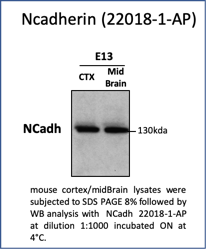
 with mouse brain tissue lysate 7000ug.")
 at dilution of 1:4000 (under 10x lens). Heat mediated antigen retrieval with Tris-EDTA buffer (pH 9.0).")
 at dilution of 1:4000 (under 40x lens). Heat mediated antigen retrieval with Tris-EDTA buffer (pH 9.0).")
 at dilution of 1:4000 (under 40x lens). Heat mediated antigen retrieval with Tris-EDTA buffer (pH 9.0).")
 at dilution of 1:4000 (under 10x lens). Heat mediated antigen retrieval with Tris-EDTA buffer (pH 9.0).")
 fixed mouse heart tissue using N-cadherin antibody (22018-1-AP) at dilution of 1:200 and CoraLite®488-Conjugated AffiniPure Goat Anti-Rabbit IgG(H+L).")
 fixed mouse heart tissue using N-cadherin antibody (22018-1-AP) at dilution of 1:200 and CoraLite®488-Conjugated AffiniPure Goat Anti-Rabbit IgG(H+L).")
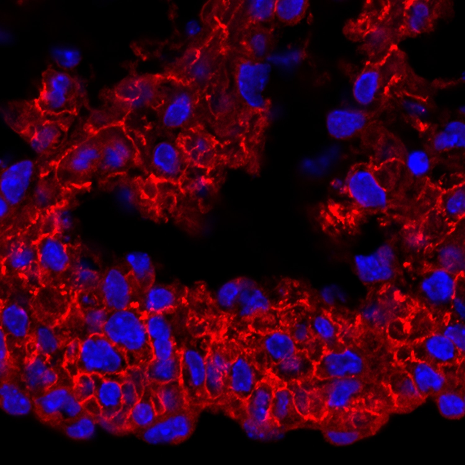
 fixed frozen OCT-embedded mouse heart tissue using 22018-1-AP (N-cadherin antibody) at dilution of 1:400 and Cy3-conjugated Affinipure Goat Anti-Rabbit IgG(H+L).")
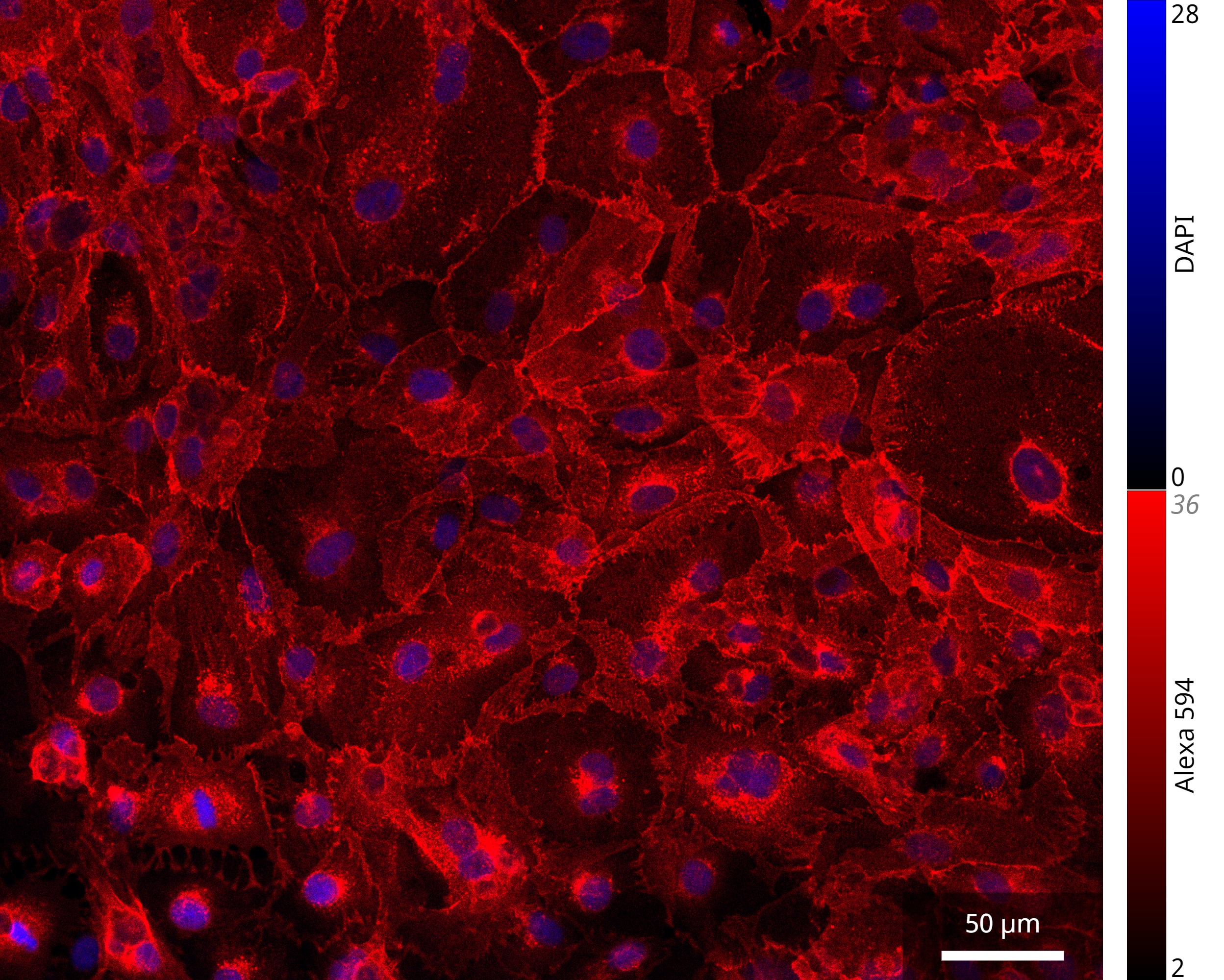
 fixed C2C12 cells using N-cadherin antibody (22018-1-AP) at dilution of 1:400 and CoraLite®488-Conjugated AffiniPure Goat Anti-Rabbit IgG(H+L) (SA00013-2).")
 fixed C2C12 cells using N-cadherin antibody (22018-1-AP) at dilution of 1:200 and CoraLite®488-Conjugated AffiniPure Goat Anti-Rabbit IgG(H+L), 594phalloidin (red).")
Geprüfte Anwendungen
| Erfolgreiche Detektion in WB | Maushirngewebe, A549-Zellen, C2C12-Zellen, C6-Zellen, HEK-293-Zellen, HeLa-Zellen, HepG2-Zellen, NCI-H1299-Zellen, PC-3-Zellen, Rattenhirngewebe, Rattenherzgewebe |
| Erfolgreiche IP | Maushirngewebe |
| Erfolgreiche Detektion in IHC | Mausherzgewebe, Maushirngewebe Hinweis: Antigendemaskierung mit TE-Puffer pH 9,0 empfohlen. (*) Wahlweise kann die Antigendemaskierung auch mit Citratpuffer pH 6,0 erfolgen. |
| Erfolgreiche Detektion in IF-P | Mausherzgewebe, C2C12-Zellen |
| Erfolgreiche Detektion in IF-Fro | Mausherzgewebe |
| Erfolgreiche Detektion in IF/ICC | C2C12-Zellen |
Empfohlene Verdünnung
| Anwendung | Verdünnung |
|---|---|
| Western Blot (WB) | WB : 1:2000-1:16000 |
| Immunpräzipitation (IP) | IP : 0.5-4.0 ug for 1.0-3.0 mg of total protein lysate |
| Immunhistochemie (IHC) | IHC : 1:2000-1:8000 |
| Immunfluoreszenz (IF)-P | IF-P : 1:50-1:500 |
| Immunfluoreszenz (IF)-FRO | IF-FRO : 1:200-1:800 |
| Immunfluoreszenz (IF)/ICC | IF/ICC : 1:200-1:800 |
| It is recommended that this reagent should be titrated in each testing system to obtain optimal results. | |
| Sample-dependent, check data in validation data gallery | |
Veröffentlichte Anwendungen
| KD/KO | See 3 publications below |
| WB | See 1340 publications below |
| IHC | See 180 publications below |
| IF | See 151 publications below |
| CoIP | See 2 publications below |
Produktinformation
22018-1-AP bindet in WB, IHC, IF/ICC, IF-P, IF-Fro, IP, CoIP, ELISA N-cadherin und zeigt Reaktivität mit human, Maus, Ratten
| Getestete Reaktivität | human, Maus, Ratte |
| In Publikationen genannte Reaktivität | human, hamster, Hund, Maus, Ratte, Rind, Gecko |
| Wirt / Isotyp | Kaninchen / IgG |
| Klonalität | Polyklonal |
| Typ | Antikörper |
| Immunogen | N-cadherin fusion protein Ag16792 |
| Vollständiger Name | cadherin 2, type 1, N-cadherin (neuronal) |
| Berechnetes Molekulargewicht | 906 aa, 100 kDa |
| Beobachtetes Molekulargewicht | 130 kDa |
| GenBank-Zugangsnummer | BC036470 |
| Gene symbol | N-cadherin |
| Gene ID (NCBI) | 1000 |
| Konjugation | Unkonjugiert |
| Form | Liquid |
| Reinigungsmethode | Antigen-Affinitätsreinigung |
| Lagerungspuffer | PBS with 0.02% sodium azide and 50% glycerol |
| Lagerungsbedingungen | Bei -20°C lagern. Nach dem Versand ein Jahr lang stabil Aliquotieren ist bei -20oC Lagerung nicht notwendig. 20ul Größen enthalten 0,1% BSA. |
Hintergrundinformationen
Neuronal cadherin (N-cadherin), also known as cadherin-2 (CDH2), is a calcium-binding protein that mediates cell-cell adhesions of neuronal and some non-neuronal cell types.
What is the molecular weight of N-cadherin? Is N-cadherin post-translationally modified?
The molecular weight of mature N-cadherin is 127 kDa. N-cadherin is synthesized in a precursor form that undergoes proteolytic cleavage by furin at the Golgi apparatus. Additionally, it can be phosphorylated by casein kinase II and N-glycosylated, which affects its stability (PMID: 12604612 and 19846557).
What is the subcellular localization of N-cadherin? What is the tissue expression pattern of N-cadherin?
N-cadherin is an integral membrane protein present at the plasma membrane, forming adherens junctions. It is widely expressed in the nervous system, where it flanks the active zone of synapses and is important for synapse formation and remodeling. It is also present in the lens, skeletal, and cardiac muscles (PMID: 3857614). In the muscle, N-cadherin plays a role in myoblast differentiation, while in the heart it is required for the formation of intercalated discs. Additionally, N-cadherin is present in blood vessels, promoting angiogenesis by forming adhesive complexes between endothelial cells and pericytes (PMID: 24521477).
What is the role of N-cadherin during the epithelial-mesenchymal transition (EMT)?
EMT is a crucial process during gastrulation that leads to the formation of mesenchymal cells. It is marked by decreased expression of E-cadherin and upregulation of N-cadherin, which promotes cell migration (PMID: 23481201). Similarly, upregulation of N-cadherin is observed in many cancer cell types and is associated with increased invasiveness and metastasis.
Protokolle
| PRODUKTSPEZIFISCHE PROTOKOLLE | |
|---|---|
| WB protocol for N-cadherin antibody 22018-1-AP | Protokoll herunterladen |
| IHC protocol for N-cadherin antibody 22018-1-AP | Protokoll herunterladenl |
| IF protocol for N-cadherin antibody 22018-1-AP | Protokoll herunterladen |
| IP protocol for N-cadherin antibody 22018-1-AP | Protokoll herunterladen |
| STANDARD-PROTOKOLLE | |
|---|---|
| Klicken Sie hier, um unsere Standardprotokolle anzuzeigen |
Publikationen
| Species | Application | Title |
|---|---|---|
Mol Cancer lncRNA ZNRD1-AS1 promotes malignant lung cell proliferation, migration, and angiogenesis via the miR-942/TNS1 axis and is positively regulated by the m6A reader YTHDC2 | ||
ACS Nano Cancer-Erythrocyte Hybrid Membrane-Camouflaged Magnetic Nanoparticles with Enhanced Photothermal-Immunotherapy for Ovarian Cancer. | ||
ACS Nano Biomimetic Nanomedicine Targeting Orchestrated Metabolism Coupled with Regulatory Factors to Disrupt the Metabolic Plasticity of Breast Cancer | ||
Nat Commun Parvimonas micra promotes oral squamous cell carcinoma metastasis through TmpC-CKAP4 axis | ||
Nat Commun Schwann cells regulate tumor cells and cancer-associated fibroblasts in the pancreatic ductal adenocarcinoma microenvironment | ||
Mol Cancer CircGPR137B/miR-4739/FTO feedback loop suppresses tumorigenesis and metastasis of hepatocellular carcinoma. |
Rezensionen
The reviews below have been submitted by verified Proteintech customers who received an incentive for providing their feedback.
FH Dhanwini (Verified Customer) (09-24-2025) | GOOD
|
FH Zeeshan (Verified Customer) (09-18-2025) | Work very good, bands are sharp
|
FH Michael (Verified Customer) (09-18-2025) | Great with western blot!
|
FH CHI (Verified Customer) (11-25-2024) | The antibody works great for IF
 |
FH Greta (Verified Customer) (02-08-2024) | Good antibody for WB
|
FH Sarah (Verified Customer) (01-04-2024) | Signal at 1:500 was very weak and grainy by immunofluorescence of fixed HCT116 cells.
|
FH Sarah (Verified Customer) (02-09-2023) | Worked well for western blot of mouse brain for 1hr at room temp
|
FH Ralph (Verified Customer) (05-17-2022) | The antibody works well in indirect immunofluorescence, stains the cell membrane.
|
FH Saba (Verified Customer) (02-21-2022) | The band intensity of the antibody is so sharp even in very diluted concentration.
|
FH Lianjie (Verified Customer) (07-26-2019) | Works very well.
 |
FH Aurelie (Verified Customer) (06-13-2019) | Great antibody, no background, works also in human U2OS and primary fibroblasts with the same efficiency.
 |

