NeuN Polyklonaler Antikörper
NeuN Polyklonal Antikörper für WB, IHC, IF-P, IF-Fro, FC (Intra), ELISA
Wirt / Isotyp
Kaninchen / IgG
Getestete Reaktivität
Hausschwein, human, Maus, Ratte und mehr (2)
Anwendung
WB, IHC, IF-P, IF-Fro, FC (Intra), Dot blot, ELISA
Konjugation
Unkonjugiert
Kat-Nr. : 26975-1-AP
Synonyme
 at dilution of 1:80000 incubated at room temperature for 1.5 hours.")
 at dilution of 1:30000 incubated at room temperature for 1.5 hours.")
 at dilution of 1:5000 incubated at room temperature for 1.5 hours.")
 at dilution of 1:30000 (under 40x lens). Heat mediated antigen retrieval with Tris-EDTA buffer (pH 9.0).")
 at dilution of 1:30000 (under 10x lens). Heat mediated antigen retrieval with Tris-EDTA buffer (pH 9.0).")
 at dilution of 1:30000 (under 40x lens). Heat mediated antigen retrieval with Tris-EDTA buffer (pH 9.0).")
 fixed paraffin-embedded rat brain tissue using NeuN antibody (26975-1-AP) at dilution of 1:400 and CoraLite®594-Conjugated Goat Anti-Rabbit IgG(H+L) (SA00013-4), GFAP antibody (60190-1-Ig, Clone: 4B2E10, green). Heat mediated antigen retrieval with Tris-EDTA buffer (pH 9.0).")
 fixed rat cerebellum tissue using 26975-1-AP (NeuN antibody, green), at dilution of 1:100 and CoraLite®488-Conjugated AffiniPure Goat Anti-Rabbit IgG(H+L). The section was co-stained with 60190-1-Ig (GFAP antibody, red).")
 fixed paraffin-embedded mouse cerebellum tissue using NeuN antibody (26975-1-AP) at dilution of 1:200 and CoraLite®488-Conjugated AffiniPure Goat Anti-Rabbit IgG(H+L) (SA00013-2). Heat mediated antigen retrieval with Tris-EDTA buffer (pH 9.0).")
 fixed mouse cerebellum tissue using NeuN antibody (26975-1-AP) at dilution of 1:5000 and CoraLite®488-Conjugated AffiniPure Goat Anti-Rabbit IgG(H+L).")
 fixed rat cerebellum tissue using 26975-1-AP (NeuN antibody, green), at dilution of 1:100 and CoraLite®488-Conjugated AffiniPure Goat Anti-Rabbit IgG(H+L). The section was co-stained with 60190-1-Ig (GFAP antibody, red).")
 fixed rat cerebellum tissue using 26975-1-AP (NeuN antibody, green), at dilution of 1:100 and CoraLite®488-Conjugated AffiniPure Goat Anti-Rabbit IgG(H+L). The section was co-stained with 60190-1-Ig (GFAP antibody, red).")
 fixed rat brain tissue using 26975-1-AP (NeuN antibody), at dilution of 1:100 and CoraLite®594-Conjugated AffiniPure Goat Anti-Rabbit IgG(H+L). The section was co-stained with 66375-1-Ig (TUBB3 antibody, green).")
 fixed frozen OCT-embedded mouse brain tissue using NeuN antibody (26975-1-AP) at dilution of 1:200 and CoraLite®594-Conjugated AffiniPure Goat Anti-Rabbit IgG(H+L) (SA00013-4).")
 and CoraLite®488-Conjugated AffiniPure Goat Anti-Rabbit IgG(H+L) at dilution 1:1000 (red), or 0.2 ug rabbit IgG isotype control (blue). Cells were fixed and permeabilized with True-Nuclear Transcription Factor Buffer Set.")
Geprüfte Anwendungen
| Erfolgreiche Detektion in WB | Maushirngewebe, Maus-Cerebellum-Gewebe, Rattenhirngewebe, Ratten-Cerebellum-Gewebe |
| Erfolgreiche Detektion in IHC | Maus-Cerebellum-Gewebe, Ratten-Cerebellum-Gewebe Hinweis: Antigendemaskierung mit TE-Puffer pH 9,0 empfohlen. (*) Wahlweise kann die Antigendemaskierung auch mit Citratpuffer pH 6,0 erfolgen. |
| Erfolgreiche Detektion in IF-P | Rattenhirngewebe, Maus-Cerebellum-Gewebe, Ratten-Cerebellum-Gewebe |
| Erfolgreiche Detektion in IF-Fro | Maushirngewebe |
| Erfolgreiche Detektion in FC (Intra) | U-87 MG-Zellen |
Empfohlene Verdünnung
| Anwendung | Verdünnung |
|---|---|
| Western Blot (WB) | WB : 1:20000-1:100000 |
| Immunhistochemie (IHC) | IHC : 1:5000-1:30000 |
| Immunfluoreszenz (IF)-P | IF-P : 1:200-1:800 |
| Immunfluoreszenz (IF)-FRO | IF-FRO : 1:50-1:500 |
| Durchflusszytometrie (FC) (INTRA) | FC (INTRA) : 0.20 ug per 10^6 cells in a 100 µl suspension |
| It is recommended that this reagent should be titrated in each testing system to obtain optimal results. | |
| Sample-dependent, check data in validation data gallery | |
Veröffentlichte Anwendungen
| WB | See 48 publications below |
| IHC | See 54 publications below |
| IF | See 286 publications below |
Produktinformation
26975-1-AP bindet in WB, IHC, IF-P, IF-Fro, FC (Intra), Dot blot, ELISA NeuN und zeigt Reaktivität mit Hausschwein, human, Maus, Ratten
| Getestete Reaktivität | Hausschwein, human, Maus, Ratte |
| In Publikationen genannte Reaktivität | human, Affe, Hausschwein, Maus, Ratte, Zebrafisch |
| Wirt / Isotyp | Kaninchen / IgG |
| Klonalität | Polyklonal |
| Typ | Antikörper |
| Immunogen | NeuN fusion protein Ag25689 |
| Vollständiger Name | hexaribonucleotide binding protein 3 |
| Beobachtetes Molekulargewicht | 46-52 kDa |
| GenBank-Zugangsnummer | NM_001082575 |
| Gene symbol | NeuN |
| Gene ID (NCBI) | 146713 |
| Konjugation | Unkonjugiert |
| Form | Liquid |
| Reinigungsmethode | Antigen-Affinitätsreinigung |
| Lagerungspuffer | PBS with 0.02% sodium azide and 50% glycerol |
| Lagerungsbedingungen | Bei -20°C lagern. Nach dem Versand ein Jahr lang stabil Aliquotieren ist bei -20oC Lagerung nicht notwendig. 20ul Größen enthalten 0,1% BSA. |
Hintergrundinformationen
Function
NeuN, also known as FOX3 or RBFOX3, belongs to a family of tissue-specific splicing regulators and is involved in neural circuitry balance, as well as neurogenesis and synaptogenesis (PMID: 26619789).
Tissue specificity
NeuN is exclusively present in post-mitotic neurons and is absent from neural progenitors, oligodendrocytes, astrocytes, and glia (PMID: 1483388).
Involvement in disease
· NeuN cytoplasmic localization is increased in the neurons of patients with HIV-associated neurocognitive disorders (PMID: 24215932).
Isoforms
There are 4 isoforms of NeuN, migrating in the 45-50 kDa range (PMID: 21747913).
Post-translational modifications
Currently not known.
Cellular localization
NeuN predominantly localizes to the nucleus but can also be present in the cytoplasm. Isoforms of NeuN differ in their cytoplasmic/nucleus localization (PMID: 21747913).
Protokolle
| PRODUKTSPEZIFISCHE PROTOKOLLE | |
|---|---|
| WB protocol for NeuN antibody 26975-1-AP | Protokoll herunterladen |
| IHC protocol for NeuN antibody 26975-1-AP | Protokoll herunterladenl |
| IF protocol for NeuN antibody 26975-1-AP | Protokoll herunterladen |
| STANDARD-PROTOKOLLE | |
|---|---|
| Klicken Sie hier, um unsere Standardprotokolle anzuzeigen |
Publikationen
| Species | Application | Title |
|---|---|---|
Immunity Fate mapping of Spp1 expression reveals age-dependent plasticity of disease-associated microglia-like cells after brain injury | ||
Adv Sci (Weinh) GPR37 Activation Alleviates Bone Cancer Pain via the Inhibition of Osteoclastogenesis and Neuronal Hyperexcitability | ||
Brain Behav Immun Egln3 expression in microglia enhances the neuroinflammatory responses in Alzheimer's disease | ||
Neuron Astrocytic ApoE reprograms neuronal cholesterol metabolism and histone-acetylation-mediated memory. | ||
Sci Adv CGG repeat RNA G-quadruplexes interact with FMRpolyG to cause neuronal dysfunction in fragile X-related tremor/ataxia syndrome. | ||
Cell Death Differ Redox regulation of TRIM28 facilitates neuronal ferroptosis by promoting SUMOylation and inhibiting OPTN-selective autophagic degradation of ACSL4 |
Rezensionen
The reviews below have been submitted by verified Proteintech customers who received an incentive for providing their feedback.
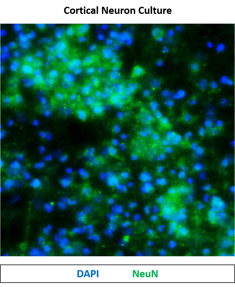
FH Víct0r (Verified Customer) (07-24-2025) | In my case, this antibody doesn't seem very specific. It binds to cells that are not neurons. However, in pure neuronal cultures, it works well, although there is a bit of background. I’ve just seen that titrating this reagent is recommended, but in this case, it wasn’t titrated.
 |
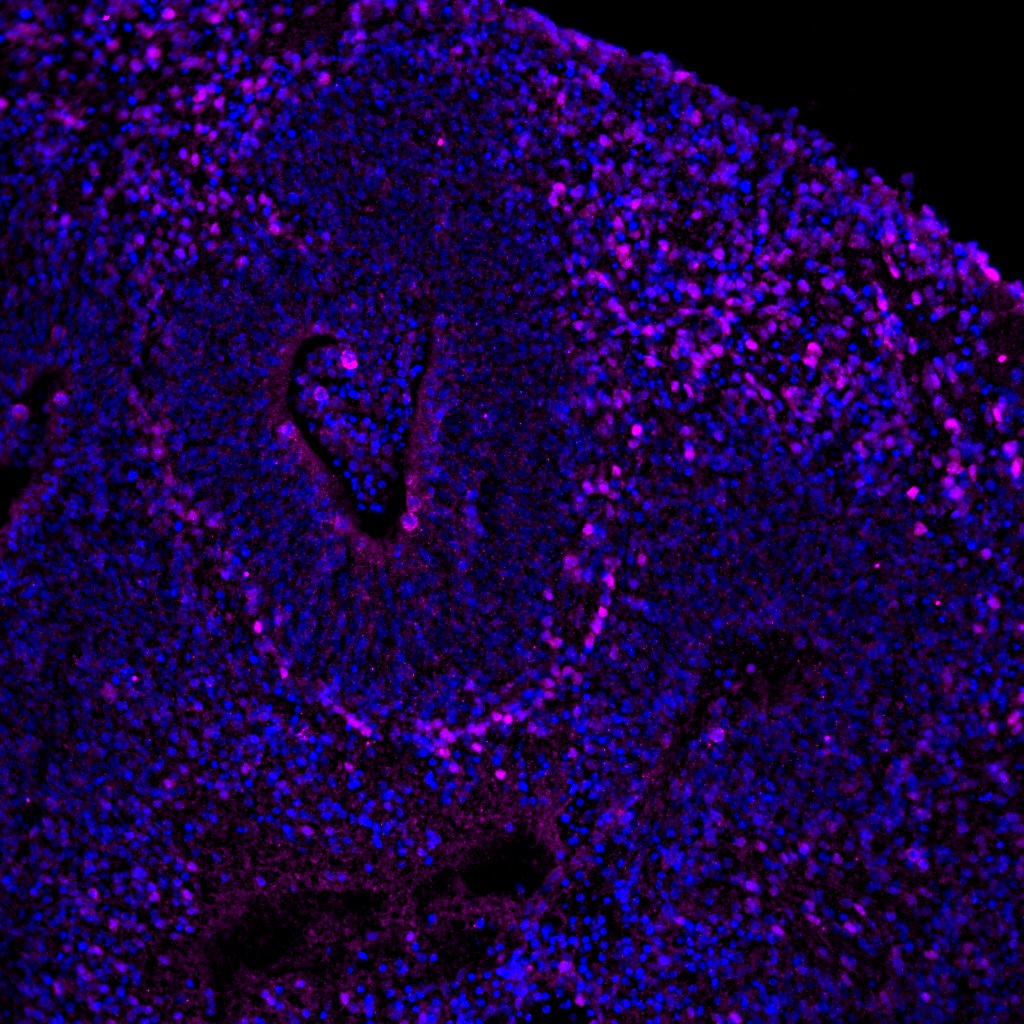
FH Raquel (Verified Customer) (07-12-2024) | Nice and clean staining. Immunofluorescence analysis in cryostat sections of 4% PFA fixed human brain organoid derived from iPSCs using 26975-1-AP NeuN antibody at dilution of 1:250 (under 20x lens). (Magenta: NeuN ; Blue: DAPI)
 |
FH Manohar (Verified Customer) (03-06-2024) |
|
FH Damien (Verified Customer) (02-15-2024) | Works well in 2D staining of induced neurons, starting at 14days. Fixed and permeabilized with 3%BSA and 0.1% Triton
|
FH Monika (Verified Customer) (06-08-2023) | Very specific and strong signal. Used without antigen retrieval. Worked good for both NBT/BCIP-colorimetry and Alexa fluor-fluorescence detection.
|
FH Russell (Verified Customer) (05-23-2023) | Antigen heat retrieval with Citrate buffer pH 6.0 at 100C for 20 min Permeabilized with 1% TX-100 Blocking Buffer PBST with 5% Donkey Serum
|
FH Irina (Verified Customer) (11-18-2021) | Good specific neuronal signal. Works nicely for double IF to look at co-localisation with other proteins.
|
FH David (Verified Customer) (01-13-2020) | Good nuclear localisation, with strong signal and low background.
|
FH Maxence (Verified Customer) (09-05-2019) | Labeling was neat without any background. Signal was quite strong and stable.
|
FH Elena (Verified Customer) (07-31-2019) | Good antibody, stained in all cell lines we used it for.
|
FH Kaspar (Verified Customer) (01-26-2018) |
|

