NeuN Monoklonaler Antikörper
NeuN Monoklonal Antikörper für IHC, IF-P, FC (Intra), ELISA
Wirt / Isotyp
Maus / IgG1
Getestete Reaktivität
human, Maus, Ratte und mehr (1)
Anwendung
IHC, IF-P, FC (Intra), ELISA
Konjugation
Unkonjugiert
CloneNo.
3A4C1
Kat-Nr. : 66836-1-Ig
Synonyme
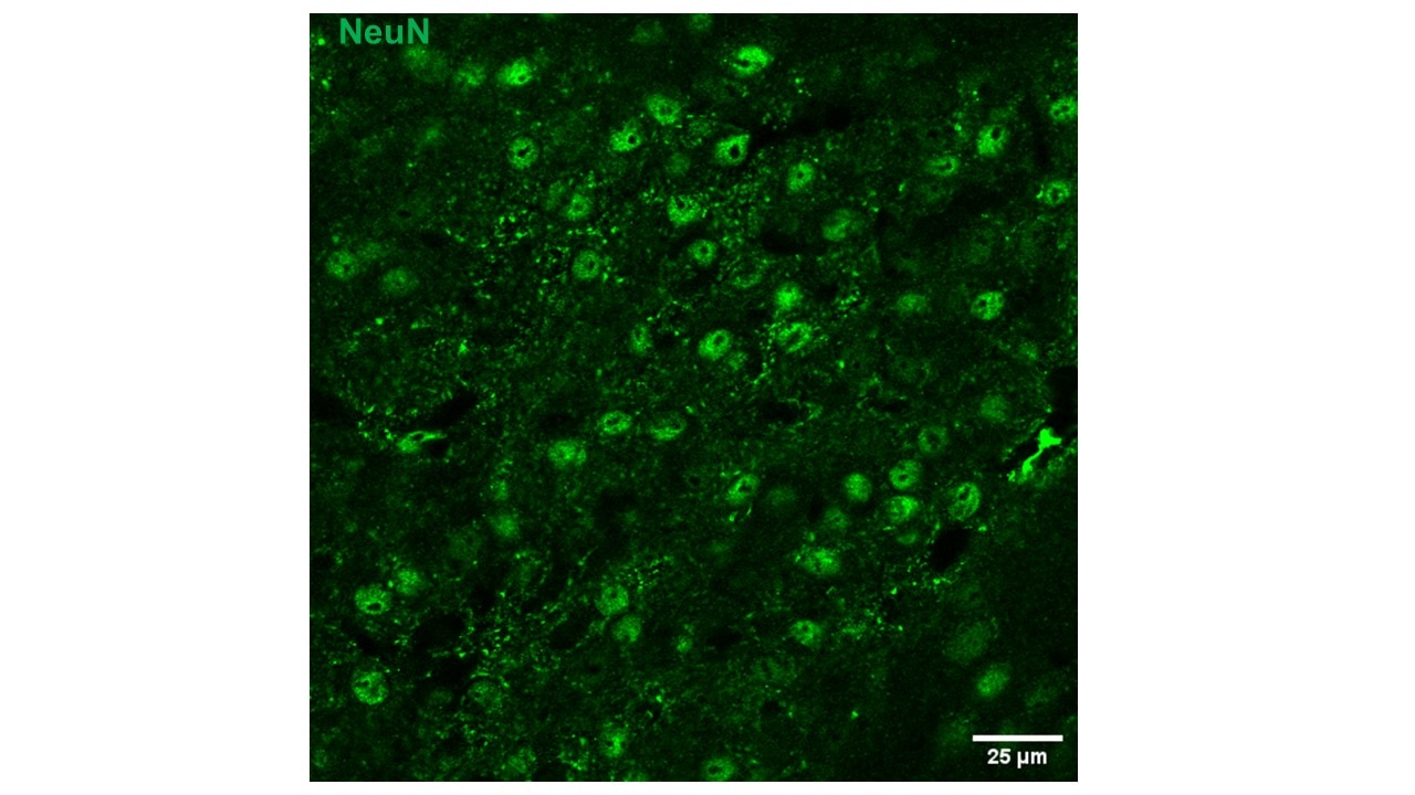
 at dilution of 1:5000 (under 10x lens). Heat mediated antigen retrieval with Sodium Citrate buffer (pH 6.0).")
 at dilution of 1:5000 (under 40x lens). Heat mediated antigen retrieval with Sodium Citrate buffer (pH 6.0).")
 at dilution of 1:20000 (under 10x lens. Heat mediated antigen retrieval with Tris-EDTA buffer (pH 9.0).")
 fixed rat cerebellum tissue using 66836-1-Ig (NeuN antibody, red), at dilution of 1:200 and CoraLite®594-Conjugated AffiniPure Goat Anti-Mouse IgG(H+L). The section was co-stained with 14479-1-AP (Calbindin-D28k antibody, green).")
 fixed mouse cerebellum tissue using 66836-1-Ig (NeuN antibody), at dilution of 1:100 and CoraLite®594-Conjugated AffiniPure Goat Anti-Mouse IgG(H+L). The section was co-stained with 14479-1-AP (Calbindin-D28k Antibody, green).")
 fixed paraffin-embedded rat cerebellum tissue using NeuN antibody (66836-1-Ig, Clone: 3A4C1 ) at dilution of 1:800 and CoraLite®647-conjugated F(ab')2 Fragment Goat Anti-Mouse IgG (H+L) (SA00014-8), CoraLite®594 GFAP antibody (CL594-60190, Clone: 4B2E10, red), NF-H/NF200 antibody (18934-1-AP, green). Heat mediated antigen retrieval with Tris-EDTA buffer (pH 9.0).")
 fixed paraffin-embedded rat brain tissue using NeuN antibody (66836-1-Ig, Clone: 3A4C1 ) at dilution of 1:800 and CoraLite®647-conjugated F(ab')2 Fragment Goat Anti-Mouse IgG (H+L) (SA00014-8), CoraLite® Plus 488 GFAP antibody (CL488-60190, Clone: 4B2E10, green), OLIG2 antibody (13999-1-AP, yellow). Heat mediated antigen retrieval with Tris-EDTA buffer (pH 9.0).")
 and CoraLite®488-Conjugated AffiniPure Goat Anti-Mouse IgG(H+L) at dilution 1:1000 (red), or 0.2 ug Control Antibody. Cells were fixed and permeabilized with Transcription Factor Staining Buffer Kit (PF00011).")
Geprüfte Anwendungen
| Erfolgreiche Detektion in IHC | Ratten-Cerebellum-Gewebe, humanes Hirngewebe Hinweis: Antigendemaskierung mit TE-Puffer pH 9,0 empfohlen. (*) Wahlweise kann die Antigendemaskierung auch mit Citratpuffer pH 6,0 erfolgen. |
| Erfolgreiche Detektion in IF-P | Ratten-Cerebellum-Gewebe, Maus-Cerebellum-Gewebe, Rattenhirngewebe |
| Erfolgreiche Detektion in FC (Intra) | SH-SY5Y-Zellen |
Empfohlene Verdünnung
| Anwendung | Verdünnung |
|---|---|
| Immunhistochemie (IHC) | IHC : 1:2500-1:10000 |
| Immunfluoreszenz (IF)-P | IF-P : 1:50-1:500 |
| Durchflusszytometrie (FC) (INTRA) | FC (INTRA) : 0.20 ug per 10^6 cells in a 100 µl suspension |
| It is recommended that this reagent should be titrated in each testing system to obtain optimal results. | |
| Sample-dependent, check data in validation data gallery | |
Veröffentlichte Anwendungen
| IHC | See 6 publications below |
| IF | See 148 publications below |
Produktinformation
66836-1-Ig bindet in IHC, IF-P, FC (Intra), ELISA NeuN und zeigt Reaktivität mit human, Maus, Ratten
| Getestete Reaktivität | human, Maus, Ratte |
| In Publikationen genannte Reaktivität | human, Maus, Ratte, Ziege |
| Wirt / Isotyp | Maus / IgG1 |
| Klonalität | Monoklonal |
| Typ | Antikörper |
| Immunogen | NeuN fusion protein Ag28016 |
| Vollständiger Name | hexaribonucleotide binding protein 3 |
| GenBank-Zugangsnummer | NM_001082575 |
| Gene symbol | NeuN |
| Gene ID (NCBI) | 146713 |
| Konjugation | Unkonjugiert |
| Form | Liquid |
| Reinigungsmethode | Protein-A-Reinigung |
| Lagerungspuffer | PBS with 0.02% sodium azide and 50% glycerol |
| Lagerungsbedingungen | Bei -20°C lagern. Nach dem Versand ein Jahr lang stabil Aliquotieren ist bei -20oC Lagerung nicht notwendig. 20ul Größen enthalten 0,1% BSA. |
Hintergrundinformationen
NeuN, encoded by FOX3, is a neuron-specific nuclear protein. Anti-NeuN stains exclusively neuronal cells in the central and peripheral nervous systems, especially postmitotic and differentiating neurons, as well as terminally differentiated neurons. Anti-NeuN has been used widely as a reliable tool to detect most postmitotic neuronal cell types. The immunohistochemical staining is primarily localized in the nucleus of the neurons with lighter staining in the cytoplasm.
Protokolle
| PRODUKTSPEZIFISCHE PROTOKOLLE | |
|---|---|
| IHC protocol for NeuN antibody 66836-1-Ig | Protokoll herunterladenl |
| IF protocol for NeuN antibody 66836-1-Ig | Protokoll herunterladen |
| STANDARD-PROTOKOLLE | |
|---|---|
| Klicken Sie hier, um unsere Standardprotokolle anzuzeigen |
Publikationen
| Species | Application | Title |
|---|---|---|
Cell Metab Acetate enables metabolic fitness and cognitive performance during sleep disruption | ||
Microbiome The microbiota-gut-brain axis participates in chronic cerebral hypoperfusion by disrupting the metabolism of short-chain fatty acids. | ||
Redox Biol LOX-mediated ECM mechanical stress induces Piezo1 activation in hypoxic-ischemic brain damage and identification of novel inhibitor of LOX | ||
Cell Death Dis ChemR23 activation attenuates cognitive impairment in chronic cerebral hypoperfusion by inhibiting NLRP3 inflammasome-induced neuronal pyroptosis | ||
Cell Death Dis Astrocyte-derived exosomal nicotinamide phosphoribosyltransferase (Nampt) ameliorates ischemic stroke injury by targeting AMPK/mTOR signaling to induce autophagy | ||
Diabetes Regulatory Role of NF-κB on HDAC2 and Tau Hyperphosphorylation in Diabetic Encephalopathy and the Therapeutic Potential of Luteolin |
Rezensionen
The reviews below have been submitted by verified Proteintech customers who received an incentive for providing their feedback.
FH Deng (Verified Customer) (08-14-2025) | it works, but has some background (see image in mouse PVN region)
 |
FH Carla (Verified Customer) (02-03-2025) | We didn't have any good results
|
FH Kenzo (Verified Customer) (01-05-2024) | This monoclonal NeuN worked well for mouse tissues.
|
FH Silvia (Verified Customer) (08-11-2022) | it works well for Immunofluorescence on mature neurons
|
FH Delphine (Verified Customer) (07-25-2022) | Immunohistochemistry with the NeuN antibody on frozen spinal cord worked but the labeling must be optimized because there is a lot of background noise.
|
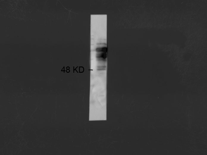
FH q (Verified Customer) (01-05-2022) | It is OK to use it in WB, but several non-specific bands above the expected MW.
 |

