OLIG2 Polyklonaler Antikörper
OLIG2 Polyklonal Antikörper für WB, IHC, IF-P, IF-Fro, IP, ELISA
Wirt / Isotyp
Kaninchen / IgG
Getestete Reaktivität
human, Maus, Ratte und mehr (1)
Anwendung
WB, IHC, IF-P, IF-Fro, IP, ELISA
Konjugation
Unkonjugiert
Kat-Nr. : 13999-1-AP
Synonyme
 at dilution of 1:4000 incubated at room temperature for 1.5 hours.")
 with mouse brain tissue lysate 1600 ug.")
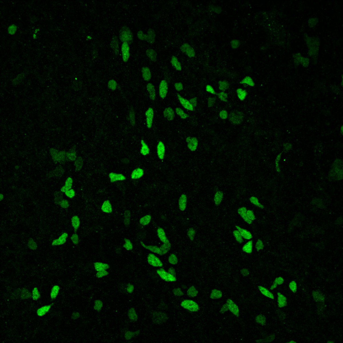
 at dilution of 1:1000 (under 40x lens). Heat mediated antigen retrieval with Tris-EDTA buffer (pH 9.0).")
 at dilution of 1:1000 (under 10x lens). Heat mediated antigen retrieval with Tris-EDTA buffer (pH 9.0).")
 at dilution of 1:200 (under 40x lens). Heat mediated antigen retrieval with Tris-EDTA buffer (pH 9.0).")
 at dilution of 1:200 (under 10x lens). Heat mediated antigen retrieval with Tris-EDTA buffer (pH 9.0).")
 at dilution of 1:800 (under 40x lens. Heat mediated antigen retrieval with Tris-EDTA buffer (pH 9.0).")
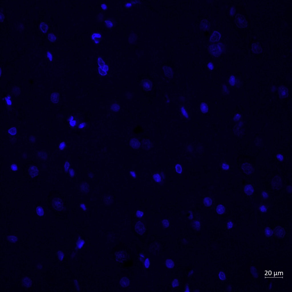
 fixed paraffin-embedded rat brain tissue using OLIG2 antibody (13999-1-AP) at dilution of 1:400 and Rhodamine (TRITC)-conjugated Goat Anti-Rabbit IgG(H+L) (SA00007-2), NeuN antibody (66836-1-Ig, Clone: 3A4C1, Magenta), CoraLite® Plus 488 GFAP antibody (CL488-60190, Clone: 4B2E10, green). Heat mediated antigen retrieval with Tris-EDTA buffer (pH 9.0).")
 fixed paraffin-embedded rat brain tissue using OLIG2 antibody (13999-1-AP) at dilution of 1:400 and Rhodamine (TRITC)-conjugated Goat Anti-Rabbit IgG(H+L) (SA00007-2), NeuN antibody (66836-1-Ig, Clone: 3A4C1, Magenta), CoraLite® Plus 488 GFAP antibody (CL488-60190, Clone: 4B2E10, green). Heat mediated antigen retrieval with Tris-EDTA buffer (pH 9.0).")
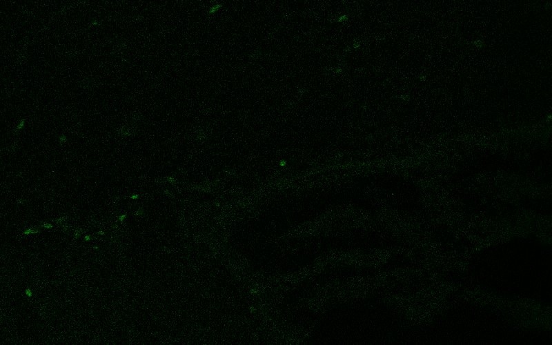
 fixed frozen OCT-embedded mouse brain tissue using OLIG2 antibody (13999-1-AP) at dilution of 1:400 and CoraLite®594-Conjugated Goat Anti-Rabbit IgG(H+L) (SA00013-4).")
Geprüfte Anwendungen
| Erfolgreiche Detektion in WB | Maushirngewebe, rat brain |
| Erfolgreiche IP | Maushirngewebe |
| Erfolgreiche Detektion in IHC | Maushirngewebe, humanes Gliomgewebe, Rattenhirngewebe Hinweis: Antigendemaskierung mit TE-Puffer pH 9,0 empfohlen. (*) Wahlweise kann die Antigendemaskierung auch mit Citratpuffer pH 6,0 erfolgen. |
| Erfolgreiche Detektion in IF-P | Rattenhirngewebe |
| Erfolgreiche Detektion in IF-Fro | Maushirngewebe |
Empfohlene Verdünnung
| Anwendung | Verdünnung |
|---|---|
| Western Blot (WB) | WB : 1:1000-1:8000 |
| Immunpräzipitation (IP) | IP : 0.5-4.0 ug for 1.0-3.0 mg of total protein lysate |
| Immunhistochemie (IHC) | IHC : 1:500-1:2000 |
| Immunfluoreszenz (IF)-P | IF-P : 1:200-1:800 |
| Immunfluoreszenz (IF)-FRO | IF-FRO : 1:200-1:800 |
| It is recommended that this reagent should be titrated in each testing system to obtain optimal results. | |
| Sample-dependent, check data in validation data gallery | |
Veröffentlichte Anwendungen
| WB | See 21 publications below |
| IHC | See 19 publications below |
| IF | See 57 publications below |
Produktinformation
13999-1-AP bindet in WB, IHC, IF-P, IF-Fro, IP, ELISA OLIG2 und zeigt Reaktivität mit human, Maus, Ratten
| Getestete Reaktivität | human, Maus, Ratte |
| In Publikationen genannte Reaktivität | human, Maus, Ratte, Rind |
| Wirt / Isotyp | Kaninchen / IgG |
| Klonalität | Polyklonal |
| Typ | Antikörper |
| Immunogen | OLIG2 fusion protein Ag5089 |
| Vollständiger Name | oligodendrocyte lineage transcription factor 2 |
| Berechnetes Molekulargewicht | 32 kDa |
| Beobachtetes Molekulargewicht | 32-36 kDa |
| GenBank-Zugangsnummer | BC047511 |
| Gene symbol | OLIG2 |
| Gene ID (NCBI) | 10215 |
| Konjugation | Unkonjugiert |
| Form | Liquid |
| Reinigungsmethode | Antigen-Affinitätsreinigung |
| Lagerungspuffer | PBS with 0.02% sodium azide and 50% glycerol |
| Lagerungsbedingungen | Bei -20°C lagern. Nach dem Versand ein Jahr lang stabil Aliquotieren ist bei -20oC Lagerung nicht notwendig. 20ul Größen enthalten 0,1% BSA. |
Hintergrundinformationen
What is the specificity of Olig2?
Oligodendrocyte transcription factor 2 (OLIG2) is expressed by cells found in the central nervous system (CNS) called oligodendrocyte precursor cells (OPCs), which form the myelin sheaths wrapping the axons of neurons in the brain and spinal cord. OPCs differentiate into oligodendrocytes that form the myelin, providing metabolic support and saltatory conduction. OLIG2 is expressed broadly throughout their development to OPCs. This protein can be used to identify many cells of the oligodendrocyte lineage. It is found mostly in the nucleoplasm but also in the cytoplasm.
What is the function of OLIG2?
The basic helix-loop-helix structure of OLIG2 allows it to function as a transcription factor, determining cell fate in the development of neural tissue, where it is located at the pMN domain in the embryonic spinal cord. Expression of OLIG2 causes neural precursors to develop into oligodendrocytes or into motor neurons and expression is then maintained postnatally. OLIG2 co-operates with other factors to cause this differentiation from precursors, although overexpression alone can cause differentiation to the oligodendrocyte lineage.1 The continued expression of OLIG2 in OPCs indicates an ongoing role in the maintenance of their stemness.
What is the involvement of OLIG2 in disease?
The expression of OLIG2 in glioblastoma, the most common type of malignant brain tumor in adults, is well characterized, where the pathological function is an extension of the normal function. Stem-like cells that propagate the tumor growth have been shown to be OLIG2-expressing, and are one of the key transcription factors involved in the re-programming of differentiated cells of the tumor to stem-like cells.2 OLIG2 has also been associated with demyelinating diseases like multiple sclerosis (MS), as OPCs have been shown to be involved in the process of remyelination.3
1. Liu, Z. et al. Induction of oligodendrocyte differentiation by Olig2 and Sox10: Evidence for reciprocal interactions and dosage-dependent mechanisms. Dev. Biol. 302, 683-693 (2007).
2. Wegener, A. et al. Gain of Olig2 function in oligodendrocyte progenitors promotes remyelination. Brain 138, 120-35 (2015).
3. Ettle, B., Schlachetzki, J. C. M. & Winkler, J. Oligodendroglia and Myelin in Neurodegenerative Diseases: More Than Just Bystanders? Mol. Neurobiol. 53, 3046-3062 (2016).
Protokolle
| PRODUKTSPEZIFISCHE PROTOKOLLE | |
|---|---|
| WB protocol for OLIG2 antibody 13999-1-AP | Protokoll herunterladen |
| IHC protocol for OLIG2 antibody 13999-1-AP | Protokoll herunterladenl |
| IF protocol for OLIG2 antibody 13999-1-AP | Protokoll herunterladen |
| IP protocol for OLIG2 antibody 13999-1-AP | Protokoll herunterladen |
| STANDARD-PROTOKOLLE | |
|---|---|
| Klicken Sie hier, um unsere Standardprotokolle anzuzeigen |
Publikationen
| Species | Application | Title |
|---|---|---|
Immunity Disruption of the Na+/K+-ATPase-purinergic P2X7 receptor complex in microglia promotes stress-induced anxiety | ||
Nat Biomed Eng Variants of the adeno-associated virus serotype 9 with enhanced penetration of the blood-brain barrier in rodents and primates | ||
Cell Metab Acetate enables metabolic fitness and cognitive performance during sleep disruption | ||
Cell Stem Cell Non-canonical Targets of HIF1a Impair Oligodendrocyte Progenitor Cell Function. | ||
Mol Neurodegener Contribution of amyloid deposition from oligodendrocytes in a mouse model of Alzheimer's disease |
Rezensionen
The reviews below have been submitted by verified Proteintech customers who received an incentive for providing their feedback.
FH Luqing (Verified Customer) (11-21-2024) | looks great using free-floating brain slices
 |
FH Reyes (Verified Customer) (03-01-2024) | Olig2 (in red) marked the nuclei of tiny cells (oligodendrocytes) around the neurons (in green)
 |
FH Clarisse (Verified Customer) (08-29-2022) | This antibody works very well (clear nuclear signal).
 |
FH An (Verified Customer) (09-17-2020) | Tried a free aliquot of this antibody to perform IHC on retinal cryosections of zebrafish. Did not work, although it was to be expected as the sequence homology was not great.
|
FH Alexander (Verified Customer) (08-05-2019) | This is a great product that has a clear nuclear stain at a 1:200 dilution and 1 hour primary incubation at room temp and 1 hour secondary incubation at room temp.
|

