- Featured Product
- KD/KO Validated
PARP1 Polyklonaler Antikörper
PARP1 Polyklonal Antikörper für WB, IHC, IF/ICC, IF-P, FC (Intra), IP, ELISA
Wirt / Isotyp
Kaninchen / IgG
Getestete Reaktivität
human, Maus, Ratte und mehr (6)
Anwendung
WB, IHC, IF/ICC, IF-P, FC (Intra), IP, CoIP, ChIP, ELISA
Konjugation
Unkonjugiert
Kat-Nr. : 13371-1-AP
Synonyme
 were subjected to SDS PAGE followed by western blot with 13371-1-AP (PARP1 antibody) at dilution of 1:1000 incubated at room temperature for 1.5 hours.")
 at dilution of 1:10000 incubated at room temperature for 1.5 hours.")
 at dilution of 1:1000 incubated at 4 degree celsius over night.")
 at dilution of 1:4000 incubated at room temperature for 1.5 hours.")
 at dilution of 1:1000 incubated at room temperature for 1.5 hours.")
 at dilution of 1:600 incubated at room temperature for 1.5 hours.")
 with K-562 cells lysate 5000ug.")
 at dilution of 1:2000 (under 40x lens). Heat mediated antigen retrieval with Tris-EDTA buffer (pH 9.0).")
 at dilution of 1:2000 (under 40x lens). Heat mediated antigen retrieval with Tris-EDTA buffer (pH 9.0).")
 at dilution of 1:500 (under 40x lens). Heat mediated antigen retrieval with Tris-EDTA buffer (pH 9.0).")
 at dilution of 1:200 (under 40x lens). Heat mediated antigen retrieval with Tris-EDTA buffer (pH 9.0).")
 at dilution of 1:2000 (under 40x lens). Heat mediated antigen retrieval with Tris-EDTA buffer (pH 9.0).")
 at dilution of 1:200 (under 10x lens. Heat mediated antigen retrieval with Tris-EDTA buffer (pH 9.0).")
 at dilution of 1:200 (under 40x lens. Heat mediated antigen retrieval with Tris-EDTA buffer (pH 9.0).")
 fixed mouse testis tissue using PARP1 antibody (13371-1-AP) at dilution of 1:200 and CoraLite®488-Conjugated AffiniPure Goat Anti-Rabbit IgG(H+L).")
 fixed mouse testis tissue using PARP1 antibody (13371-1-AP) at dilution of 1:200 and CoraLite®488-Conjugated AffiniPure Goat Anti-Rabbit IgG(H+L).")
 fixed MCF-7 cells using PARP1 antibody (13371-1-AP) at dilution of 1:200 and CoraLite®488-Conjugated AffiniPure Goat Anti-Rabbit IgG(H+L).")
 fixed HEK-293 cells using PARP1 antibody (13371-1-AP) at dilution of 1:200 and CoraLite®488-Conjugated AffiniPure Goat Anti-Rabbit IgG(H+L).")
 and CoraLite®488-Conjugated AffiniPure Goat Anti-Rabbit IgG(H+L) at dilution 1:1000 (red), or 0.4 ug Control Antibody. Cells were fixed and permeabilized with Transcription Factor Staining Buffer Kit (PF00011).")
Geprüfte Anwendungen
| Erfolgreiche Detektion in WB | HeLa-Zellen, C6-Zellen, mit Cobaltchlorid behandelte HeLa-Zellen, mit Fas-Antikörper behandelte HeLa-Zellen, Jurkat-Zellen, THP-1-Zellen |
| Erfolgreiche IP | K-562-Zellen |
| Erfolgreiche Detektion in IHC | Maus-Kolongewebe, humanes Mammakarzinomgewebe, humanes Lungenkarzinomgewebe, Maushodengewebe Hinweis: Antigendemaskierung mit TE-Puffer pH 9,0 empfohlen. (*) Wahlweise kann die Antigendemaskierung auch mit Citratpuffer pH 6,0 erfolgen. |
| Erfolgreiche Detektion in IF-P | Maushodengewebe |
| Erfolgreiche Detektion in IF/ICC | HEK-293-Zellen, MCF-7-Zellen |
| Erfolgreiche Detektion in FC (Intra) | K-562-Zellen |
Empfohlene Verdünnung
| Anwendung | Verdünnung |
|---|---|
| Western Blot (WB) | WB : 1:1000-1:8000 |
| Immunpräzipitation (IP) | IP : 0.5-4.0 ug for 1.0-3.0 mg of total protein lysate |
| Immunhistochemie (IHC) | IHC : 1:1000-1:4000 |
| Immunfluoreszenz (IF)-P | IF-P : 1:50-1:500 |
| Immunfluoreszenz (IF)/ICC | IF/ICC : 1:50-1:500 |
| Durchflusszytometrie (FC) (INTRA) | FC (INTRA) : 0.40 ug per 10^6 cells in a 100 µl suspension |
| It is recommended that this reagent should be titrated in each testing system to obtain optimal results. | |
| Sample-dependent, check data in validation data gallery | |
Veröffentlichte Anwendungen
| KD/KO | See 5 publications below |
| WB | See 701 publications below |
| IHC | See 29 publications below |
| IF | See 22 publications below |
| IP | See 9 publications below |
| CoIP | See 5 publications below |
| ChIP | See 2 publications below |
Produktinformation
13371-1-AP bindet in WB, IHC, IF/ICC, IF-P, FC (Intra), IP, CoIP, ChIP, ELISA PARP1 und zeigt Reaktivität mit human, Maus, Ratten
| Getestete Reaktivität | human, Maus, Ratte |
| In Publikationen genannte Reaktivität | human, Affe, Hausschwein, Huhn, Hund, Maus, Ratte, Rind, Pilz |
| Wirt / Isotyp | Kaninchen / IgG |
| Klonalität | Polyklonal |
| Typ | Antikörper |
| Immunogen | PARP1 fusion protein Ag4193 |
| Vollständiger Name | poly (ADP-ribose) polymerase 1 |
| Berechnetes Molekulargewicht | 1014 aa, 113 kDa |
| Beobachtetes Molekulargewicht | 113-116 kDa, 89 kDa |
| GenBank-Zugangsnummer | BC037545 |
| Gene symbol | PARP1 |
| Gene ID (NCBI) | 142 |
| Konjugation | Unkonjugiert |
| Form | Liquid |
| Reinigungsmethode | Antigen-Affinitätsreinigung |
| Lagerungspuffer | PBS with 0.02% sodium azide and 50% glycerol |
| Lagerungsbedingungen | Bei -20°C lagern. Nach dem Versand ein Jahr lang stabil Aliquotieren ist bei -20oC Lagerung nicht notwendig. 20ul Größen enthalten 0,1% BSA. |
Hintergrundinformationen
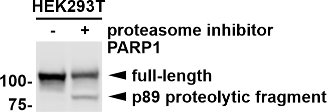
PARP1 (poly(ADP-ribose) polymerase 1) is a nuclear enzyme catalyzing the poly(ADP-ribosyl)ation of many key proteins in vivo. The normal function of PARP1 is the routine repair of DNA damage. Activated by DNA strand breaks, the PARP1 is cleaved into an 85 to 89-kDa COOH-terminal fragment and a 24-kDa NH2-terminal peptide by caspases during the apoptotic process. The appearance of PARP fragments is commonly considered as an important biomarker of apoptosis. In addition to caspases, other proteases like calpains, cathepsins, granzymes and matrix metalloproteinases (MMPs) have also been reported to cleave PARP1 and gave rise to fragments ranging from 42-89-kDa. This antibody was generated against the C-terminal region of human PARP1 and it recognizes the full-length as well as the cleavage of the PARP1.
Protokolle
| PRODUKTSPEZIFISCHE PROTOKOLLE | |
|---|---|
| WB protocol for PARP1 antibody 13371-1-AP | Protokoll herunterladen |
| IHC protocol for PARP1 antibody 13371-1-AP | Protokoll herunterladenl |
| IF protocol for PARP1 antibody 13371-1-AP | Protokoll herunterladen |
| IP protocol for PARP1 antibody 13371-1-AP | Protokoll herunterladen |
| STANDARD-PROTOKOLLE | |
|---|---|
| Klicken Sie hier, um unsere Standardprotokolle anzuzeigen |
Publikationen
| Species | Application | Title |
|---|---|---|
Cell Res Mannose antagonizes GSDME-mediated pyroptosis through AMPK activated by metabolite GlcNAc-6P | ||
Mol Cancer YTHDF2 mediates the mRNA degradation of the tumor suppressors to induce AKT phosphorylation in N6-methyladenosine-dependent way in prostate cancer. | ||
Exp Mol Med Protective effect of hepatocyte-enriched lncRNA-Mir122hg by promoting hepatocyte proliferation in acute liver injury | ||
Nat Commun The ATM and ATR kinases regulate centrosome clustering and tumor recurrence by targeting KIFC1 phosphorylation. | ||
Rezensionen
The reviews below have been submitted by verified Proteintech customers who received an incentive for providing their feedback.
FH Jianneng (Verified Customer) (07-24-2025) | It works as its description! Size is right, signal is good!
|
FH K. (Verified Customer) (10-26-2023) | this antibody worked well, but some times we need to add it at high concentrations
|
FH Priscila (Verified Customer) (04-25-2023) | Very clean detection on western blotting. Sharp bands, and the p89 proteolytic fragment of PARP1 is detectable (tested conditions: 25µg of protein lysate and 1:2,500 dilution for endogenous PARP1 in HEK293T cells).
 |
FH Ruchi (Verified Customer) (12-09-2022) | The Ab was used for IF at a dilution of 1:100 for staining mice skin wounds. This is a nuclear protein and so required an additional time for fixation (30 minutes in 4% paraformaldehyde) and permeabilization by chilled methanol for 10 minutes. The blocking buffer used was 4% BSA.
|
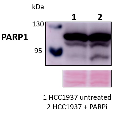
FH Marina (Verified Customer) (05-30-2022) | In the blot we could detect native PARP1 as well as its cleaved 89 kDa form after PARP inhibitor treatment
 |
FH Huanzhou (Verified Customer) (04-08-2022) | This product works well for WB.
|
FH Azita (Verified Customer) (06-02-2021) | Western blot analysis using PARP1 polyclonal antibody in NSC34 cell line at dilution of 1:500.
|
FH Brandon (Verified Customer) (02-01-2019) | Detected Full-length and cleaved PARP-1. Also detected a smaller fragment in mouse mouse liver tissue.
|

