• Featured Product
  • KD/KO Validated

PCNA Polyklonaler Antikörper

PCNA Polyklonal Antikörper für FC, IF, IHC, IP, WB, ELISA

Wirt / Isotyp

Kaninchen / IgG

Getestete Reaktivität

human, Maus, Ratte und mehr (5)

Anwendung

WB, IP, IHC, IF, CoIP, Cell treatment, ELISA

Konjugation

Unkonjugiert

Kat-Nr. : 10205-2-AP

Synonyme

PCNA


Filter:
  • Western Blot (WB) analysis of HEK293 cells using PCNA Polyclonal antibody (10205-2-AP)

    WB analysis of HEK293 cells using 10205-2-AP

    WB result of PCNA antibody (10205-2-AP, 1:10000) with si-control and si-PCNA transfected HEK293 cells.

  • Western Blot (WB) analysis of various lysates using PCNA Polyclonal antibody (10205-2-AP)

    WB analysis using 10205-2-AP

    Various lysates were subjected to SDS PAGE followed by western blot with 10205-2-AP (PCNA antibody) at dilution of 1:10000 incubated at room temperature for 1.5 hours.

  • Western Blot (WB) analysis of various lysates using PCNA Polyclonal antibody (10205-2-AP)

    WB analysis using 10205-2-AP

    Various lysates were subjected to SDS PAGE followed by western blot with 10205-2-AP (PCNA antibody) at dilution of 1:50000 incubated at room temperature for 1.5 hours.

  • Western Blot (WB) analysis of rat testis tissue using PCNA Polyclonal antibody (10205-2-AP)

    WB analysis of rat testis using 10205-2-AP

    rat testis tissue were subjected to SDS PAGE followed by western blot with 10205-2-AP (PCNA Antibody) at dilution of 1:1000 incubated at room temperature for 1.5 hours.

  • Western Blot (WB) analysis of Jurkat cells using PCNA Polyclonal antibody (10205-2-AP)

    WB analysis of Jurkat using 10205-2-AP

    Jurkat cells were subjected to SDS PAGE followed by western blot with 10205-2-AP (PCNA antibody) at dilution of 1:2000 incubated at room temperature for 1.5 hours.

  • Western Blot (WB) analysis of mouse liver tissue using PCNA Polyclonal antibody (10205-2-AP)

    WB analysis of mouse liver using 10205-2-AP

    mouse liver tissue were subjected to SDS PAGE followed by western blot with 10205-2-AP (PCNA antibody) at dilution of 1:1000 incubated at room temperature for 1.5 hours.

  • Western Blot (WB) analysis of A431 cells using PCNA Polyclonal antibody (10205-2-AP)

    WB analysis of A431 using 10205-2-AP

    A431 cells were subjected to SDS PAGE followed by western blot with 10205-2-AP (PCNA antibody) at dilution of 1:1000 incubated at room temperature for 1.5 hours.

  • Western Blot (WB) analysis of NIH/3T3 cells using PCNA Polyclonal antibody (10205-2-AP)

    WB analysis of NIH/3T3 using 10205-2-AP

    NIH/3T3 cells were subjected to SDS PAGE followed by western blot with 10205-2-AP (PCNA antibody) at dilution of 1:1000 incubated at room temperature for 1.5 hours.

  • Western Blot (WB) analysis of HepG2 cells using PCNA Polyclonal antibody (10205-2-AP)

    WB analysis of HepG2 using 10205-2-AP

    HepG2 cells were subjected to SDS PAGE followed by western blot with 10205-2-AP (PCNA antibody) at dilution of 1:1000 incubated at room temperature for 1.5 hours.

  • Western Blot (WB) analysis of mouse testis tissue using PCNA Polyclonal antibody (10205-2-AP)

    WB analysis of mouse testis using 10205-2-AP

    mouse testis tissue were subjected to SDS PAGE followed by western blot with 10205-2-AP (PCNA antibody) at dilution of 1:1000 incubated at room temperature for 1.5 hours.

  • Western Blot (WB) analysis of Raji cells using PCNA Polyclonal antibody (10205-2-AP)

    WB analysis of Raji using 10205-2-AP

    Raji cells were subjected to SDS PAGE followed by western blot with 10205-2-AP (PCNA antibody) at dilution of 1:2000 incubated at room temperature for 1.5 hours.

  • Western Blot (WB) analysis of rat liver tissue using PCNA Polyclonal antibody (10205-2-AP)

    WB analysis of rat liver using 10205-2-AP

    rat liver tissue were subjected to SDS PAGE followed by western blot with 10205-2-AP (PCNA Antibody) at dilution of 1:1000 incubated at room temperature for 1.5 hours.

  • Immunoprecipitation (IP) experiment of MCF-7 cells using PCNA Polyclonal antibody (10205-2-AP)

    IP experiment of MCF-7 using 10205-2-AP

    IP result of anti-PCNA(IP:10205-2-AP, 4ug; Detection:10205-2-AP 1:20000) with MCF-7 cells lysate 1880 ug.

  • Immunoprecipitation (IP) experiment using PCNA Polyclonal antibody (10205-2-AP)

    IP experiment using 10205-2-AP

    IP result of anti-PCNA (10205-2-AP for IP and Detection) with MCF7 cell.

  • Immunohistochemistry (IHC) staining of human stomach cancer tissue using PCNA Polyclonal antibody (10205-2-AP)

    IHC staining of human stomach cancer using 10205-2-AP

    Immunohistochemical analysis of paraffin-embedded human stomach cancer tissue slide using 10205-2-AP (PCNA antibody) at dilution of 1:3000 (under 10x lens). Heat mediated antigen retrieval with Tris-EDTA buffer (pH 9.0).

  • Immunohistochemistry (IHC) staining of human stomach cancer tissue using PCNA Polyclonal antibody (10205-2-AP)

    IHC staining of human stomach cancer using 10205-2-AP

    Immunohistochemical analysis of paraffin-embedded human stomach cancer tissue slide using 10205-2-AP (PCNA antibody) at dilution of 1:3000 (under 40x lens). Heat mediated antigen retrieval with Tris-EDTA buffer (pH 9.0).

  • Immunohistochemistry (IHC) staining of human liver cancer tissue using PCNA Polyclonal antibody (10205-2-AP)

    IHC staining of human liver cancer using 10205-2-AP

    Immunohistochemical analysis of paraffin-embedded human liver cancer tissue slide using 10205-2-AP (PCNA antibody) at dilution of 1:2000 (under 40x lens). Heat mediated antigen retrieval with Tris-EDTA buffer (pH 9.0).

  • Immunohistochemistry (IHC) staining of mouse heart tissue using PCNA Polyclonal antibody (10205-2-AP)

    IHC staining of mouse heart using 10205-2-AP

    Immunohistochemical analysis of paraffin-embedded mouse heart tissue slide using 10205-2-AP (PCNA antibody) at dilution of 1:200 (under 10x lens). Heat mediated antigen retrieval with Tris-EDTA buffer (pH 9.0).

  • Immunohistochemistry (IHC) staining of human colon cancer tissue using PCNA Polyclonal antibody (10205-2-AP)

    IHC staining of human colon cancer using 10205-2-AP

    Immunohistochemical analysis of paraffin-embedded human colon cancer tissue slide using 10205-2-AP (PCNA antibody) at dilution of 1:200 (under 10x lens). Heat mediated antigen retrieval with Tris-EDTA buffer (pH 9.0).

  • Immunohistochemistry (IHC) staining of human malignant melanoma tissue using PCNA Polyclonal antibody (10205-2-AP)

    IHC staining of human malignant melanoma using 10205-2-AP

    Immunohistochemical analysis of paraffin-embedded human malignant melanoma tissue slide using 10205-2-AP (PCNA antibody) at dilution of 1:200 (under 10x lens)..

  • Immunohistochemistry (IHC) staining of human malignant melanoma tissue using PCNA Polyclonal antibody (10205-2-AP)

    IHC staining of human malignant melanoma using 10205-2-AP

    Immunohistochemical analysis of paraffin-embedded human malignant melanoma tissue slide using 10205-2-AP (PCNA antibody) at dilution of 1:200 (under 40x lens)..

  • Immunohistochemistry (IHC) staining of human liver cancer tissue using PCNA Polyclonal antibody (10205-2-AP)

    IHC staining of human liver cancer using 10205-2-AP

    Immunohistochemical analysis of paraffin-embedded human liver cancer tissue slide using 10205-2-AP (PCNA antibody) at dilution of 1:200 (under 40x lens). Heat mediated antigen retrieval with Tris-EDTA buffer (pH 9.0).

  • Immunohistochemistry (IHC) staining of human liver cancer tissue using PCNA Polyclonal antibody (10205-2-AP)

    IHC staining of human liver cancer using 10205-2-AP

    Immunohistochemical analysis of paraffin-embedded human liver cancer tissue slide using 10205-2-AP (PCNA antibody) at dilution of 1:200 (under 10x lens). Heat mediated antigen retrieval with Tris-EDTA buffer (pH 9.0).

  • Immunohistochemistry (IHC) staining of human liver cancer tissue using PCNA Polyclonal antibody (10205-2-AP)

    IHC staining of human liver cancer using 10205-2-AP

    Immunohistochemical analysis of paraffin-embedded human liver cancer tissue slide using 10205-2-AP (PCNA antibody) at dilution of 1:800 (under 10x lens). Heat mediated antigen retrieval with Tris-EDTA buffer (pH 9.0).

  • Immunohistochemistry (IHC) staining of human liver cancer tissue using PCNA Polyclonal antibody (10205-2-AP)

    IHC staining of human liver cancer using 10205-2-AP

    Immunohistochemical analysis of paraffin-embedded human liver cancer tissue slide using 10205-2-AP (PCNA antibody) at dilution of 1:800 (under 40x lens). Heat mediated antigen retrieval with Tris-EDTA buffer (pH 9.0).

  • Immunohistochemistry (IHC) staining of human breast cancer tissue using PCNA Polyclonal antibody (10205-2-AP)

    IHC staining of human breast cancer using 10205-2-AP

    Immunohistochemical analysis of paraffin-embedded human breast cancer tissue slide using 10205-2-AP (PCNA antibody) at dilution of 1:800 (under 40x lens). Heat mediated antigen retrieval with Tris-EDTA buffer (pH 9.0).

  • Immunohistochemistry (IHC) staining of human breast cancer tissue using PCNA Polyclonal antibody (10205-2-AP)

    IHC staining of human breast cancer using 10205-2-AP

    Immunohistochemical analysis of paraffin-embedded human breast cancer tissue slide using 10205-2-AP (PCNA antibody) at dilution of 1:800 (under 10x lens). Heat mediated antigen retrieval with Tris-EDTA buffer (pH 9.0).

  • Immunofluorescence (IF) / fluorescent staining of HepG2 cells using PCNA Polyclonal antibody (10205-2-AP)

    IF Staining of HepG2 using 10205-2-AP

    Immunofluorescent analysis of (4% PFA) fixed HepG2 cells using PCNA antibody (10205-2-AP) at dilution of 1:600 and CoraLite®488-Conjugated AffiniPure Goat Anti-Rabbit IgG(H+L), CL94-phalloidin (red), DAPI (Blue).

  • Immunofluorescence (IF) / fluorescent staining of HepG2 cells using PCNA Polyclonal antibody (10205-2-AP)

    IF Staining of HepG2 using 10205-2-AP

    Immunofluorescent analysis of (4% PFA) fixed HepG2 cells using 10205-2-AP (PCNA antibody), at dilution of 1:200 and CoraLite®488-Conjugated AffiniPure Goat Anti-Rabbit IgG(H+L).

  • Immunofluorescence (IF) / fluorescent staining of mouse testis tissue using PCNA Polyclonal antibody (10205-2-AP)

    IF Staining of mouse testis using 10205-2-AP

    Immunofluorescent analysis of (4% PFA) 
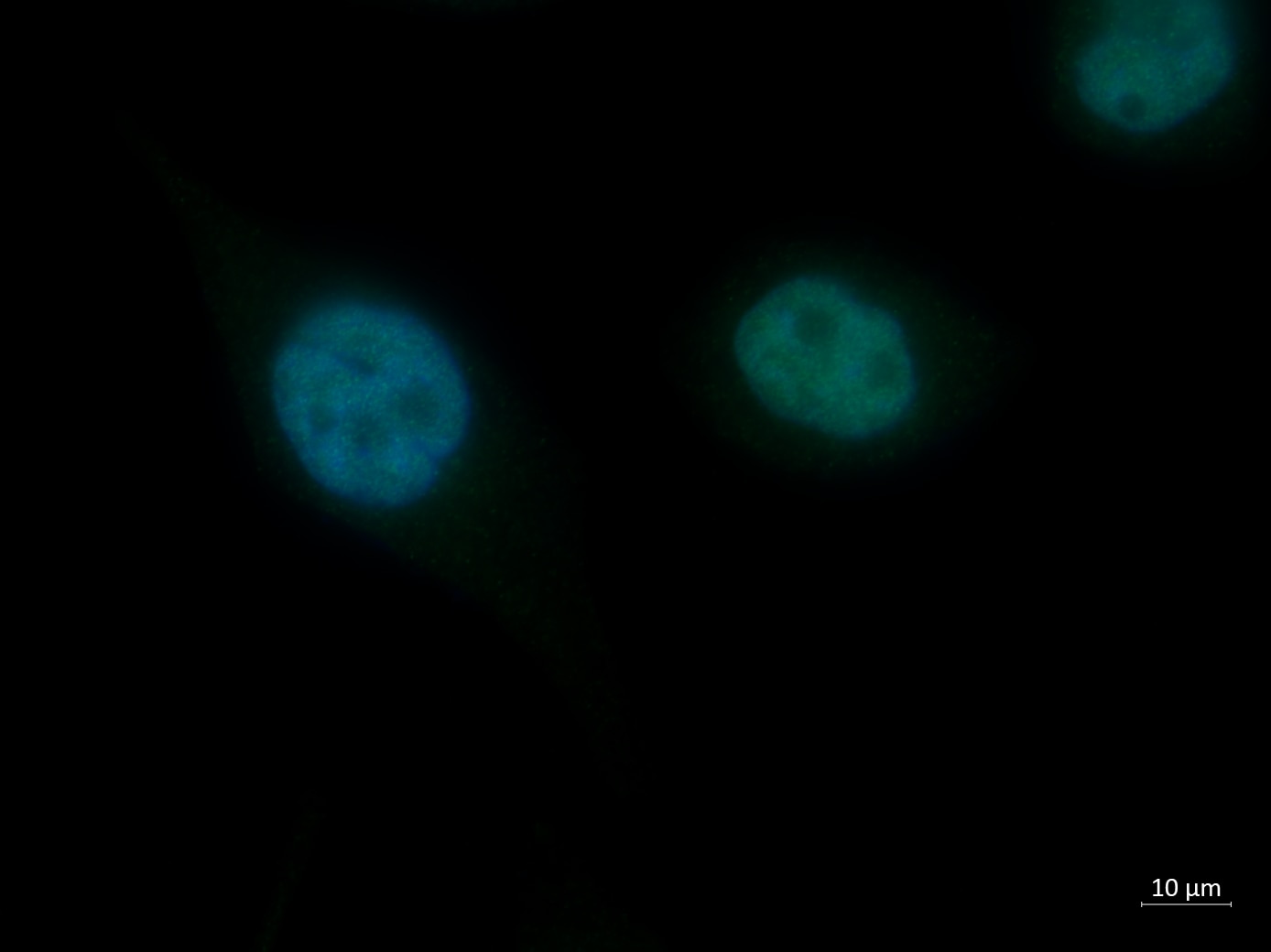
fixed mouse testis tissue using PCNA antibody (10205-2-AP) at dilution of 1:200 and CoraLite®488-Conjugated AffiniPure Goat Anti-Rabbit IgG(H+L).

  • Immunofluorescence (IF) / fluorescent staining of MCF-7 cells using PCNA Polyclonal antibody (10205-2-AP)

    IF Staining of MCF-7 using 10205-2-AP

    Immunofluorescent analysis of (4% PFA) fixed MCF-7 cells using PCNA antibody (10205-2-AP) at dilution of 1:400 and CoraLite®488-Conjugated AffiniPure Goat Anti-Rabbit IgG(H+L).

  • Immunofluorescence (IF) / fluorescent staining of Neuro-2a cells using PCNA Polyclonal antibody (10205-2-AP)

    IF Staining of Neuro-2a using 10205-2-AP

    Immunofluorescent analysis of (-20°C Ethanol) fixed Neuro-2a cells using 10205-2-AP (PCNA antibody), at dilution of 1:200 and CoraLite®488-Conjugated AffiniPure Goat Anti-Rabbit IgG(H+L).

  • Immunofluorescence (IF) / fluorescent staining of human breast cancer tissue using PCNA Polyclonal antibody (10205-2-AP)

    IF Staining of human breast cancer using 10205-2-AP

    Immunofluorescent analysis of (4% PFA) fixed human breast cancer tissue using 10205-2-AP (PCNA antibody), at dilution of 1:100 and CoraLite®488-Conjugated AffiniPure Goat Anti-Rabbit IgG(H+L).

  • Flow cytometry (FC) experiment of Jurkat cells using PCNA Polyclonal antibody (10205-2-PBS)

    FC experiment of Jurkat using 10205-2-PBS

    1X10^6 Jurkat cells were intracellularly stained with 0.2 ug Anti-Human PCNA (10205-2-AP) and CoraLite®488-Conjugated AffiniPure Goat Anti-Rabbit IgG(H+L) at dilution 1:1000 (red), and 0.2 ug Control Antibody. Cells were fixed and permeabilized with Transcription Factor Staining Buffer Kit (PF00011).

"PCNA Antibodies" Comparison

View side-by-side comparison of PCNA antibodies from other vendors to find the one that best suits your research needs.

Geprüfte Anwendungen

Erfolgreiche Detektion in WBA431-Zellen, C2C12-Zellen, HeLa-Zellen, HepG2-Zellen, Jurkat-Zellen, Maushodengewebe, Mauslebergewebe, Mausmilzgewebe, MCF-7-Zellen, NIH/3T3-Zellen, PC-12-Zellen, Raji-Zellen, Rattenhodengewebe, Rattenlebergewebe
Erfolgreiche IPMCF-7-Zellen, N/A
Erfolgreiche Detektion in IHChumanes Magenkrebsgewebe, humanes Kolonkarzinomgewebe, humanes Leberkarzinomgewebe, humanes malignes Melanomgewebe, humanes Mammakarzinomgewebe
Hinweis: Antigendemaskierung mit TE-Puffer pH 9,0 empfohlen. (*) Wahlweise kann die Antigendemaskierung auch mit Citratpuffer pH 6,0 erfolgen.
Erfolgreiche Detektion in IFHepG2-Zellen, humanes Mammakarzinomgewebe, Maushodengewebe, MCF-7-Zellen, Neuro-2a-Zellen

Empfohlene Verdünnung

AnwendungVerdünnung
Western Blot (WB)WB : 1:5000-1:50000
Immunpräzipitation (IP)IP : 0.5-4.0 ug for 1.0-3.0 mg of total protein lysate
Immunhistochemie (IHC)IHC : 1:1500-1:6000
Immunfluoreszenz (IF)IF : 1:300-1:1200
It is recommended that this reagent should be titrated in each testing system to obtain optimal results.
Sample-dependent, check data in validation data gallery

Produktinformation

10205-2-AP bindet in WB, IP, IHC, IF, CoIP, Cell treatment, ELISA PCNA und zeigt Reaktivität mit human, Maus, Ratten

Getestete Reaktivität human, Maus, Ratte
In Publikationen genannte Reaktivitäthuman, Fisch, Huhn, Kaninchen, Maus, Ratte, Ziege, Medaka-Embryos
Wirt / Isotyp Kaninchen / IgG
Klonalität Polyklonal
Typ Antikörper
Immunogen PCNA fusion protein Ag0277
Vollständiger Name proliferating cell nuclear antigen
Berechnetes Molekulargewicht 29 kDa/31 kDa
Beobachtetes Molekulargewicht 36-38 kDa
GenBank-ZugangsnummerBC000491
Gene symbol PCNA
Gene ID (NCBI) 5111
Konjugation Unkonjugiert
Form Liquid
Reinigungsmethode Antigen-Affinitätsreinigung
Lagerungspuffer PBS mit 0.02% Natriumazid und 50% Glycerin pH 7.3.
LagerungsbedingungenBei -20°C lagern. Nach dem Versand ein Jahr lang stabil Aliquotieren ist bei -20oC Lagerung nicht notwendig. 20ul Größen enthalten 0,1% BSA.

Hintergrundinformationen

Proliferating Cell Nuclear Antigen, commonly known as PCNA, is a protein that acts as a processivity factor for DNA polymerase δ in eukaryotic cells. This protein is an auxiliary protein of DNA polymerase delta and is involved in the control of eukaryotic DNA replication by increasing the polymerase's processibility during elongation of the leading strand. PCNA induces a robust stimulatory effect on the 3'-5' exonuclease and 3'-phosphodiesterase, but not apurinic-apyrimidinic (AP) endonuclease, APEX2 activities. It has to be loaded onto DNA in order to be able to stimulate APEX2. PCNA protein is highly conserved during evolution; the deduced amino acid sequences of rat and human differ by only 4 of 261 amino acids. PCNA has been used as loading control for proliferating cells. This antibody is a rabbit polyclonal antibody raised against an internal region of human PCNA. The calculated molecular weight of PCNA is 29 kDa, but modified PCNA is 36kDa (PMID: 1358458).

Protokolle

Produktspezifische Protokolle
WB protocol for PCNA antibody 10205-2-APProtokoll herunterladen
IHC protocol for PCNA antibody 10205-2-APProtokoll herunterladen
IF protocol for PCNA antibody 10205-2-APProtokoll herunterladen
IP protocol for PCNA antibody 10205-2-APProtokoll herunterladen
Standard-Protokolle
Klicken Sie hier, um unsere Standardprotokolle anzuzeigen

Publikationen

SpeciesApplicationTitle
humanWB

Mil Med Res

Aspartoacylase suppresses prostate cancer progression by blocking LYN activation

Authors - Hong Weng
mouseIF

Brain Behav Immun

An enriched environment restores hepatitis B vaccination-mediated impairments in synaptic function through IFN-γ/Arginase1 signaling.

Authors - Fangfang Qi
humanWB

Nat Commun

The RNA-binding protein LRPPRC promotes resistance to CDK4/6 inhibition in lung cancer

Authors - Wei Zhou
rabbitIF

Nat Commun

Four-dimensional hydrogel dressing adaptable to the urethral microenvironment for scarless urethral reconstruction

Authors - Yujie Hua
ratWB

Acta Pharm Sin B

Protocatechuic aldehyde protects cardiomycoytes against ischemic injury via regulation of nuclear pyruvate kinase M2.

Authors - Xunxun Wu
mouseWB

Hepatology

Liver-specific deficiency of unc-51 like kinase 1 and 2 protects mice from acetaminophen-induced liver injury.

Authors - Yu Sun

Rezensionen

The reviews below have been submitted by verified Proteintech customers who received an incentive forproviding their feedback.


FH

Mi (Verified Customer) (06-25-2023)

It works well in human brown adipocyte cells.

  • Applications: Western Blot
  • Primary Antibody Dilution: 1:1000
  • Cell Tissue Type: human brown adipocytes
FH

Emma (Verified Customer) (11-29-2021)

Nice antibody which worked well on PC3 cells fixed with 4% PFA. Antibody used at 1:50

  • Applications: Immunofluorescence
  • Primary Antibody Dilution: 1:50
  • Cell Tissue Type: PC3 cells
PCNA Antibody Immunofluorescence validation (1:50 dilution) in PC3 cells (Cat no:10205-2-AP)