- Featured Product
- KD/KO Validated
PD-L1/CD274 Monoklonaler Antikörper
PD-L1/CD274 Monoklonal Antikörper für WB, IHC, IF/ICC, IF-P, ELISA
Wirt / Isotyp
Maus / IgG1
Getestete Reaktivität
Hausschwein, human, Maus, Ratte
Anwendung
WB, IHC, IF/ICC, IF-P, IP, CoIP, ChIP, ELISA
Konjugation
Unkonjugiert
CloneNo.
2B11D11
Kat-Nr. : 66248-1-Ig
Synonyme
 at dilution of 1:4000 incubated at room temperature for 1.5 hours.")
 at dilution of 1:5000 incubated at room temperature for 1.5 hours.")
 at dilution of 1:1000 incubated at room temperature for 1.5 hours.")
 at dilution of 1:1000 incubated at room temperature for 1.5 hours.")
 at dilution of 1:1000 incubated at room temperature for 1.5 hours.")
 at dilution of 1:1000 incubated at room temperature for 1.5 hours.")
 with si-Control and si-PDL1 transfected HepG2 cells.")
 at dilution of 1:5000 incubated at room temperature for 1.5 hours.")
 at dilution of 1:5000 incubated at room temperature for 1.5 hours. PNGase F was obtained from Atagenix (cat.NO. ata808).")
 at dilution of 1:10000 (under 4x lens. Heat mediated antigen retrieval with Tris-EDTA buffer (pH 9.0).")
 at dilution of 1:10000 (under 10x lens. Heat mediated antigen retrieval with Tris-EDTA buffer (pH 9.0).")
 at dilution of 1:20000 (under 10x lens). Heat mediated antigen retrieval with Tris-EDTA buffer (pH 9.0). Slide was treated with PNGase F before staining.")
 at dilution of 1:20000 (under 40x lens). Heat mediated antigen retrieval with Tris-EDTA buffer (pH 9.0). Slide was treated with PNGase F before staining.")
 at dilution of 1:500 (under 10x lens).")
 at dilution of 1:500 (under 40x lens).")
 at dilution of 1:1000 (under 10x lens). Heat mediated antigen retrieval with Tris-EDTA buffer (pH 9.0).")
 at dilution of 1:1000 (under 40x lens). Heat mediated antigen retrieval with Tris-EDTA buffer (pH 9.0).")
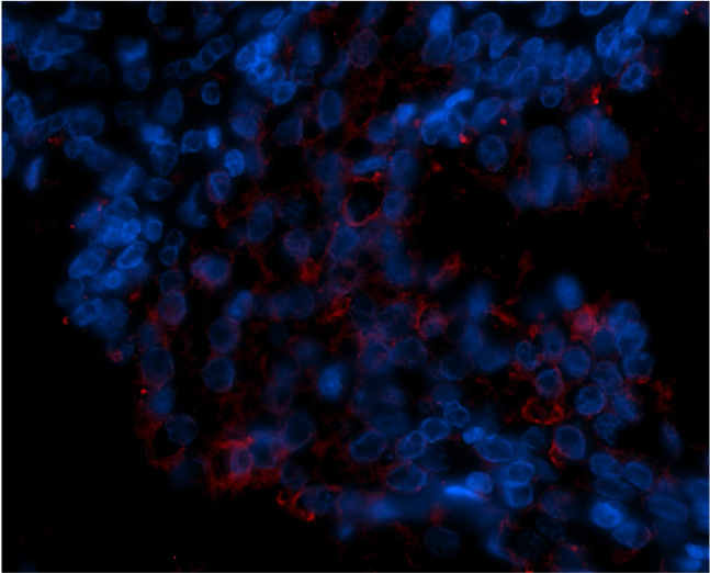
 fixed paraffin-embedded human placenta tissue using PD-L1/CD274 antibody (66248-1-Ig, Clone: 2B11D11 ) at dilution of 1:800 and CoraLite®488-Conjugated Goat Anti-Mouse IgG(H+L) (SA00013-1). Heat mediated antigen retrieval with Tris-EDTA buffer (pH 9.0).")
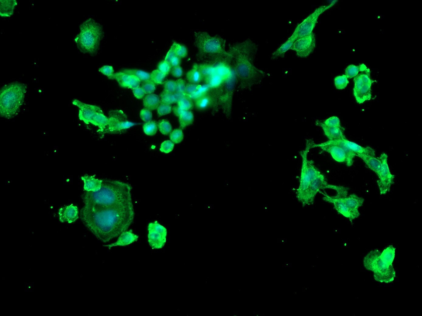
 fixed HeLa cells using 66248-1-Ig(PD-L1/CD274 antibody) at dilution of 1:300 and Alexa Fluor 488-conjugated AffiniPure Goat Anti-Mouse IgG(H+L).")
Geprüfte Anwendungen
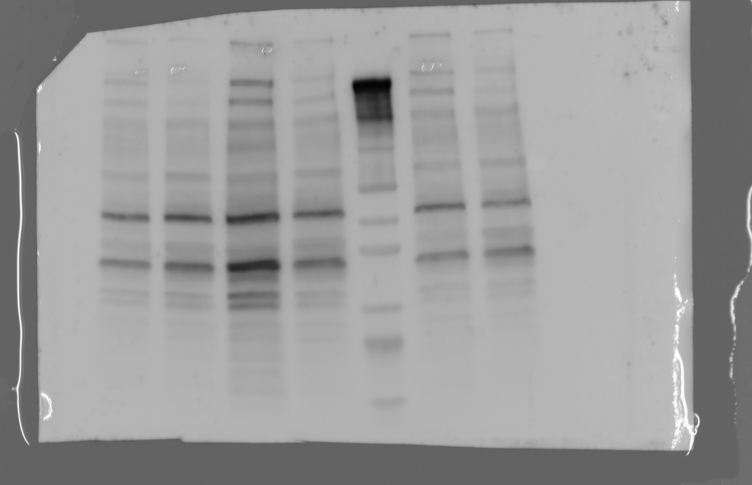
| Erfolgreiche Detektion in WB | A375-Zellen, A549-Zellen, HepG2-Zellen, humanes Plazenta-Gewebe, humanes Skelettmuskelgewebe, K-562-Zellen, Hausschwein-Lungengewebe, RAW 264.7-Zellen, THP-1-Zellen |
| Erfolgreiche Detektion in IHC | humanes Tonsillitisgewebe, humanes Herzgewebe, humanes Lungenkarzinomgewebe, humanes Plazenta-Gewebe, Mausherzgewebe Hinweis: Antigendemaskierung mit TE-Puffer pH 9,0 empfohlen. (*) Wahlweise kann die Antigendemaskierung auch mit Citratpuffer pH 6,0 erfolgen. |
| Erfolgreiche Detektion in IF-P | humanes Plazenta-Gewebe |
| Erfolgreiche Detektion in IF/ICC | HeLa-Zellen |
Empfohlene Verdünnung
| Anwendung | Verdünnung |
|---|---|
| Western Blot (WB) | WB : 1:2000-1:10000 |
| Immunhistochemie (IHC) | IHC : 1:5000-1:20000 |
| Immunfluoreszenz (IF)-P | IF-P : 1:400-1:1600 |
| Immunfluoreszenz (IF)/ICC | IF/ICC : 1:50-1:500 |
| It is recommended that this reagent should be titrated in each testing system to obtain optimal results. | |
| Sample-dependent, check data in validation data gallery | |
Veröffentlichte Anwendungen
| KD/KO | See 7 publications below |
| WB | See 274 publications below |
| IHC | See 168 publications below |
| IF | See 115 publications below |
| IP | See 8 publications below |
| CoIP | See 5 publications below |
| ChIP | See 1 publications below |
Produktinformation
66248-1-Ig bindet in WB, IHC, IF/ICC, IF-P, IP, CoIP, ChIP, ELISA PD-L1/CD274 und zeigt Reaktivität mit Hausschwein, human, Maus, Ratten
| Getestete Reaktivität | Hausschwein, human, Maus, Ratte |
| In Publikationen genannte Reaktivität | human, Hausschwein, Maus, Ratte |
| Wirt / Isotyp | Maus / IgG1 |
| Klonalität | Monoklonal |
| Typ | Antikörper |
| Immunogen | PD-L1/CD274 fusion protein Ag12443 |
| Vollständiger Name | CD274 molecule |
| Berechnetes Molekulargewicht | 290 aa, 33 kDa |
| Beobachtetes Molekulargewicht | 45-50 kDa, 33 kDa |
| GenBank-Zugangsnummer | BC074984 |
| Gene symbol | PD-L1 |
| Gene ID (NCBI) | 29126 |
| Konjugation | Unkonjugiert |
| Form | Liquid |
| Reinigungsmethode | Protein-G-Reinigung |
| Lagerungspuffer | PBS with 0.02% sodium azide and 50% glycerol |
| Lagerungsbedingungen | Bei -20°C lagern. Nach dem Versand ein Jahr lang stabil Aliquotieren ist bei -20oC Lagerung nicht notwendig. 20ul Größen enthalten 0,1% BSA. |
Hintergrundinformationen
PD-L1, also known as CD274 or B7H1, stands for programmed cell death ligand 1. It is a type I transmembrane protein that is thought to repress immune responses by binding to its receptor (PD1), thus inhibiting T-cell activation, proliferation, and cytokine production. It contains V-like and C-like immunoglobulin domains. PD-L1 expression is regulated by various cytokines, such as TNF-α or LPS (ISSN: 1848-7718). Increased expression of this protein in certain types of cancers, e.g., renal cell carcinoma or colon cancer, correlates with poor prognosis.
What is the molecular weight of PD-L1?
Depending on the isoform, the calculated molecular weight of the protein varies between 20 and 33 kDa (176-290 aa).
What are the isoforms of PD-L1?
According to NCBI, three different isoforms have been identified. There are significant differences in the untranslated and protein coding regions.
What is the subcellular localization and tissue specificity of PD-L1?
It is predicted to localize in the plasma membrane of various cell types, with a particularly high expression in placental trophoblast and subsets of immune cells. High levels of PD-L1 protein have also been detected in lung and colon tissues.
What is the function of PD-L1 in immune responses?
PD-L1 is critical for the induction and maintenance of immune self-tolerance during infection or inflammation in normal tissues. The interaction of PD-L1 and its receptors is responsible for preventing auto-immune phenotypes and balancing the overall immune response in situations such as pregnancy or tissue allografts. The interaction between PD-L1 and PD-1 or B7.1 starts an inhibitory signaling cascade, which results in the decreased proliferation of antigen-specific T-cells and increased survival of regulatory T-cells (PMID: 15240681).
How can PD-L1's implication in cancer be used as a drug target?
In certain tumors, high expression of PD-L1 serves as a stop-sign to inhibit the recognition of cancer cells by T-cells (PMID: 23087408). The interaction between PD-L1 and its receptors (PD1 and B7.1) is a mechanism for the tumor to evade the host immune response (PMID: 29357948). Several mAbs have been developed to target that interaction and thus prevent the inactivation of cytotoxic T-cells by the tumor (PMIDs: 23890059, 18173375).
Protokolle
| PRODUKTSPEZIFISCHE PROTOKOLLE | |
|---|---|
| WB protocol for PD-L1/CD274 antibody 66248-1-Ig | Protokoll herunterladen |
| IHC protocol for PD-L1/CD274 antibody 66248-1-Ig | Protokoll herunterladenl |
| IF protocol for PD-L1/CD274 antibody 66248-1-Ig | Protokoll herunterladen |
| STANDARD-PROTOKOLLE | |
|---|---|
| Klicken Sie hier, um unsere Standardprotokolle anzuzeigen |
Publikationen
| Species | Application | Title |
|---|---|---|
Cell Res Targeting ATAD3A-PINK1-mitophagy axis overcomes chemoimmunotherapy resistance by redirecting PD-L1 to mitochondria | ||
Adv Mater A Bifunctional Lysosome-Targeting Chimera Nanoplatform for Tumor-Selective Protein Degradation and Enhanced Cancer Immunotherapy | ||
Cell Metab Dual impacts of serine/glycine-free diet in enhancing antitumor immunity and promoting evasion via PD-L1 lactylation | ||
Cancer Cell ADORA1 Inhibition Promotes Tumor Immune Evasion by Regulating the ATF3-PD-L1 Axis. | ||
Cell Metab NAD+ Metabolism Maintains Inducible PD-L1 Expression to Drive Tumor Immune Evasion. |
Rezensionen
The reviews below have been submitted by verified Proteintech customers who received an incentive for providing their feedback.
FH Margarita (Verified Customer) (11-20-2024) | Secondary antibody: AF 488
 |
FH hala (Verified Customer) (01-27-2023) | there is 2 bands one at the specific size and the other one is higher
 |
FH Emma (Verified Customer) (11-29-2021) | Used for IF on FFPE prostate tissue at 1:50 with Tris-EDTA antigen retrieval (pH 9.0).
 |
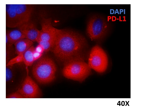
FH Marina (Verified Customer) (06-14-2021) | HCC1937 breast cancer cells were fixed with 4% PFA, permeabilized and stained overnight at 4ºC with anti-PD-L1 Ig diluted 1:300. Nuclei were stained with DAPI.
 |

