PGAM5 Polyklonaler Antikörper
PGAM5 Polyklonal Antikörper für WB, IHC, IF/ICC, ELISA
Wirt / Isotyp
Kaninchen / IgG
Getestete Reaktivität
human und mehr (3)
Anwendung
WB, IHC, IF/ICC, CoIP, ELISA
Konjugation
Unkonjugiert
Kat-Nr. : 28445-1-AP
Synonyme
Geprüfte Anwendungen
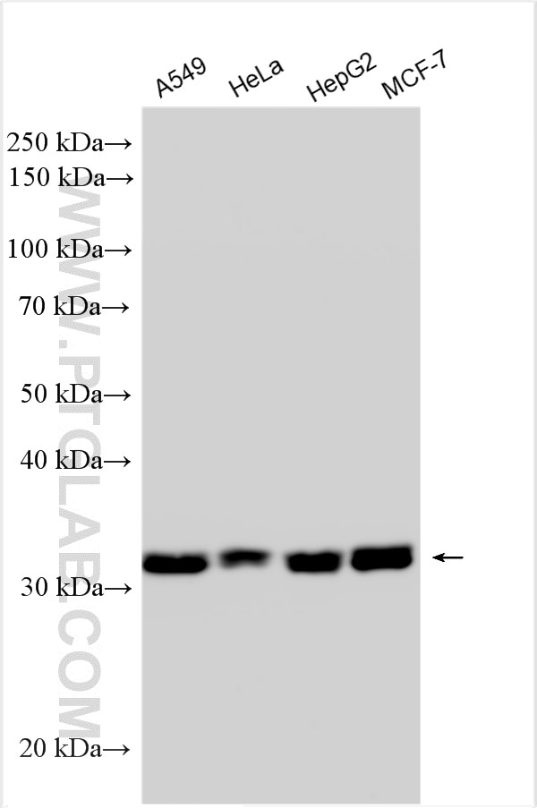
| Erfolgreiche Detektion in WB | A549-Zellen, HeLa-Zellen, HepG2-Zellen, MCF-7-Zellen |
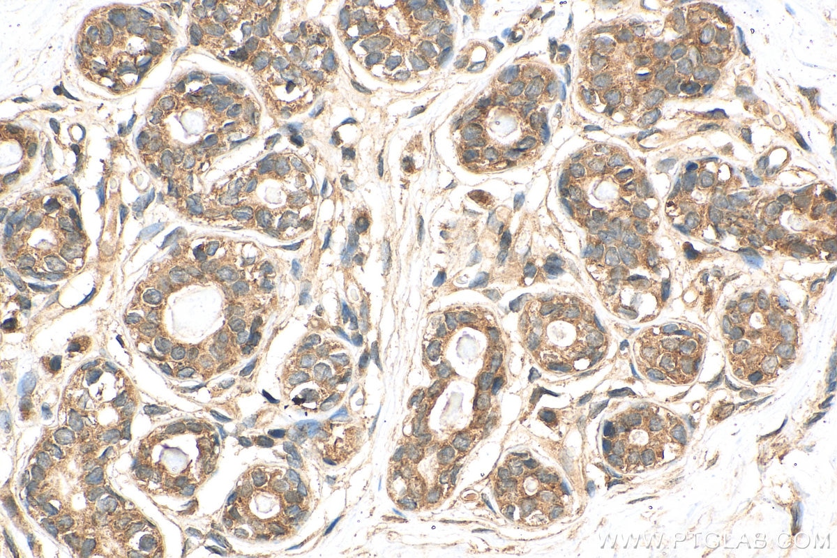
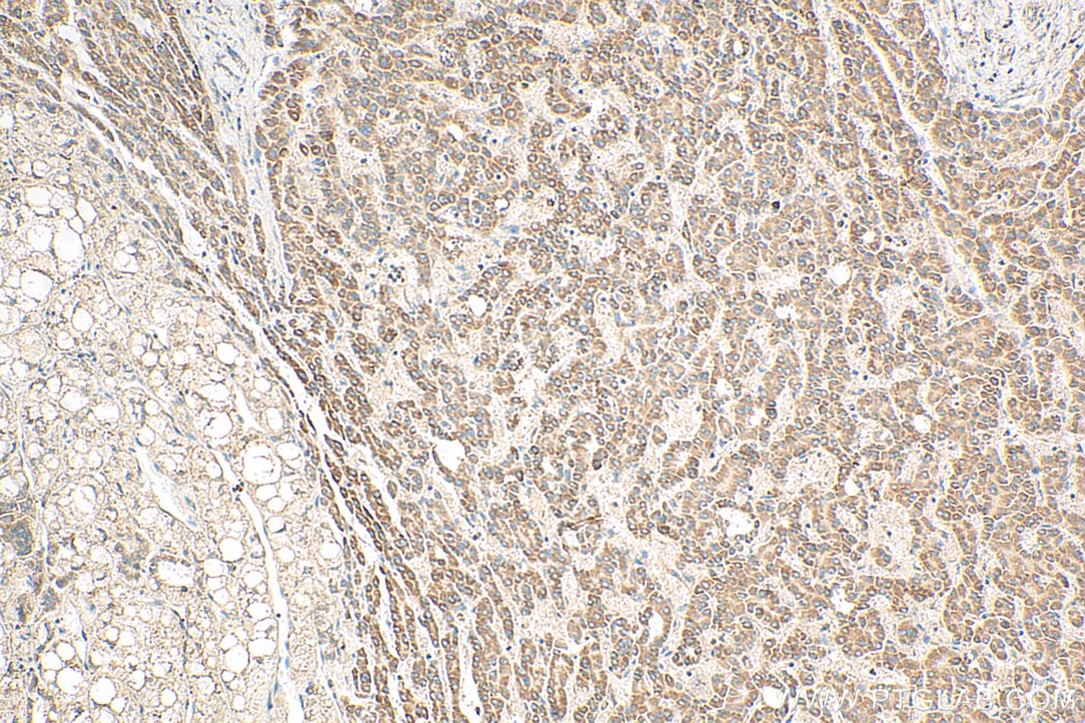
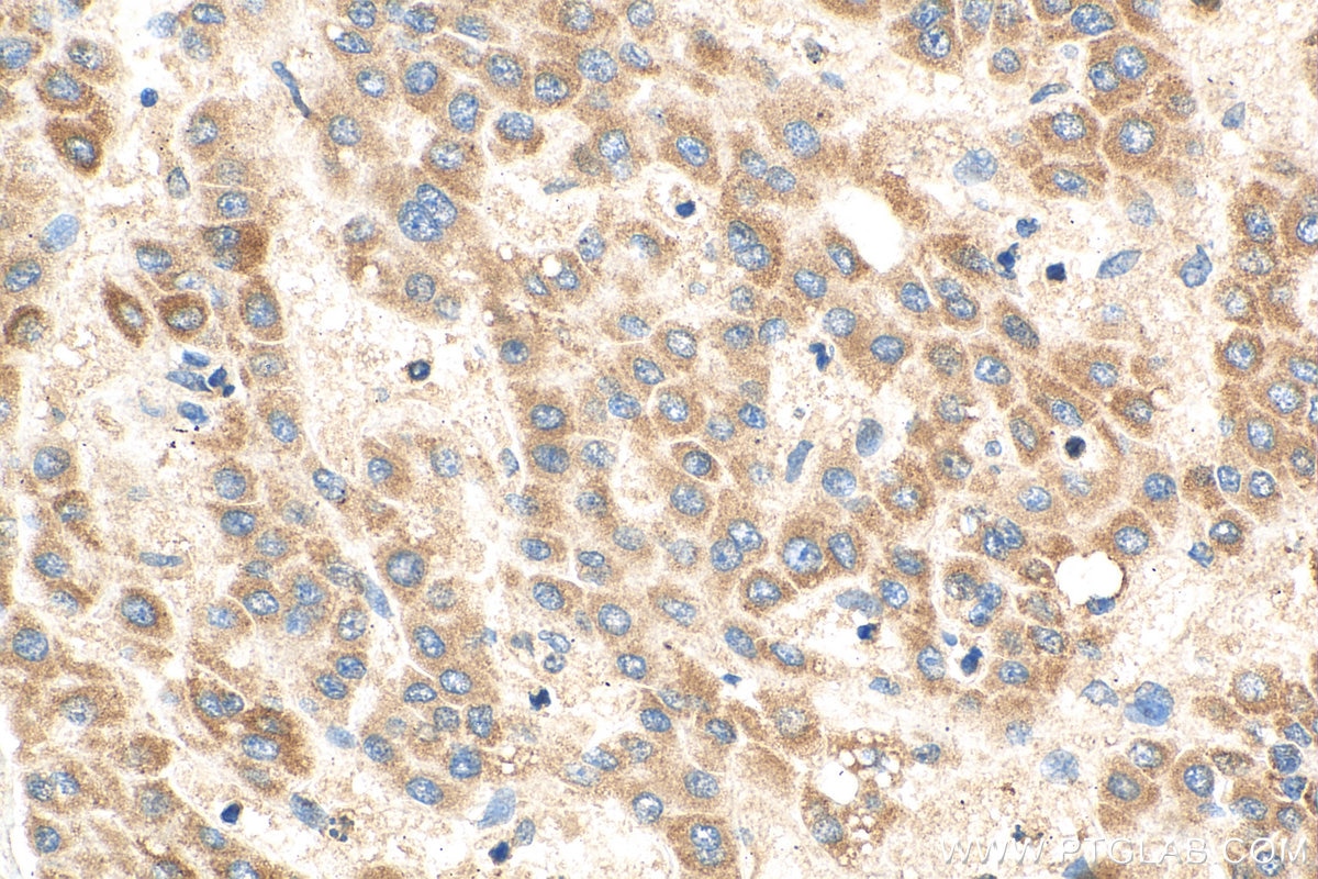
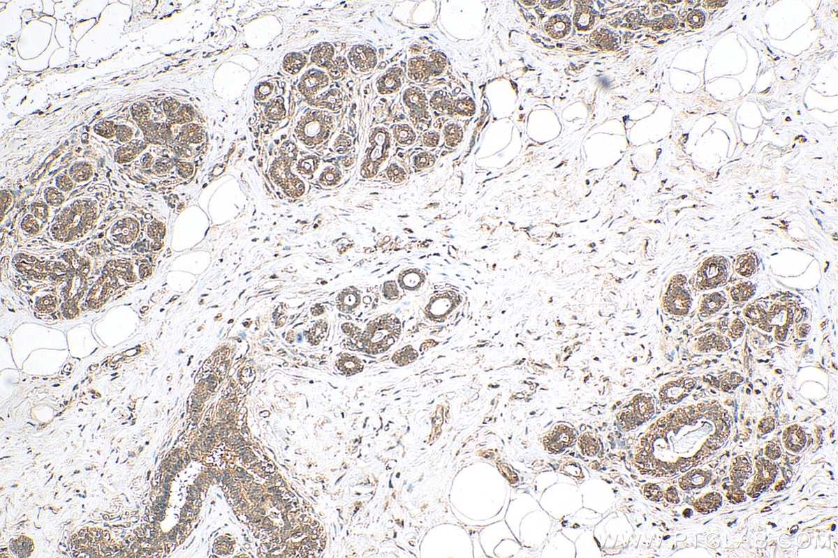
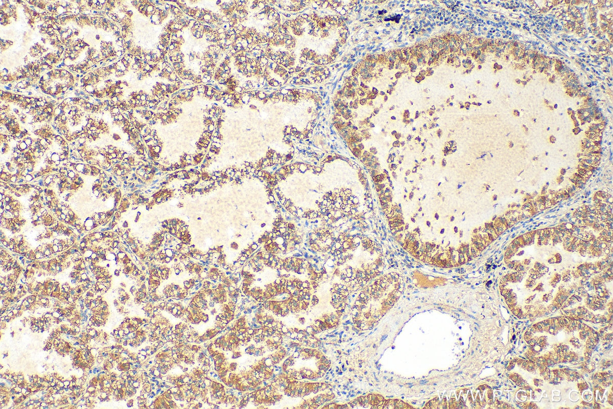
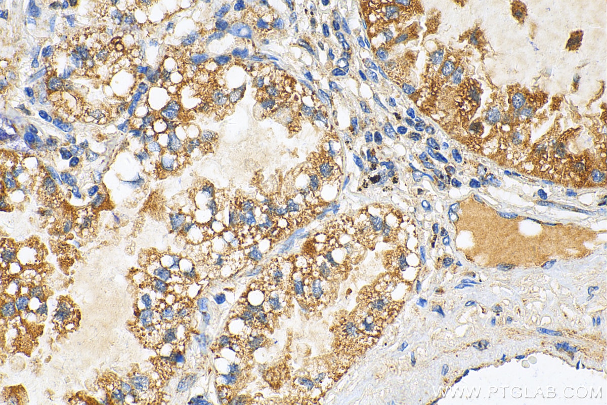
| Erfolgreiche Detektion in IHC | humanes Lungenkarzinomgewebe, humanes Mammakarzinomgewebe, humanes Leberkarzinomgewebe Hinweis: Antigendemaskierung mit TE-Puffer pH 9,0 empfohlen. (*) Wahlweise kann die Antigendemaskierung auch mit Citratpuffer pH 6,0 erfolgen. |
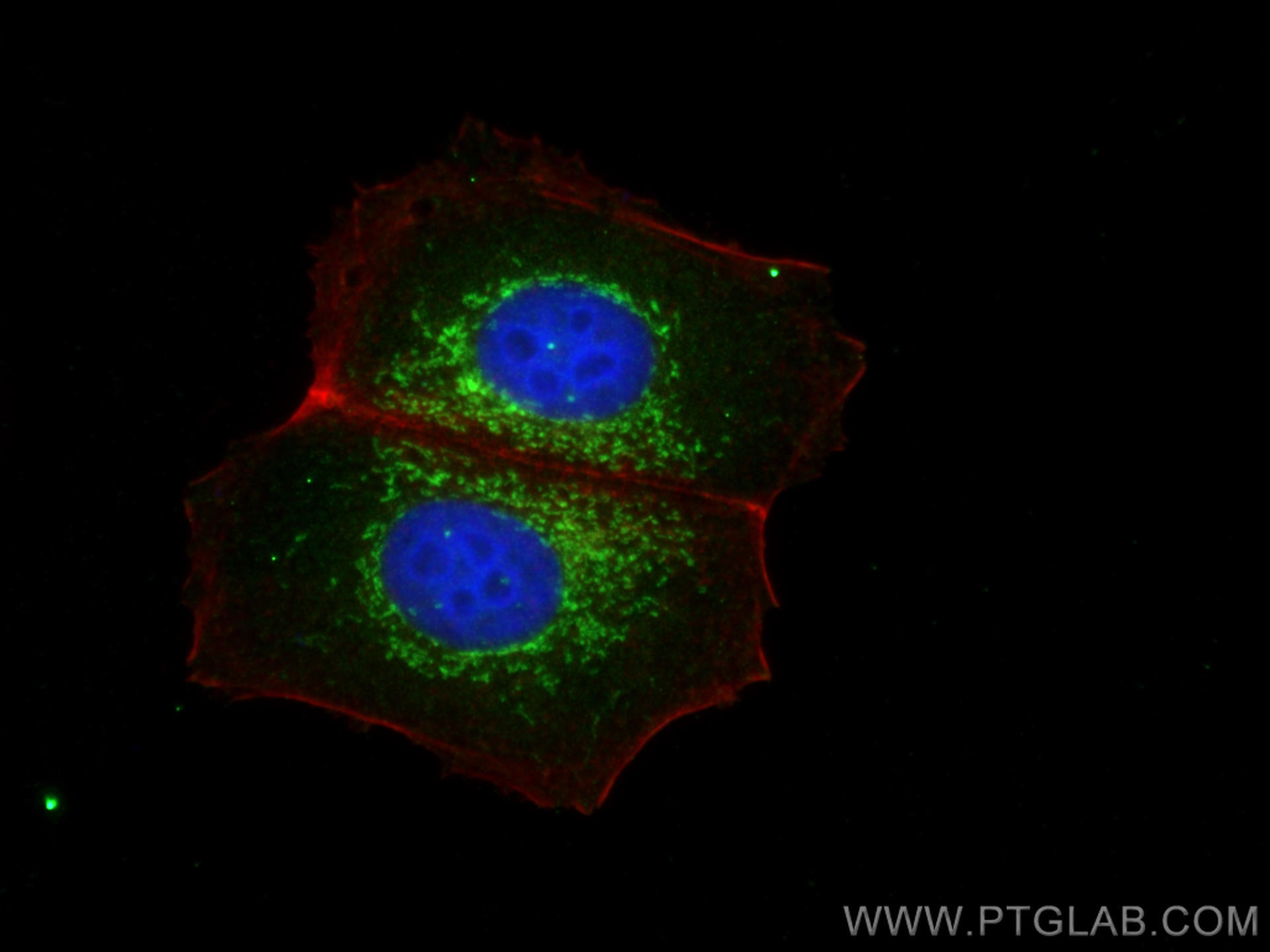
| Erfolgreiche Detektion in IF/ICC | MCF-7-Zellen |
Empfohlene Verdünnung
| Anwendung | Verdünnung |
|---|---|
| Western Blot (WB) | WB : 1:2000-1:14000 |
| Immunhistochemie (IHC) | IHC : 1:1000-1:4000 |
| Immunfluoreszenz (IF)/ICC | IF/ICC : 1:200-1:800 |
| It is recommended that this reagent should be titrated in each testing system to obtain optimal results. | |
| Sample-dependent, check data in validation data gallery | |
Veröffentlichte Anwendungen
| WB | See 17 publications below |
| IHC | See 4 publications below |
| IF | See 5 publications below |
| CoIP | See 1 publications below |
Produktinformation
28445-1-AP bindet in WB, IHC, IF/ICC, CoIP, ELISA PGAM5 und zeigt Reaktivität mit human
| Getestete Reaktivität | human |
| In Publikationen genannte Reaktivität | human, Hausschwein, Maus, Ziege |
| Wirt / Isotyp | Kaninchen / IgG |
| Klonalität | Polyklonal |
| Typ | Antikörper |
| Immunogen | PGAM5 fusion protein Ag28195 |
| Vollständiger Name | phosphoglycerate mutase family member 5 |
| Berechnetes Molekulargewicht | 32 kDa |
| Beobachtetes Molekulargewicht | 32 kDa |
| GenBank-Zugangsnummer | BC008196 |
| Gene symbol | PGAM5 |
| Gene ID (NCBI) | 192111 |
| Konjugation | Unkonjugiert |
| Form | Liquid |
| Reinigungsmethode | Antigen-Affinitätsreinigung |
| Lagerungspuffer | PBS with 0.02% sodium azide and 50% glycerol |
| Lagerungsbedingungen | Bei -20°C lagern. Nach dem Versand ein Jahr lang stabil Aliquotieren ist bei -20oC Lagerung nicht notwendig. 20ul Größen enthalten 0,1% BSA. |
Hintergrundinformationen
Phosphoglycerate mutase 5 (PGAM5) is a mitochondrial Serine (Ser)/Threonine (Thr) phosphatase normally located in the inner mitochondrial membrane. Upon mitochondrial dysfunction, PGAM5 recruits and dephosphorylates Drp1 at Ser-637, triggers its GTPase activity and promotes mitochondrial fission. PGAM5 regulates mitophagy by stabilizing PINK1 under stress conditions, which recruits E3 ubiquitin ligase PARKIN for degradation of the damaged mitochondria. PGAM5 can be cleaved and released to the cytoplasm through PARKIN, which activates Wnt signaling and induces mitochondrial biogenesis (PMID: 32439975). PGAM5 has 2 isoforms with the molecular mass of 28 and 32 kDa.
Protokolle
| PRODUKTSPEZIFISCHE PROTOKOLLE | |
|---|---|
| WB protocol for PGAM5 antibody 28445-1-AP | Protokoll herunterladen |
| IHC protocol for PGAM5 antibody 28445-1-AP | Protokoll herunterladenl |
| IF protocol for PGAM5 antibody 28445-1-AP | Protokoll herunterladen |
| STANDARD-PROTOKOLLE | |
|---|---|
| Klicken Sie hier, um unsere Standardprotokolle anzuzeigen |
Publikationen
| Species | Application | Title |
|---|---|---|
Cell Biosci Differential effects of PGAM5 knockout on high fat high fructose diet and methionine choline-deficient diet induced non-alcoholic steatohepatitis (NASH) in mice | ||
Int J Mol Sci The Mitochondrial PHB2/OMA1/DELE1 Pathway Cooperates with Endoplasmic Reticulum Stress to Facilitate the Response to Chemotherapeutics in Ovarian Cancer. | ||
Int Immunopharmacol 4-Octyl itaconate inhibits inflammation to attenuate psoriasis as an agonist of oxeiptosis | ||
J Virol PGAM5 degrades PDCoV N protein and activates type I interferon to antagonize viral replication | ||
Biochim Biophys Acta Mol Cell Res Tau phosphorylation and OPA1 proteolysis are unrelated events: Implications for Alzheimer's Disease. | ||
Heliyon LFHP-1c improves cognitive function after TBI in mice by reducing oxidative stress through the PGAM5-NRF2-KEAP1 ternary complex |
Rezensionen
The reviews below have been submitted by verified Proteintech customers who received an incentive for providing their feedback.
FH Iram (Verified Customer) (07-03-2020) | 1:1000 dilution in 1%BSA giving a very clear results
|
