PHGDH Monoklonaler Antikörper
PHGDH Monoklonal Antikörper für WB, IHC, IF/ICC, IP, ELISA
Wirt / Isotyp
Maus / IgG1
Getestete Reaktivität
human, Maus, Ratte
Anwendung
WB, IHC, IF/ICC, IP, ELISA
Konjugation
Unkonjugiert
CloneNo.
1E8B8
Kat-Nr. : 67591-1-Ig
Synonyme
 at dilution of 1:10000 incubated at room temperature for 1.5 hours.")
 with HeLa cells lysate 980 ug.")
 at dilution of 1:4000 (under 10x lens). Heat mediated antigen retrieval with Tris-EDTA buffer (pH 9.0).")
 fixed HeLa cells using PHGDH antibody (67591-1-Ig, Clone: 1E8B8 ) at dilution of 1:800 and CoraLite®488-Conjugated Goat Anti-Mouse IgG(H+L) (SA00013-1).")
Geprüfte Anwendungen
| Erfolgreiche Detektion in WB | HeLa-Zellen, HEK-293-Zellen, HepG2-Zellen, Jurkat-Zellen, K-562-Zellen, NIH/3T3-Zellen |
| Erfolgreiche IP | HeLa-Zellen |
| Erfolgreiche Detektion in IHC | humanes Urothelkarzinomgewebe Hinweis: Antigendemaskierung mit TE-Puffer pH 9,0 empfohlen. (*) Wahlweise kann die Antigendemaskierung auch mit Citratpuffer pH 6,0 erfolgen. |
| Erfolgreiche Detektion in IF/ICC | HeLa-Zellen |
Empfohlene Verdünnung
| Anwendung | Verdünnung |
|---|---|
| Western Blot (WB) | WB : 1:5000-1:50000 |
| Immunpräzipitation (IP) | IP : 0.5-4.0 ug for 1.0-3.0 mg of total protein lysate |
| Immunhistochemie (IHC) | IHC : 1:2000-1:8000 |
| Immunfluoreszenz (IF)/ICC | IF/ICC : 1:400-1:1600 |
| It is recommended that this reagent should be titrated in each testing system to obtain optimal results. | |
| Sample-dependent, check data in validation data gallery | |
Veröffentlichte Anwendungen
| WB | See 4 publications below |
Produktinformation
67591-1-Ig bindet in WB, IHC, IF/ICC, IP, ELISA PHGDH und zeigt Reaktivität mit human, Maus, Ratten
| Getestete Reaktivität | human, Maus, Ratte |
| In Publikationen genannte Reaktivität | human, Maus |
| Wirt / Isotyp | Maus / IgG1 |
| Klonalität | Monoklonal |
| Typ | Antikörper |
| Immunogen | PHGDH fusion protein Ag6877 |
| Vollständiger Name | phosphoglycerate dehydrogenase |
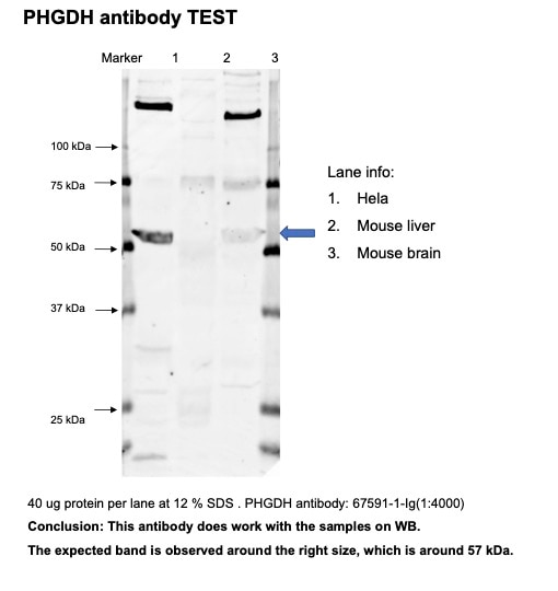
| Berechnetes Molekulargewicht | 57 kDa |
| Beobachtetes Molekulargewicht | 57 kDa |
| GenBank-Zugangsnummer | BC000303 |
| Gene symbol | PHGDH |
| Gene ID (NCBI) | 26227 |
| Konjugation | Unkonjugiert |
| Form | Liquid |
| Reinigungsmethode | Protein-G-Reinigung |
| Lagerungspuffer | PBS with 0.02% sodium azide and 50% glycerol |
| Lagerungsbedingungen | Bei -20°C lagern. Nach dem Versand ein Jahr lang stabil Aliquotieren ist bei -20oC Lagerung nicht notwendig. 20ul Größen enthalten 0,1% BSA. |
Hintergrundinformationen
PHGDH(D-3-phosphoglycerate dehydrogenase) is also named as 3-PGDH, PGDH3 and belongs to the D-isomer specific 2-hydroxyacid dehydrogenase family. It catalyzes the transition of 3-phosphoglycerate into 3-phosphohydroxypyruvate, which is the first and rate-limiting step in the phosphorylated pathway of serine biosynthesis, using NAD+/NADH as a cofactor. 3-PGDH deficiency is a rare recessive inborn error in the biosynthesis of the amino acid L-serine characterized clinically by congenital microcephaly, psychomotor retardation, and intractable seizures(PMID:19235232 ).
Protokolle
| PRODUKTSPEZIFISCHE PROTOKOLLE | |
|---|---|
| WB protocol for PHGDH antibody 67591-1-Ig | Protokoll herunterladen |
| IHC protocol for PHGDH antibody 67591-1-Ig | Protokoll herunterladenl |
| IF protocol for PHGDH antibody 67591-1-Ig | Protokoll herunterladen |
| IP protocol for PHGDH antibody 67591-1-Ig | Protokoll herunterladen |
| STANDARD-PROTOKOLLE | |
|---|---|
| Klicken Sie hier, um unsere Standardprotokolle anzuzeigen |
Publikationen
| Species | Application | Title |
|---|---|---|
Mol Cell The long noncoding RNA glycoLINC assembles a lower glycolytic metabolon to promote glycolysis. | ||
Int Immunopharmacol Artesunate attenuates serum amyloid A-induced M1 macrophage differentiation through the promotion of PHGDH | ||
Oncogene Interaction of PHGDH with IGF2BP1 facilitates m6A-dependent stabilization of TCF7L2 mRNA to confer multidrug resistance in gastric cancer | ||
Adv Sci (Weinh) RFWD3 Reprograms Nucleotide Metabolism Through PHGDH to Induce Chemoresistance In Osteosarcoma |
Rezensionen
The reviews below have been submitted by verified Proteintech customers who received an incentive for providing their feedback.
FH Hua (Verified Customer) (11-30-2022) | Good antibody for WB. Strong signal.
 |

