Syndecan-3 Polyklonaler Antikörper
Syndecan-3 Polyklonal Antikörper für WB, IHC, IF/ICC, IP, ELISA
Wirt / Isotyp
Kaninchen / IgG
Getestete Reaktivität
human, Maus, Ratte
Anwendung
WB, IHC, IF/ICC, IP, ELISA
Konjugation
Unkonjugiert
Kat-Nr. : 10886-1-AP
Synonyme
 at dilution of 1:1000 incubated at room temperature for 1.5 hours.")
 at dilution of 1:500 incubated at room temperature for 1.5 hours.")
 at dilution of 1:500 incubated at room temperature for 1.5 hours.")
 at dilution of 1:500 incubated at room temperature for 1.5 hours.")
 at dilution of 1:500 incubated at room temperature for 1.5 hours.")
 at dilution of 1:500 incubated at room temperature for 1.5 hours.")
 with mouse lung tissue lysate 4000ug.")
 with A549 cells lysate 2800ug.")
. Heat mediated antigen retrieval with Tris-EDTA buffer (pH 9.0).")
. Heat mediated antigen retrieval with Tris-EDTA buffer (pH 9.0).")
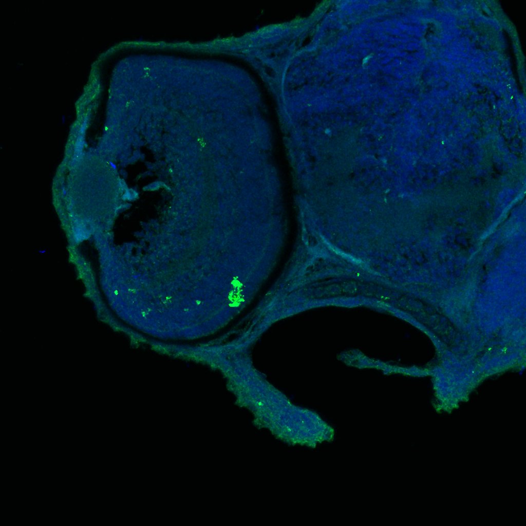
 fixed PC-12 cells using Syndecan-3 antibody (10886-1-AP) at dilution of 1:200 and CoraLite®488-Conjugated Goat Anti-Rabbit IgG(H+L).")
.")
Geprüfte Anwendungen
| Erfolgreiche Detektion in WB | A549-Zellen, Jurkat-Zellen, L02-Zellen, MCF-7-Zellen, Mausnierengewebe, Mauslungengewebe, Rattennierengewebe, Rattenlungengewebe, SH-SY5Y-Zellen |
| Erfolgreiche IP | Mauslungengewebe, A549-Zellen |
| Erfolgreiche Detektion in IHC | humanes Kolonkarzinomgewebe Hinweis: Antigendemaskierung mit TE-Puffer pH 9,0 empfohlen. (*) Wahlweise kann die Antigendemaskierung auch mit Citratpuffer pH 6,0 erfolgen. |
| Erfolgreiche Detektion in IF/ICC | PC-12-Zellen, MCF-7-Zellen |
Empfohlene Verdünnung
| Anwendung | Verdünnung |
|---|---|
| Western Blot (WB) | WB : 1:500-1:2000 |
| Immunpräzipitation (IP) | IP : 0.5-4.0 ug for 1.0-3.0 mg of total protein lysate |
| Immunhistochemie (IHC) | IHC : 1:50-1:500 |
| Immunfluoreszenz (IF)/ICC | IF/ICC : 1:50-1:500 |
| It is recommended that this reagent should be titrated in each testing system to obtain optimal results. | |
| Sample-dependent, check data in validation data gallery | |
Veröffentlichte Anwendungen
| WB | See 2 publications below |
| IHC | See 4 publications below |
| IF | See 1 publications below |
Produktinformation
10886-1-AP bindet in WB, IHC, IF/ICC, IP, ELISA Syndecan-3 und zeigt Reaktivität mit human, Maus, Ratten
| Getestete Reaktivität | human, Maus, Ratte |
| In Publikationen genannte Reaktivität | human, Maus |
| Wirt / Isotyp | Kaninchen / IgG |
| Klonalität | Polyklonal |
| Typ | Antikörper |
| Immunogen | Syndecan-3 fusion protein Ag1317 |
| Vollständiger Name | syndecan 3 |
| Berechnetes Molekulargewicht | 38 kDa |
| Beobachtetes Molekulargewicht | 60-70 kDa |
| GenBank-Zugangsnummer | BC013974 |
| Gene symbol | Syndecan-3 |
| Gene ID (NCBI) | 9672 |
| Konjugation | Unkonjugiert |
| Form | Liquid |
| Reinigungsmethode | Antigen-Affinitätsreinigung |
| Lagerungspuffer | PBS with 0.02% sodium azide and 50% glycerol |
| Lagerungsbedingungen | Bei -20°C lagern. Nach dem Versand ein Jahr lang stabil Aliquotieren ist bei -20oC Lagerung nicht notwendig. 20ul Größen enthalten 0,1% BSA. |
Hintergrundinformationen
Syndecan-3 is a member o f the Syndecan proteoglycan family. It plays a role in the organization of cell shape by affecting the actin cytoskeleton, possibly by transferring signals from the cell surface in a sugar-dependent mechanism.
Protokolle
| PRODUKTSPEZIFISCHE PROTOKOLLE | |
|---|---|
| WB protocol for Syndecan-3 antibody 10886-1-AP | Protokoll herunterladen |
| IHC protocol for Syndecan-3 antibody 10886-1-AP | Protokoll herunterladenl |
| IF protocol for Syndecan-3 antibody 10886-1-AP | Protokoll herunterladen |
| IP protocol for Syndecan-3 antibody 10886-1-AP | Protokoll herunterladen |
| STANDARD-PROTOKOLLE | |
|---|---|
| Klicken Sie hier, um unsere Standardprotokolle anzuzeigen |
Publikationen
| Species | Application | Title |
|---|---|---|
Nucleic Acids Res Metabolic and chemical regulation of tRNA modification associated with taurine deficiency and human disease. | ||
Am J Pathol No haploinsufficiency but loss of heterozygosity for EXT in multiple osteochondromas. | ||
Am J Pathol Screening for potential targets for therapy in mesenchymal, clear cell, and dedifferentiated chondrosarcoma reveals Bcl-2 family members and TGFβ as potential targets. | ||
J Cell Sci Electrophoresis of cell membrane heparan sulfate regulates galvanotaxis in glial cells. | ||
BMC Cancer Prognostic significance of the expression of GFRα1, GFRα3 and syndecan-3, proteins binding ARTEMIN, in mammary carcinoma. | ||
Medicine (Baltimore) Two novel predictive biomarkers for osteosarcoma and glycolysis pathways: A profiling study on HS2ST1 and SDC3 |
Rezensionen
The reviews below have been submitted by verified Proteintech customers who received an incentive for providing their feedback.
FH Kamal (Verified Customer) (02-15-2024) | Mouse liver lysates were subjected to SDS PAGE followed by western blot with 10886-1-AP (Syndecan-3 antibody) at dilution of 1:2000 in 1X TBST incubated overnight at 4 degree C. Syndecan-3 appeared at 65 kDa.
|
FH Ryan (Verified Customer) (02-28-2019) | Tissue was fixed in PFA with tris-HCl antigen retrieval ph=6. Co-localisation with microglia based on known markers (not shown).
 |

