

Tested Applications
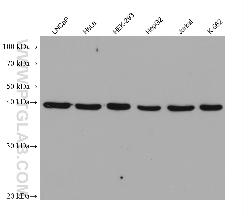
| Positive WB detected in | LNCaP cells, HeLa cells, HEK-293 cells, HepG2 cells, Jurkat cells, K-562 cells |
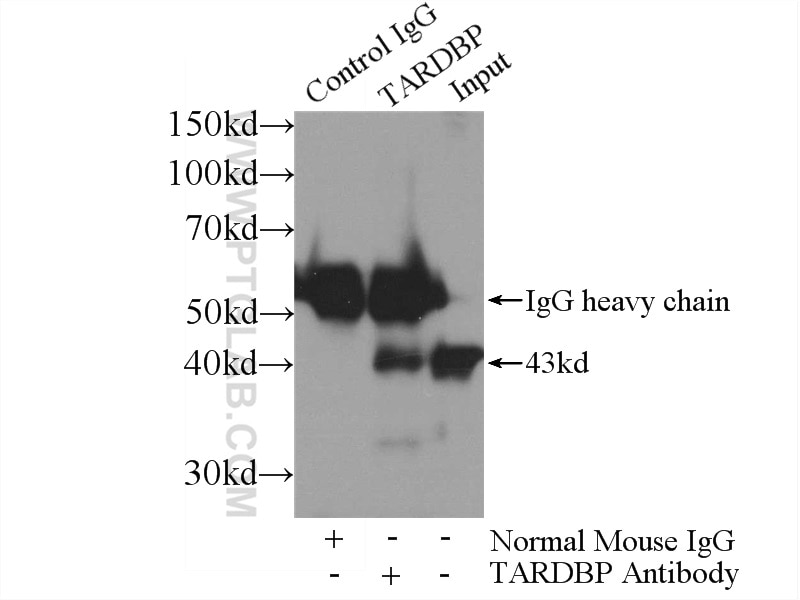
| Positive IP detected in | K-562 cells |
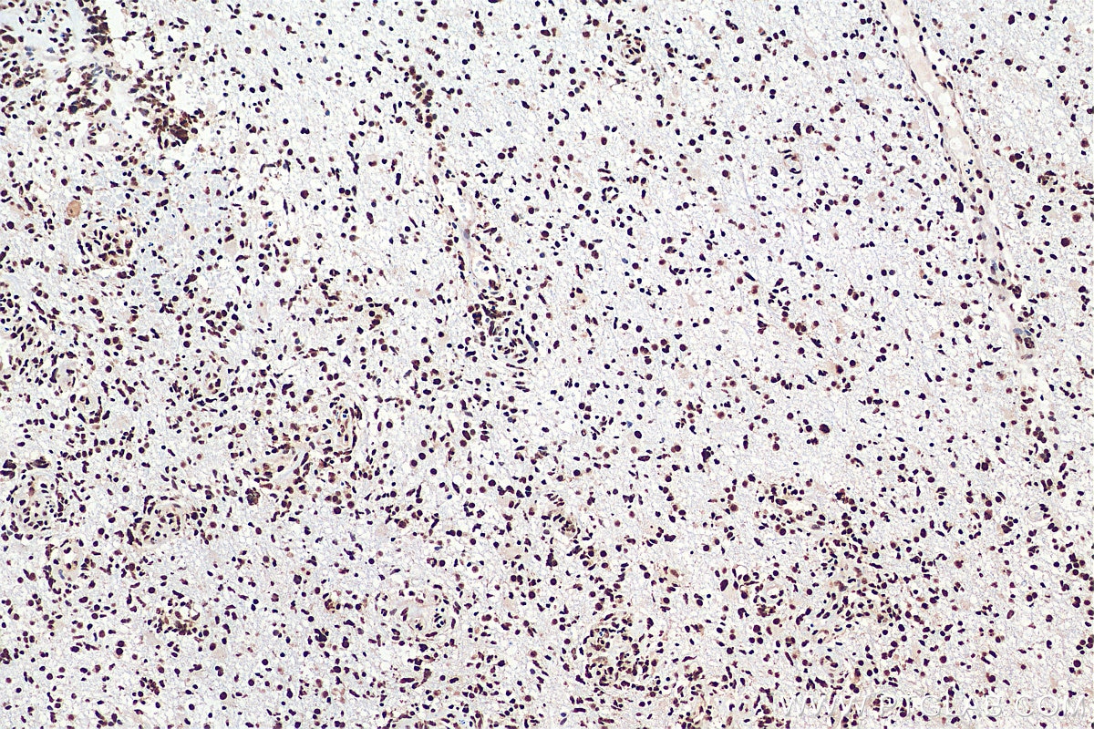
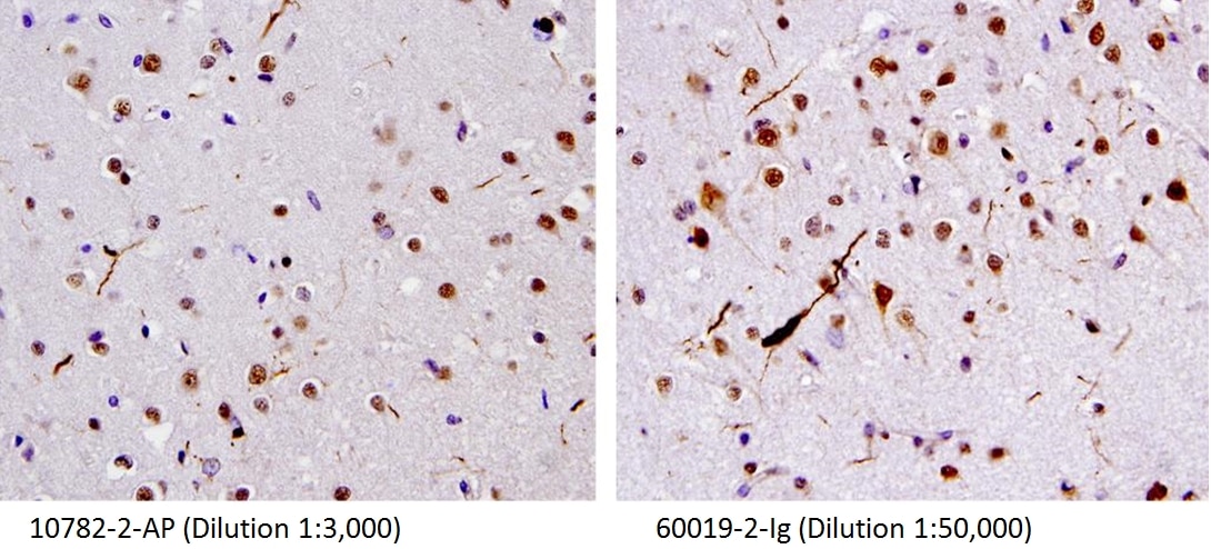
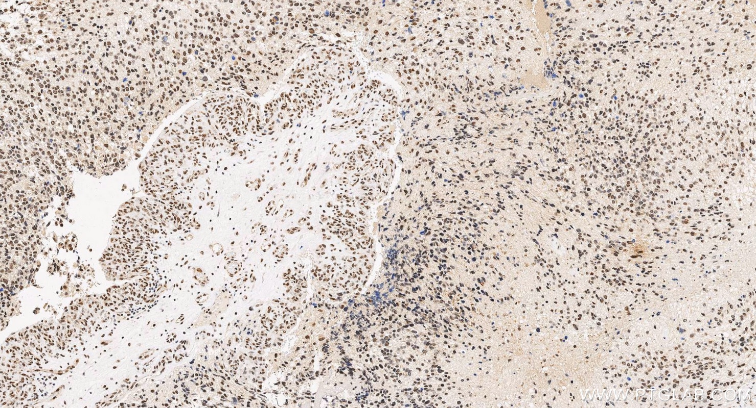
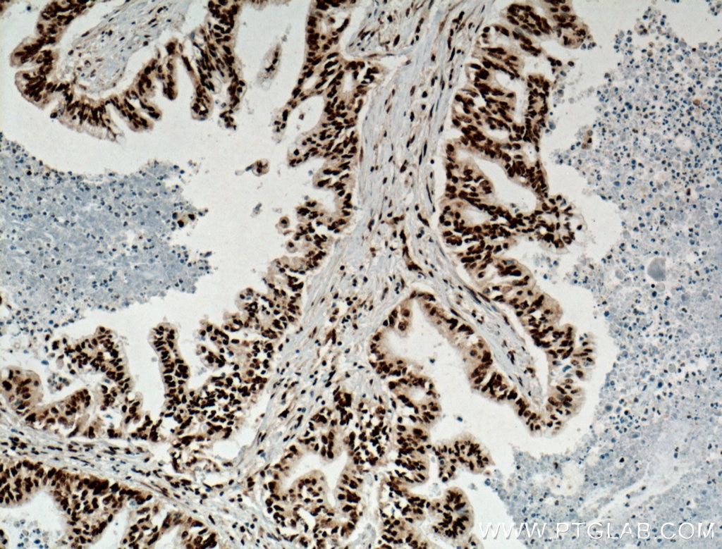
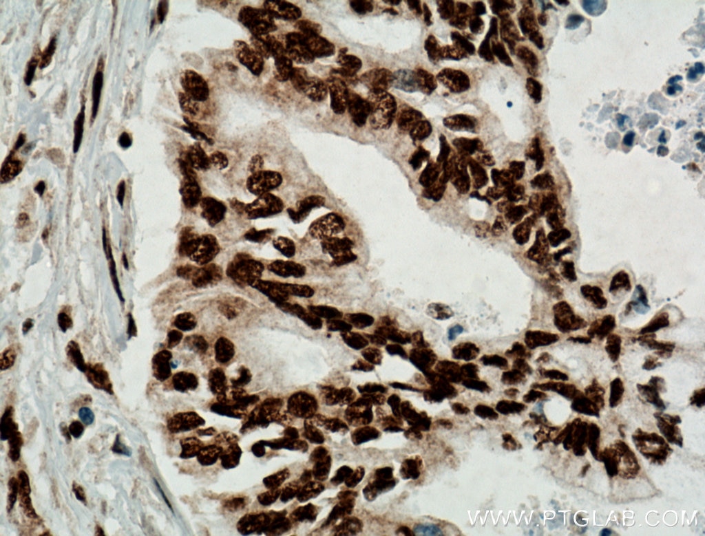
| Positive IHC detected in | human gliomas tissue, human brain(FTLD-U) tissue, human pancreas cancer tissue Note: suggested antigen retrieval with TE buffer pH 9.0; (*) Alternatively, antigen retrieval may be performed with citrate buffer pH 6.0 |
Recommended dilution
| Application | Dilution |
|---|---|
| Western Blot (WB) | WB : 1:5000-1:50000 |
| Immunoprecipitation (IP) | IP : 0.5-4.0 ug for 1.0-3.0 mg of total protein lysate |
| Immunohistochemistry (IHC) | IHC : 1:5000-1:20000 |
| It is recommended that this reagent should be titrated in each testing system to obtain optimal results. | |
| Sample-dependent, Check data in validation data gallery. | |
Product Information
60019-2-Ig targets TDP-43 (human specific) in WB, IHC, IP, CoIP, RIP, ELISA applications and shows reactivity with human samples.
| Tested Reactivity | human |
| Cited Reactivity | human, yeast |
| Host / Isotype | Mouse / IgG1 |
| Class | Monoclonal |
| Type | Antibody |
| Immunogen |
Recombinant protein Predict reactive species |
| Full Name | TAR DNA binding protein |
| Calculated Molecular Weight | 43 kDa |
| Observed Molecular Weight | 43 kDa |
| GenBank Accession Number | BC001487 |
| Gene Symbol | TDP-43 |
| Gene ID (NCBI) | 23435 |
| RRID | AB_2200520 |
| Conjugate | Unconjugated |
| Form | Liquid |
| Purification Method | Protein G purification |
| UNIPROT ID | Q13148 |
| Storage Buffer | PBS with 0.02% sodium azide and 50% glycerol, pH 7.3. |
| Storage Conditions | Store at -20°C. Stable for one year after shipment. Aliquoting is unnecessary for -20oC storage. 20ul sizes contain 0.1% BSA. |
Background Information
Transactivation response (TAR) DNA-binding protein of 43 kDa (also known as TARDBP or TDP-43) was first isolated as a transcriptional inactivator binding to the TAR DNA element of the HIV-1 virus. Neumann et al. (2006) found that a hyperphosphorylated, ubiquitinated, and cleaved form of TARDBP, known as pathologic TDP-43, is the major component of the tau-negative and ubiquitin-positive inclusions that characterize amyotrophic lateral sclerosis (ALS) and the most common pathological subtype of frontotemporal lobar degeneration (FTLD-U). Various forms of TDP-43 exist, including 18-35 kDa of cleaved C-terminal fragments, 45-50 kDa phospho-protein, 55 kDa glycosylated form, 75 kDa hyperphosphorylated form, and 90-300 kDa cross-linked form. (PMID: 17023659,19823856, 21666678, 22193176). 60019-2-Ig is a mouse monoclonal antibody recognizing the cleavage product of 20-30 kDa in addition to the native and phosphorylated forms of TDP-43. Immunohistochemical analyses of TDP-43 using this antibody detect both normal diffuse nuclear staining and insoluble inclusions in pathologic tissues. Notably this antibody only recognizes human TDP-43 but not reacts with mouse or rat TDP-43.
Protocols
| Product Specific Protocols | |
|---|---|
| IHC protocol for TDP-43 (human specific) antibody 60019-2-Ig | Download protocol |
| IP protocol for TDP-43 (human specific) antibody 60019-2-Ig | Download protocol |
| WB protocol for TDP-43 (human specific) antibody 60019-2-Ig | Download protocol |
| Standard Protocols | |
|---|---|
| Click here to view our Standard Protocols |
Publications
What published studies show
Antibody: Anti-TDP-43 (60019-2-Ig)
Total Citations: 133
Journals (selected): Nature, Nature Medicine, Nature Neuroscience, Neuron, EMBO J, Brain, Acta Neuropathol, Nat Commun, Sci Adv, Cell Rep, J Biol Chem, Hum Mol Genet, Neurobiol Dis, Mol Neurodegener, iScience, Front Neurosci, and others.
Research Fields: ALS/FTLD pathogenesis, TDP-43 aggregation and misfolding, RNA splicing regulation, neuroinflammation, biomarker discovery, autophagy/proteostasis, and prion-like propagation mechanisms.
| Species | Application | Title |
|---|---|---|
Nature Mutations in UBQLN2 cause dominant X-linked juvenile and adult-onset ALS and ALS/dementia. | ||
Nat Med The inhibition of TDP-43 mitochondrial localization blocks its neuronal toxicity. | ||
Nat Neurosci TDP-43 extracted from frontotemporal lobar degeneration subject brains displays distinct aggregate assemblies and neurotoxic effects reflecting disease progression rates. |
Reviews
What customers say
Both reviewers gave this antibody a perfect 5-star rating. One user successfully applied it for Western Blot (1:2000) in Neuroblastoma cell lines (SK-N-BE(2)C and IMR32) as well as Proximity Ligation Assay (1:50). The other user confirmed excellent performance across multiple applications including Western Blot (1:1000), Immunofluorescence (1:500), Immunoprecipitation, and RNA Immunoprecipitation (30 µg) in SH-SY5Y cells. Overall, users report reliable results and versatility across various techniques.
The reviews below have been submitted by verified Proteintech customers who received an incentive for providing their feedback.
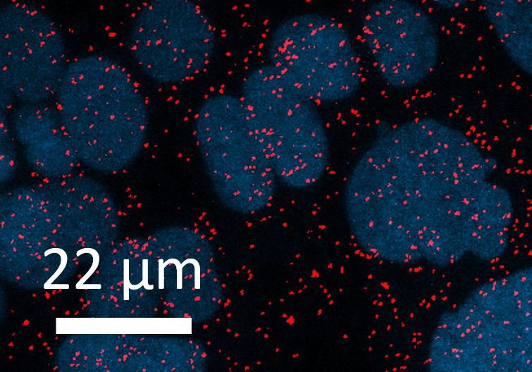
Lidiya (Verified Customer) (05-26-2022) | The antibody works great! Antibody was used for WB in Neuroblastoma cell lines (SK-N-BE(2)C and IMR32) with HRP-conjugated secondary. Antibody was also used for a Proximity Ligation Assay to assess interaction with another protein in SK-N-BE(2)C cells. 1:50 of antibody was used for this assay.
 |
David (Verified Customer) (01-13-2020) | Great antibody for several applications. Works nicely for immunoblot and immunofluourescence. Can also be used for RNA immunoprecipitation.
|









