- Featured Product
- KD/KO Validated
TBC1D5 Polyklonaler Antikörper
TBC1D5 Polyklonal Antikörper für WB, IHC, IP, ELISA
Wirt / Isotyp
Kaninchen / IgG
Getestete Reaktivität
human, Maus
Anwendung
WB, IHC, IF, IP, ELISA
Konjugation
Unkonjugiert
Kat-Nr. : 17078-1-AP
Synonyme
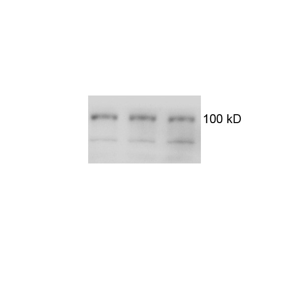
 at dilution of 1:5000 incubated at room temperature for 1.5 hours.")
 with Jurkat cells lysate 1360 ug.")
 at dilution of 1:200 (under 40x lens). Heat mediated antigen retrieval with Tris-EDTA buffer (pH 9.0).")
 at dilution of 1:200 (under 10x lens). Heat mediated antigen retrieval with Tris-EDTA buffer (pH 9.0).")
Geprüfte Anwendungen
| Erfolgreiche Detektion in WB | A431-Zellen, HEK-293T-Zellen, Jurkat-Zellen, PC-3-Zellen |
| Erfolgreiche IP | Jurkat-Zellen |
| Erfolgreiche Detektion in IHC | Maushodengewebe Hinweis: Antigendemaskierung mit TE-Puffer pH 9,0 empfohlen. (*) Wahlweise kann die Antigendemaskierung auch mit Citratpuffer pH 6,0 erfolgen. |
Empfohlene Verdünnung
| Anwendung | Verdünnung |
|---|---|
| Western Blot (WB) | WB : 1:2000-1:10000 |
| Immunpräzipitation (IP) | IP : 0.5-4.0 ug for 1.0-3.0 mg of total protein lysate |
| Immunhistochemie (IHC) | IHC : 1:50-1:500 |
| It is recommended that this reagent should be titrated in each testing system to obtain optimal results. | |
| Sample-dependent, check data in validation data gallery | |
Veröffentlichte Anwendungen
| KD/KO | See 5 publications below |
| WB | See 13 publications below |
| IF | See 6 publications below |
Produktinformation
17078-1-AP bindet in WB, IHC, IF, IP, ELISA TBC1D5 und zeigt Reaktivität mit human, Maus
| Getestete Reaktivität | human, Maus |
| In Publikationen genannte Reaktivität | human, Maus |
| Wirt / Isotyp | Kaninchen / IgG |
| Klonalität | Polyklonal |
| Typ | Antikörper |
| Immunogen | TBC1D5 fusion protein Ag10991 |
| Vollständiger Name | TBC1 domain family, member 5 |
| Berechnetes Molekulargewicht | 795 aa, 89 kDa |
| Beobachtetes Molekulargewicht | 89 kDa |
| GenBank-Zugangsnummer | BC013145 |
| Gene symbol | TBC1D5 |
| Gene ID (NCBI) | 9779 |
| Konjugation | Unkonjugiert |
| Form | Liquid |
| Reinigungsmethode | Antigen-Affinitätsreinigung |
| Lagerungspuffer | PBS with 0.02% sodium azide and 50% glycerol |
| Lagerungsbedingungen | Bei -20°C lagern. Nach dem Versand ein Jahr lang stabil Aliquotieren ist bei -20oC Lagerung nicht notwendig. 20ul Größen enthalten 0,1% BSA. |
Hintergrundinformationen
TBC1D5, a member of TBC (Tre2/Bub2/Cdc16)1 domain family, is a novel retromer-interacting protein. TBC1D5 is a Rab GTPase-activating proteins (GAPs) proteins that negatively regulates VPS35/29/26 recruitment and causes Rab7 to dissociate from the membrane. TBC1D5 could bridge the endosome and autophagosome via its C-terminal LIR motif, and Rab GAPs are implicated in the reprogramming of the endocytic trafficking events under starvation-induced autophagy. The TBC1D5 protein exists 89 kDa and 91 kDa isoforms.
Protokolle
| PRODUKTSPEZIFISCHE PROTOKOLLE | |
|---|---|
| WB protocol for TBC1D5 antibody 17078-1-AP | Protokoll herunterladen |
| IHC protocol for TBC1D5 antibody 17078-1-AP | Protokoll herunterladenl |
| IP protocol for TBC1D5 antibody 17078-1-AP | Protokoll herunterladen |
| STANDARD-PROTOKOLLE | |
|---|---|
| Klicken Sie hier, um unsere Standardprotokolle anzuzeigen |
Publikationen
| Species | Application | Title |
|---|---|---|
Sci Adv De novo macrocyclic peptides for inhibiting, stabilizing, and probing the function of the retromer endosomal trafficking complex. | ||
EMBO J Control of RAB7 activity and localization through the retromer-TBC1D5 complex enables RAB7-dependent mitophagy.
| ||
J Cell Biol Retromer and TBC1D5 maintain late endosomal RAB7 domains to enable amino acid-induced mTORC1 signaling.
| ||
EMBO Rep BioID reveals an ATG9A interaction with ATG13-ATG101 in the degradation of p62/SQSTM1-ubiquitin clusters. | ||
Int J Biol Sci TBK1 Facilitates GLUT1-Dependent Glucose Consumption by suppressing mTORC1 Signaling in Colorectal Cancer Progression.
| ||
Mol Biol Cell Trafficking defects in WASH-knockout fibroblasts originate from collapsed endosomal and lysosomal networks. |
Rezensionen
The reviews below have been submitted by verified Proteintech customers who received an incentive for providing their feedback.
FH q (Verified Customer) (06-01-2021) | It is OK to detect TBC1D5 (100 kD) in both mouse and postmortem brain lysates, although there are some non specific bands.
 |
FH Florian (Verified Customer) (04-09-2019) | This is a very strong antibody that is reasonably specific in western blot applications. We have validated it through Crispr/Cas9 mediated knockout of TBC1D5 in HeLa cells (Jimenez-Orgaz et al, EMBOJ, 2018). While the strongest band is indeed TBC1D5, as our knockout confirms, it also produces several weaker, unspecific bands on membranes blocked with 5% milk. Because of the unspecific bands, I rate it with four stars instead of five. We have not tested it for immunofluorescence. Overall, it is a very good antibody that will probably also work at much lower dilutions than 1:1000. Diluting it further may also reduce the unspecific signal.
|
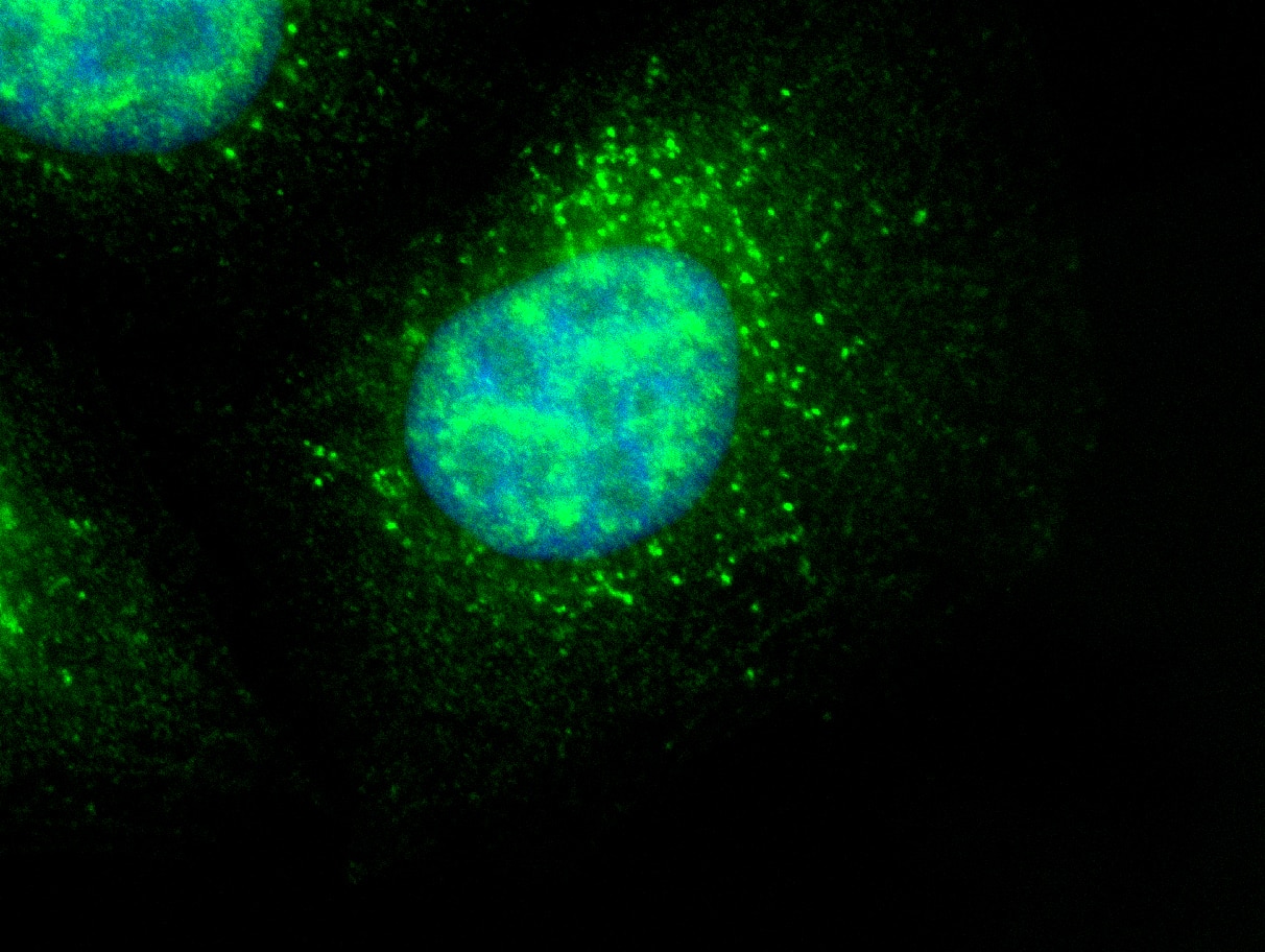
FH David (Verified Customer) (02-28-2019) | Hela cells stained with anti-TBC1D5 (green) and Hoechst (blue)
 |

