- Featured Product
- KD/KO Validated
TGF Beta 1 Polyklonaler Antikörper
TGF Beta 1 Polyklonal Antikörper für WB, IF/ICC, IF-P, ELISA
Wirt / Isotyp
Kaninchen / IgG
Getestete Reaktivität
human, Maus, Ratte und mehr (5)
Anwendung
WB, IHC, IF/ICC, IF-P, IP, CoIP, ELISA
Konjugation
Unkonjugiert
Kat-Nr. : 21898-1-AP
Synonyme
Geprüfte Anwendungen
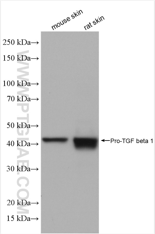
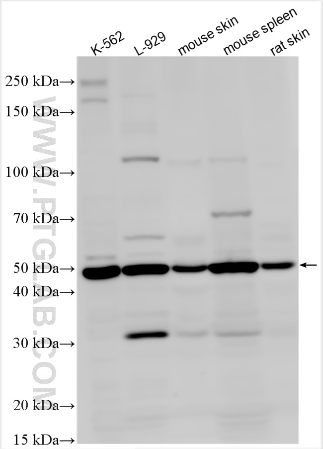
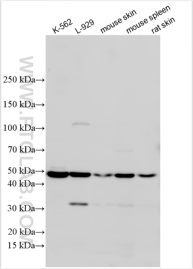
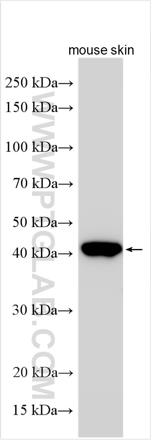
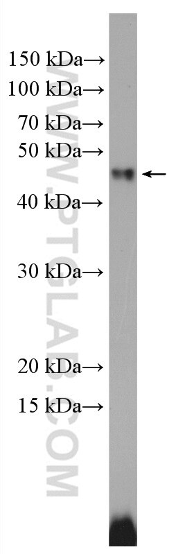
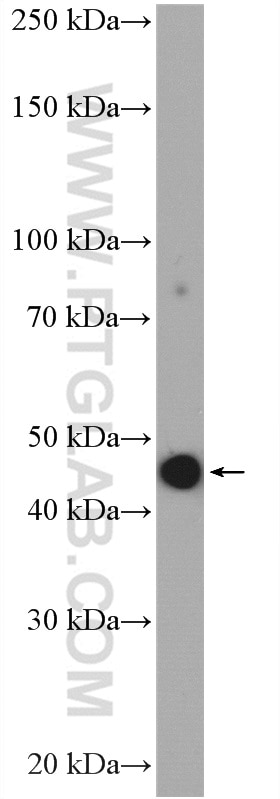
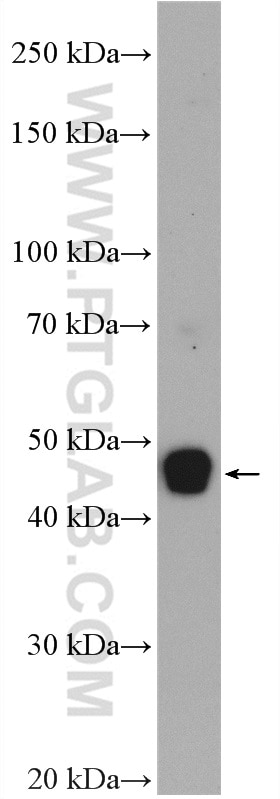
| Erfolgreiche Detektion in WB | Maushautgewebe, A549-Zellen, HeLa-Zellen, K-562-Zellen, L-929-Zellen, Mausmilzgewebe, Rattenhautgewebe |
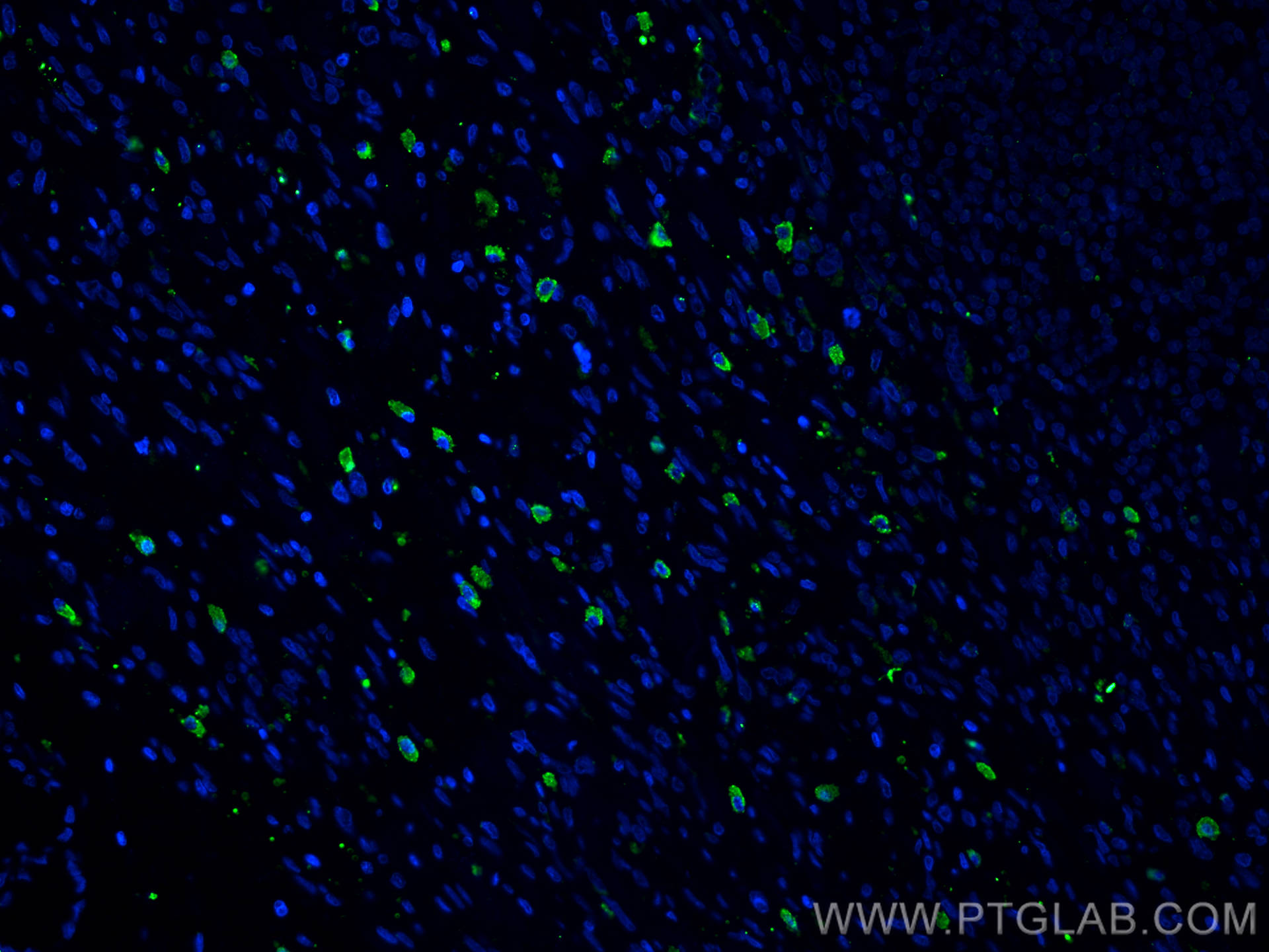
| Erfolgreiche Detektion in IF-P | humanes Magenkrebsgewebe |
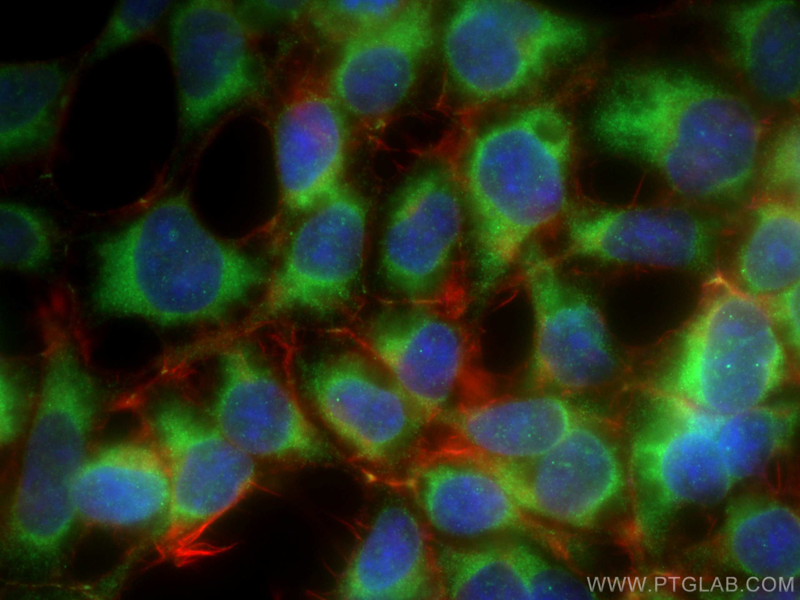
| Erfolgreiche Detektion in IF/ICC | HEK-293-Zellen |
Empfohlene Verdünnung
| Anwendung | Verdünnung |
|---|---|
| Western Blot (WB) | WB : 1:1000-1:5000 |
| Immunfluoreszenz (IF)-P | IF-P : 1:50-1:500 |
| Immunfluoreszenz (IF)/ICC | IF/ICC : 1:200-1:800 |
| It is recommended that this reagent should be titrated in each testing system to obtain optimal results. | |
| Sample-dependent, check data in validation data gallery | |
Veröffentlichte Anwendungen
| KD/KO | See 2 publications below |
| WB | See 606 publications below |
| IHC | See 171 publications below |
| IF | See 82 publications below |
| IP | See 1 publications below |
| ELISA | See 2 publications below |
| CoIP | See 1 publications below |
Produktinformation
21898-1-AP bindet in WB, IHC, IF/ICC, IF-P, IP, CoIP, ELISA TGF Beta 1 und zeigt Reaktivität mit human, Maus, Ratten
| Getestete Reaktivität | human, Maus, Ratte |
| In Publikationen genannte Reaktivität | human, Ente, Hausschwein, Hund, Kaninchen, Maus, Ratte, Zebrafisch |
| Wirt / Isotyp | Kaninchen / IgG |
| Klonalität | Polyklonal |
| Typ | Antikörper |
| Immunogen | TGF Beta 1 fusion protein Ag13591 |
| Vollständiger Name | transforming growth factor, beta 1 |
| Berechnetes Molekulargewicht | 44 kDa |
| Beobachtetes Molekulargewicht | 44 kDa |
| GenBank-Zugangsnummer | BC000125 |
| Gene symbol | TGFB1 |
| Gene ID (NCBI) | 7040 |
| Konjugation | Unkonjugiert |
| Form | Liquid |
| Reinigungsmethode | Antigen-Affinitätsreinigung |
| Lagerungspuffer | PBS with 0.02% sodium azide and 50% glycerol |
| Lagerungsbedingungen | Bei -20°C lagern. Nach dem Versand ein Jahr lang stabil Aliquotieren ist bei -20oC Lagerung nicht notwendig. 20ul Größen enthalten 0,1% BSA. |
Hintergrundinformationen
TGFB, also named as LAP and TGFB1, is a multifunctional peptide that controls proliferation, differentiation, and other functions in many cell types. TGFB acts synergistically with TGFA in inducing transformation. It also acts as a negative autocrine growth factor. Dysregulation of TGFB activation and signaling may result in apoptosis. Many cells synthesize TGFB and almost all of them have specific receptors for it. TGFB positively and negatively regulates many other growth factors. It plays an important role in bone remodeling as it is a potent stimulator of osteoblastic bone formation, causing chemotaxis, proliferation and differentiation in committed osteoblasts. It is highly expressed in bone. Mutation of TGFB are the cause of Camurati-Engelmann disease (CED) which known as progressive diaphyseal dysplasia 1 (DPD1).
This antibody detects the pro-TGF beta 1 and the cleaved fragment Latency-associated peptide.
Protokolle
| PRODUKTSPEZIFISCHE PROTOKOLLE | |
|---|---|
| WB protocol for TGF Beta 1 antibody 21898-1-AP | Protokoll herunterladen |
| IF protocol for TGF Beta 1 antibody 21898-1-AP | Protokoll herunterladen |
| STANDARD-PROTOKOLLE | |
|---|---|
| Klicken Sie hier, um unsere Standardprotokolle anzuzeigen |
Publikationen
| Species | Application | Title |
|---|---|---|
Adv Mater Neonatal Tissue-derived Extracellular Vesicle Therapy (NEXT): A Potent Strategy for Precision Regenerative Medicine | ||
ACS Nano Mesenchymal Stem Cell-Derived Extracellular Vesicles Attenuate Mitochondrial Damage and Inflammation by Stabilizing Mitochondrial DNA. | ||
Blood C1Q labels a highly aggressive macrophage-like leukemia population indicating extramedullary infiltration and relapse | ||
Acta Pharm Sin B Fucoidan-functionalized activated platelet-hitchhiking micelles simultaneously track tumor cells and remodel the immunosuppressive microenvironment for efficient metastatic cancer treatment. | ||
Adv Sci (Weinh) M2 Macrophage-Derived Extracellular Vesicles Reprogram Immature Neutrophils into Anxa1hi Neutrophils to Enhance Inflamed Bone Regeneration | ||
Dev Cell Endothelial progenitor cells control remodeling of uterine spiral arteries for the establishment of utero-placental circulation |
Rezensionen
The reviews below have been submitted by verified Proteintech customers who received an incentive for providing their feedback.
FH Javier (Verified Customer) (09-09-2025) | Good antibody for WB assay.
|
FH Juhi (Verified Customer) (07-23-2025) | Antibody works fine, giving correct band size in western blot.
|
FH Andrea (Verified Customer) (03-22-2024) | We had a good confocal signal and beautiful bands in our experimental group of high TGFb
|
FH Kenzo (Verified Customer) (08-15-2023) | This antibody works well for labeling TGF beta 1 on mouse kidney tissue section. Very reliable antibody giving reproducible results.
|
FH Celina (Verified Customer) (07-31-2023) | antibody worked well for IF of human cardiac ventricular fibroblasts
|
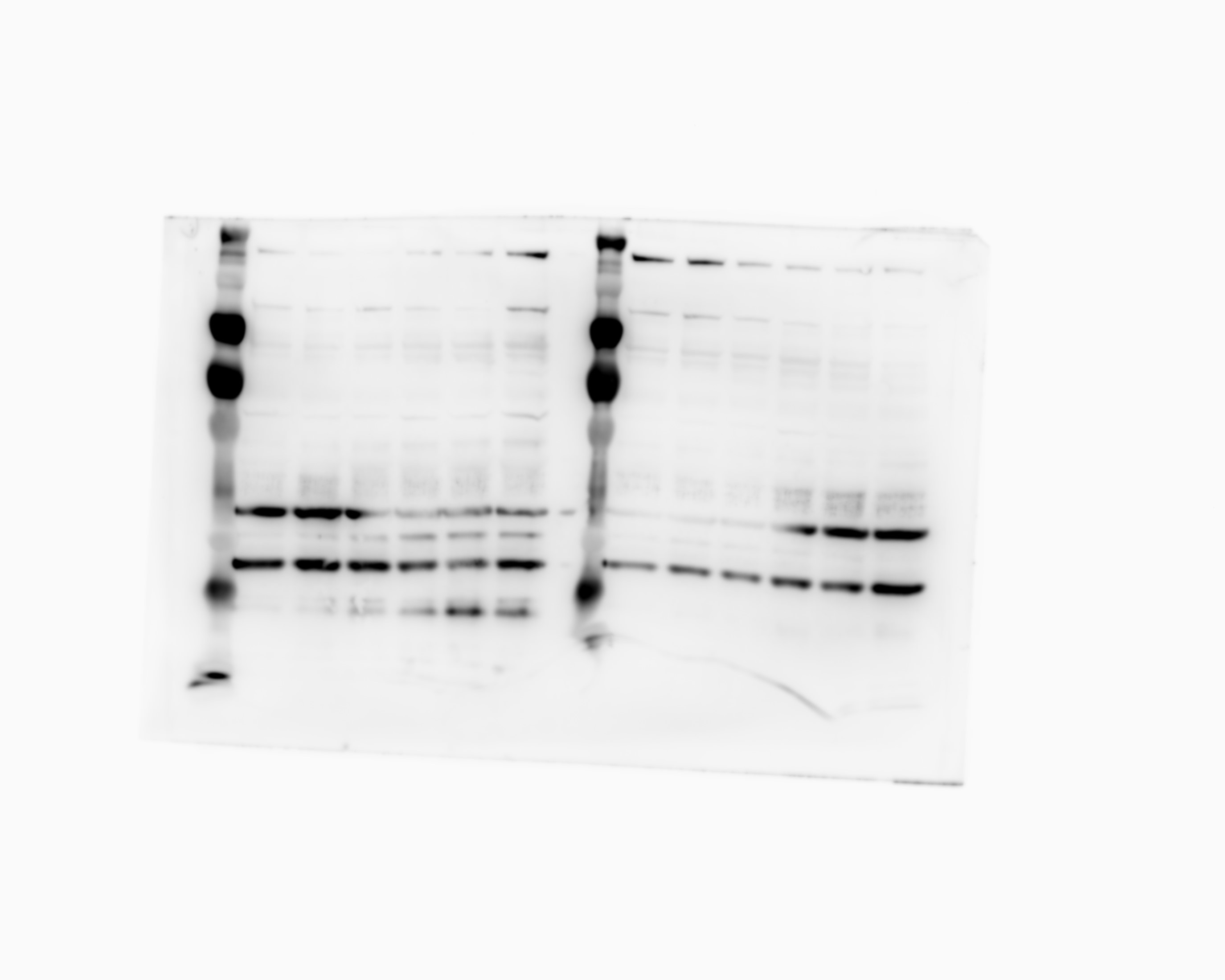
FH Udesh (Verified Customer) (12-06-2022) | The antibody worked well in Western Blot and detected bands for mature TGF-B1 as well as LAP.
 |
FH Yasuyo (Verified Customer) (05-24-2022) | Works great!!!
 |
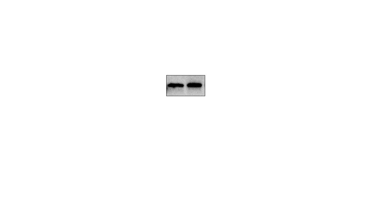
FH P (Verified Customer) (12-01-2021) | Excellent
 |
FH Iram (Verified Customer) (09-04-2020) | Very clean bands for western
|
FH Qingyuan (Verified Customer) (11-05-2019) | The antibody clearly shows one band with the right molecular weight size.
|
