VDAC1/2/3 Polyklonaler Antikörper
VDAC1/2/3 Polyklonal Antikörper für WB, IHC, ELISA
Wirt / Isotyp
Kaninchen / IgG
Getestete Reaktivität
human, Maus, Ratte und mehr (1)
Anwendung
WB, IHC, IF, CoIP, ELISA
Konjugation
Unkonjugiert
Kat-Nr. : 11663-1-AP
Synonyme
 at dilution of 1:3000 incubated at room temperature for 1.5 hours.")
 at dilution of 1:500 incubated at room temperature for 1.5 hours.")
 at dilution of 1:200 (under 10x lens). Heat mediated antigen retrieval with Tris-EDTA buffer (pH 9.0).")
 at dilution of 1:200 (under 40x lens). Heat mediated antigen retrieval with Tris-EDTA buffer (pH 9.0).")
 at dilution of 1:600 (under 20x lens). Heat mediated antigen retrieval with Tris-EDTA buffer (pH 9.0).")
 at dilution of 1:600 (under 20x lens). Heat mediated antigen retrieval with Tris-EDTA buffer (pH 9.0).")
 at dilution of 1:50 (under 10x lens).")
 at dilution of 1:50 (under 40x lens).")
Geprüfte Anwendungen
| Erfolgreiche Detektion in WB | Maushirngewebe, humanes Herzgewebe, Mausherzgewebe, Rattenhirngewebe, Rattenherzgewebe |
| Erfolgreiche Detektion in IHC | humanes Prostatakarzinomgewebe, humanes Prostatagewebe Hinweis: Antigendemaskierung mit TE-Puffer pH 9,0 empfohlen. (*) Wahlweise kann die Antigendemaskierung auch mit Citratpuffer pH 6,0 erfolgen. |
Empfohlene Verdünnung
| Anwendung | Verdünnung |
|---|---|
| Western Blot (WB) | WB : 1:1000-1:6000 |
| Immunhistochemie (IHC) | IHC : 1:200-1:1200 |
| It is recommended that this reagent should be titrated in each testing system to obtain optimal results. | |
| Sample-dependent, check data in validation data gallery | |
Veröffentlichte Anwendungen
| KD/KO | See 1 publications below |
| WB | See 27 publications below |
| IHC | See 3 publications below |
| IF | See 5 publications below |
| IP | See 2 publications below |
| CoIP | See 1 publications below |
Produktinformation
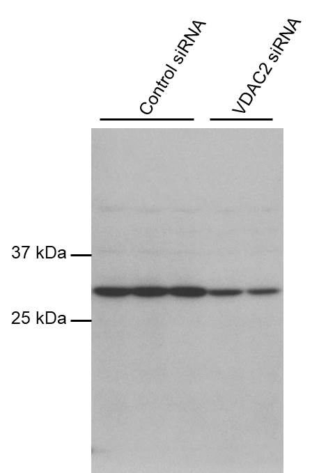
11663-1-AP bindet in WB, IHC, IF, CoIP, ELISA VDAC1/2/3 und zeigt Reaktivität mit human, Maus, Ratten
| Getestete Reaktivität | human, Maus, Ratte |
| In Publikationen genannte Reaktivität | human, Ente, Maus, Ratte |
| Wirt / Isotyp | Kaninchen / IgG |
| Klonalität | Polyklonal |
| Typ | Antikörper |
| Immunogen | VDAC1/2/3 fusion protein Ag2266 |
| Vollständiger Name | voltage-dependent anion channel 2 |
| Berechnetes Molekulargewicht | 294 aa, 32 kDa |
| Beobachtetes Molekulargewicht | 32 kDa |
| GenBank-Zugangsnummer | BC000165 |
| Gene symbol | VDAC2 |
| Gene ID (NCBI) | 7417 |
| Konjugation | Unkonjugiert |
| Form | Liquid |
| Reinigungsmethode | Antigen-Affinitätsreinigung |
| Lagerungspuffer | PBS with 0.02% sodium azide and 50% glycerol |
| Lagerungsbedingungen | Bei -20°C lagern. Nach dem Versand ein Jahr lang stabil Aliquotieren ist bei -20oC Lagerung nicht notwendig. 20ul Größen enthalten 0,1% BSA. |
Hintergrundinformationen
VDACs (Voltage Dependent Anion selective Channels), also known as mitochondrial porins, are a family of pore-forming proteins discovered in the mitochondrial outer membrane. Mammals show a conserved genetic organization of the VDAC genes. It's reported that the amount of VDAC transcripts in liver is usually lower than in the other tissues. VDAC2 and expecially VDAC3 are highly expressed in testis, while mouse VDAC1 is poorly expressed in this tissue.(PMID: 22020053)
Protokolle
| PRODUKTSPEZIFISCHE PROTOKOLLE | |
|---|---|
| WB protocol for VDAC1/2/3 antibody 11663-1-AP | Protokoll herunterladen |
| IHC protocol for VDAC1/2/3 antibody 11663-1-AP | Protokoll herunterladenl |
| STANDARD-PROTOKOLLE | |
|---|---|
| Klicken Sie hier, um unsere Standardprotokolle anzuzeigen |
Publikationen
| Species | Application | Title |
|---|---|---|
Mol Cell MLKL activity requires a splicing-regulated, druggable intramolecular interaction | ||
Nat Commun Kastor and Polluks polypeptides encoded by a single gene locus cooperatively regulate VDAC and spermatogenesis. | ||
Autophagy MYBL2 guides autophagy suppressor VDAC2 in the developing ovary to inhibit autophagy through a complex of VDAC2-BECN1-BCL2L1 in mammals. | ||
Cell Death Differ SPATA33 is an autophagy mediator for cargo selectivity in germline mitophagy. | ||
Proc Natl Acad Sci U S A SPATA33 localizes calcineurin to the mitochondria and regulates sperm motility in mice. | ||
Cell Death Dis Pathological convergence of APP and SNCA deficiency in hippocampal degeneration of young rats |
Rezensionen
The reviews below have been submitted by verified Proteintech customers who received an incentive for providing their feedback.
FH Jun (Verified Customer) (06-12-2022) | Works very well.
 |

