- Featured Product
- KD/KO Validated
VPS26A Polyklonaler Antikörper
VPS26A Polyklonal Antikörper für WB, IHC, IF/ICC, ELISA
Wirt / Isotyp
Kaninchen / IgG
Getestete Reaktivität
human, Maus, Ratte
Anwendung
WB, IHC, IF/ICC, ELISA
Konjugation
Unkonjugiert
Kat-Nr. : 12804-1-AP
Synonyme
 at dilution of 1:600 incubated at room temperature for 1.5 hours.")
 at dilution of 1:300 incubated at room temperature for 1.5 hours.")
 at dilution of 1:200 (under 10x lens). Heat mediated antigen retrieval with Tris-EDTA buffer (pH 9.0).")
 at dilution of 1:200 (under 40x lens). Heat mediated antigen retrieval with Tris-EDTA buffer (pH 9.0).")
 at dilution of 1:100 (under 10x lens).")
 at dilution of 1:200 (under 10x lens).")
 at dilution of 1:200 (under 40x lens).")
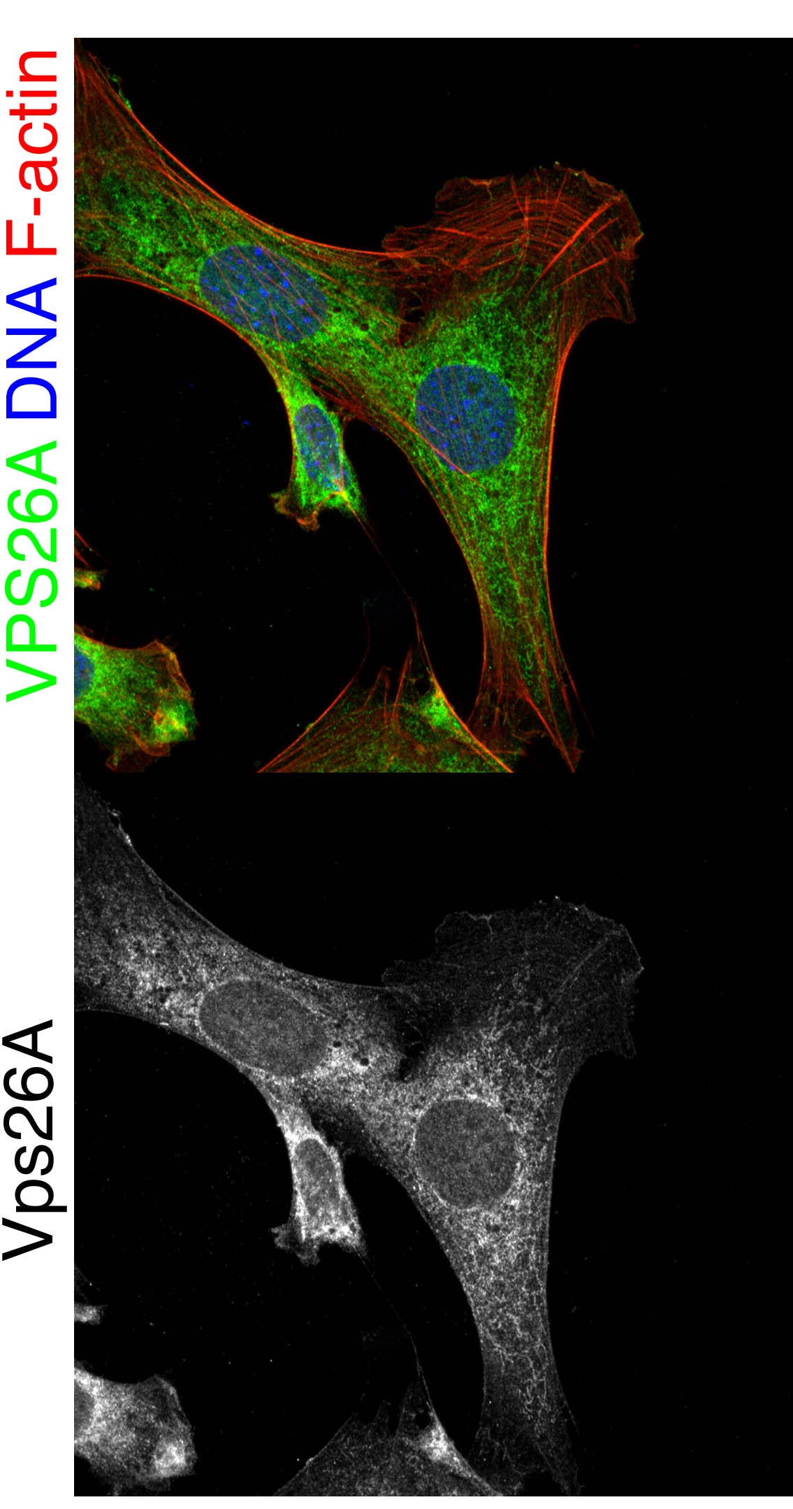
 fixed HeLa cells using 12804-1-AP (VPS26A antibody), at dilution of 1:200 and CoraLite®488-Conjugated Goat Anti-Rabbit IgG(H+L), CL555-phalloidine stains F-actin (red).")
 at dilution of 1:50 and Alexa Fluor 488-conjugated Goat Anti-Rabbit IgG(H+L).")
Geprüfte Anwendungen
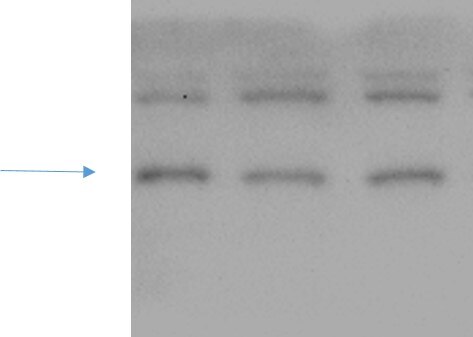
| Erfolgreiche Detektion in WB | Mausnierengewebe, HEK-293-Zellen, Rattennierengewebe |
| Erfolgreiche Detektion in IHC | humanes Mammakarzinomgewebe, humanes Zervixkarzinomgewebe, humanes Nierengewebe Hinweis: Antigendemaskierung mit TE-Puffer pH 9,0 empfohlen. (*) Wahlweise kann die Antigendemaskierung auch mit Citratpuffer pH 6,0 erfolgen. |
| Erfolgreiche Detektion in IF/ICC | HeLa-Zellen |
Empfohlene Verdünnung
| Anwendung | Verdünnung |
|---|---|
| Western Blot (WB) | WB : 1:500-1:1000 |
| Immunhistochemie (IHC) | IHC : 1:50-1:500 |
| Immunfluoreszenz (IF)/ICC | IF/ICC : 1:50-1:500 |
| It is recommended that this reagent should be titrated in each testing system to obtain optimal results. | |
| Sample-dependent, check data in validation data gallery | |
Veröffentlichte Anwendungen
| KD/KO | See 1 publications below |
| WB | See 7 publications below |
| IHC | See 1 publications below |
| IF | See 2 publications below |
Produktinformation
12804-1-AP bindet in WB, IHC, IF/ICC, ELISA VPS26A und zeigt Reaktivität mit human, Maus, Ratten
| Getestete Reaktivität | human, Maus, Ratte |
| In Publikationen genannte Reaktivität | human |
| Wirt / Isotyp | Kaninchen / IgG |
| Klonalität | Polyklonal |
| Typ | Antikörper |
| Immunogen | VPS26A fusion protein Ag3391 |
| Vollständiger Name | vacuolar protein sorting 26 homolog A (S. pombe) |
| Berechnetes Molekulargewicht | 38 kDa |
| Beobachtetes Molekulargewicht | 38 kDa |
| GenBank-Zugangsnummer | BC022505 |
| Gene symbol | VPS26A |
| Gene ID (NCBI) | 9559 |
| Konjugation | Unkonjugiert |
| Form | Liquid |
| Reinigungsmethode | Antigen-Affinitätsreinigung |
| Lagerungspuffer | PBS with 0.02% sodium azide and 50% glycerol |
| Lagerungsbedingungen | Bei -20°C lagern. Nach dem Versand ein Jahr lang stabil Aliquotieren ist bei -20oC Lagerung nicht notwendig. 20ul Größen enthalten 0,1% BSA. |
Hintergrundinformationen
In mammals, there are two paralogues of yeast Vps26, VPS26A and VPS26B (PMID: 16190980). VPS26 is a component of the retromer complex composed of VPS26 (VPS26A or VPS26B), VPS29, VPS35, SNX1, and SNX2. VPS26A and VPS26B subunits define distinct retromer complexes (PMID: 21920005). The retromer complex is important in recycling transmembrane receptors from endosomes to the trans-Golgi network (TGN).
Protokolle
| PRODUKTSPEZIFISCHE PROTOKOLLE | |
|---|---|
| WB protocol for VPS26A antibody 12804-1-AP | Protokoll herunterladen |
| IHC protocol for VPS26A antibody 12804-1-AP | Protokoll herunterladenl |
| IF protocol for VPS26A antibody 12804-1-AP | Protokoll herunterladen |
| STANDARD-PROTOKOLLE | |
|---|---|
| Klicken Sie hier, um unsere Standardprotokolle anzuzeigen |
Publikationen
| Species | Application | Title |
|---|---|---|
Cell Metab Phospholipase PLA2G6, a Parkinsonism-Associated Gene, Affects Vps26 and Vps35, Retromer Function, and Ceramide Levels, Similar to α-Synuclein Gain. | ||
bioRxiv Noncanonical roles of ATG5 and membrane atg8ylation in retromer assembly and function | ||
Elife Noncanonical roles of ATG5 and membrane atg8ylation in retromer assembly and function | ||
Autophagy RAB21 controls autophagy and cellular energy homeostasis by regulating retromer-mediated recycling of SLC2A1/GLUT1 | ||
Int J Mol Sci The Prognostic Value and the Oncogenic and Immunological Roles of Vacuolar Protein Sorting Associated Protein 26 A in Pancreatic Adenocarcinoma
|
Rezensionen
The reviews below have been submitted by verified Proteintech customers who received an incentive for providing their feedback.
FH Xin (Verified Customer) (10-10-2022) | It is Ok to detect endogenous VPS26A although there are some non-specific bands.
 |
FH Stephen (Verified Customer) (09-07-2019) | Oc-2 cells fixed with 4% PFA Perm. by 0.3% tx100 for 5 min blocked with 1% BSA in 1XPBS for 2 hours vps26a antibody incubated 1:300 overnight at 4 degrees in 1%BSA in 1x PBS. Co-stained with DAPI (blue) and Ph647 (red) to visualize DNA and F-actin respectively.
 |

