Vinculin Monoklonaler Antikörper

Vinculin Monoklonal Antikörper für WB, IHC, IF/ICC, FC (Intra), ELISA

Wirt / Isotyp

Maus / IgG1

Getestete Reaktivität

Hausschwein, human, Maus, Ratte und mehr (3)

Anwendung

WB, IHC, IF/ICC, FC (Intra), CoIP, ELISA

Konjugation

Unkonjugiert

Publikationen(203)

CloneNo.

2B5A7

Kat-Nr. : 66305-1-Ig

Synonyme

VCL, MVCL, MV, Metavinculin, CMD1W


Filter:
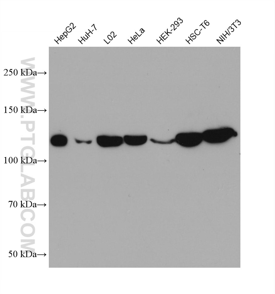
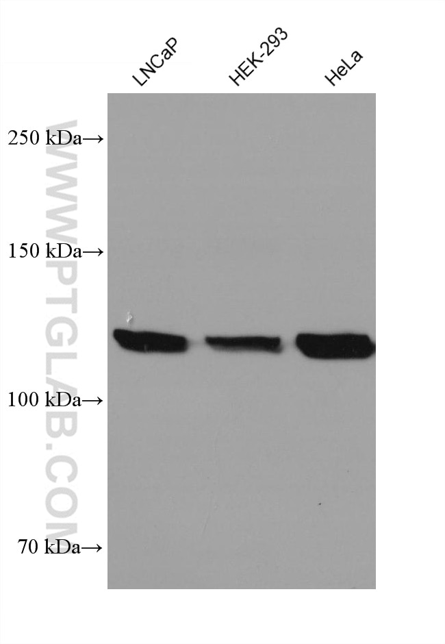
  • Western Blot (WB) analysis of various lysates using Vinculin Monoclonal antibody (66305-1-Ig)

    WB analysis using 66305-1-Ig

    Various lysates were subjected to SDS PAGE followed by western blot with 66305-1-Ig (Vinculin antibody) at dilution of 1:20000 incubated at room temperature for 1.5 hours.

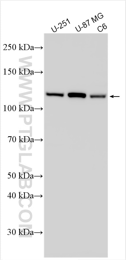
  • Western Blot (WB) analysis of various lysates using Vinculin Monoclonal antibody (66305-1-Ig)

    WB analysis using 66305-1-Ig

    Various lysates were subjected to SDS PAGE followed by western blot with 66305-1-Ig (Vinculin antibody) at dilution of 1:10000 incubated at room temperature for 1.5 hours.

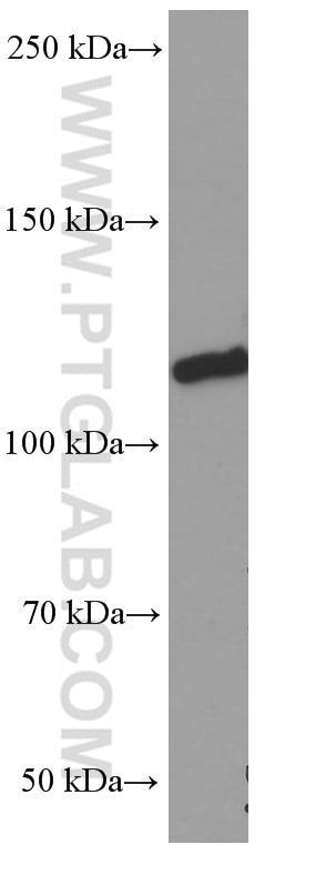
  • Western Blot (WB) analysis of pig heart tissue using Vinculin Monoclonal antibody (66305-1-Ig)

    WB analysis of pig heart using 66305-1-Ig

    pig heart tissue were subjected to SDS PAGE followed by western blot with 66305-1-Ig (Vinculin Antibody) at dilution of 1:5000 incubated at room temperature for 1.5 hours.

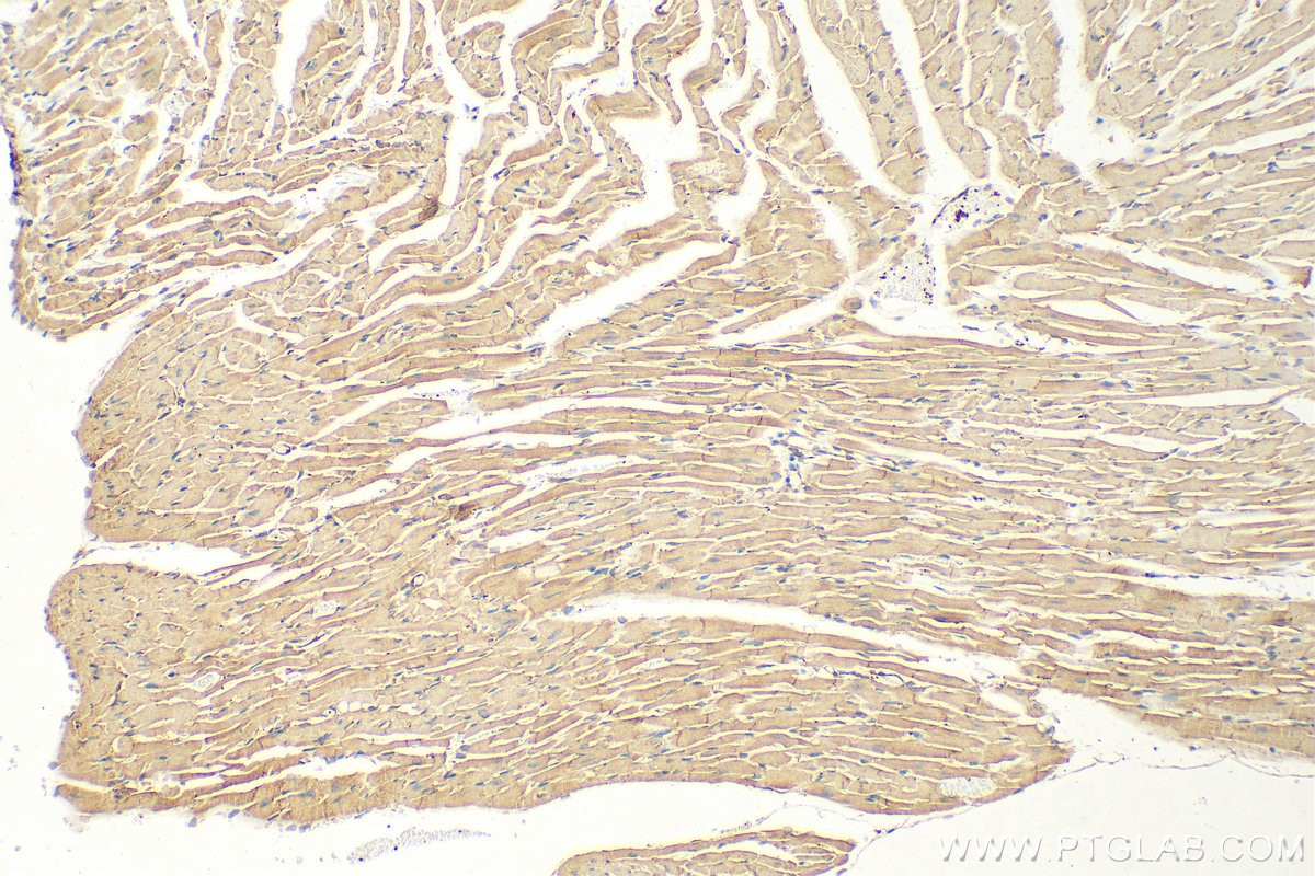
  • Immunohistochemistry (IHC) staining of mouse heart tissue using Vinculin Monoclonal antibody (66305-1-Ig)

    IHC staining of mouse heart using 66305-1-Ig

    Immunohistochemical analysis of paraffin-embedded mouse heart tissue slide using 66305-1-Ig (Vinculin antibody) at dilution of 1:400 (under 10x lens). Heat mediated antigen retrieval with Tris-EDTA buffer (pH 9.0).

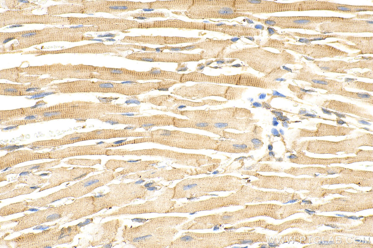
  • Immunohistochemistry (IHC) staining of mouse heart tissue using Vinculin Monoclonal antibody (66305-1-Ig)

    IHC staining of mouse heart using 66305-1-Ig

    Immunohistochemical analysis of paraffin-embedded mouse heart tissue slide using 66305-1-Ig (Vinculin antibody) at dilution of 1:400 (under 40x lens). Heat mediated antigen retrieval with Tris-EDTA buffer (pH 9.0).

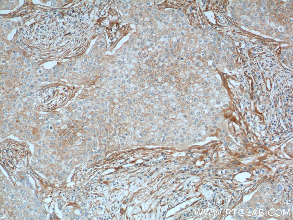
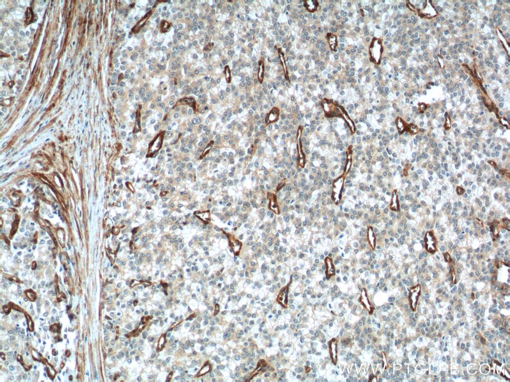
  • Immunohistochemistry (IHC) staining of human breast cancer tissue using Vinculin Monoclonal antibody (66305-1-Ig)

    IHC staining of human breast cancer using 66305-1-Ig

    Immunohistochemical analysis of paraffin-embedded human breast cancer tissue slide using 66305-1-Ig (Vinculin Antibody) at dilution of 1:200 (under 10x lens). Heat mediated antigen retrieval with Tris-EDTA buffer (pH 9.0).

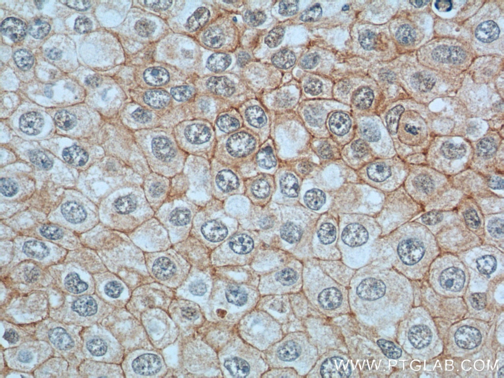
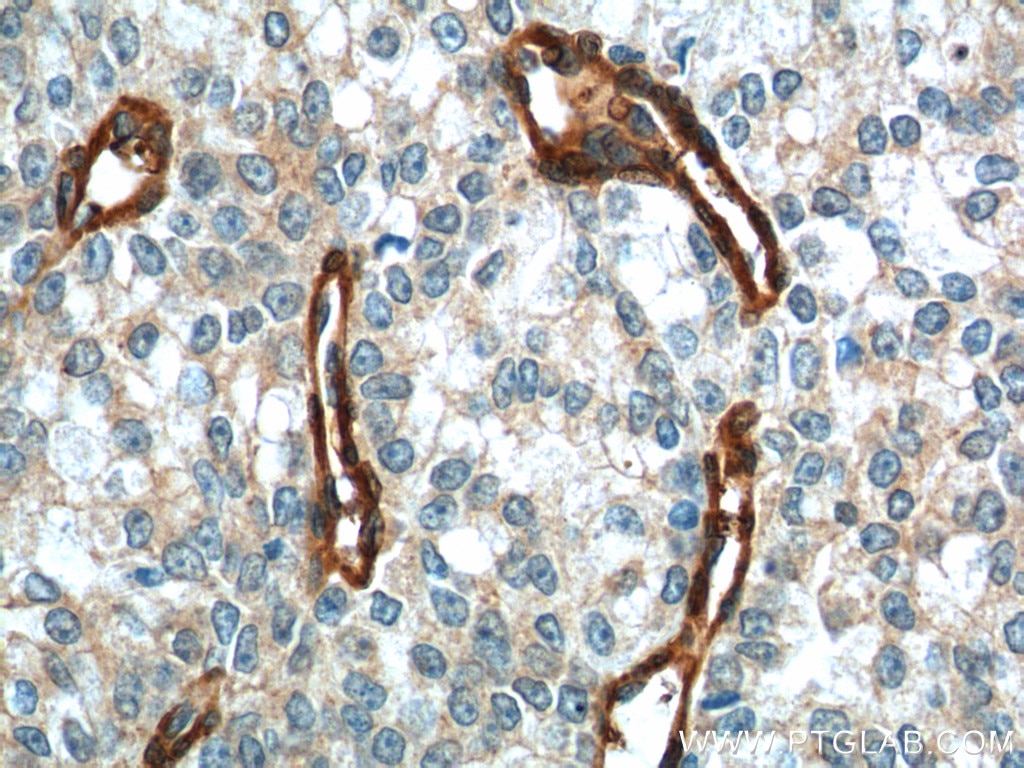
  • Immunohistochemistry (IHC) staining of human breast cancer tissue using Vinculin Monoclonal antibody (66305-1-Ig)

    IHC staining of human breast cancer using 66305-1-Ig

    Immunohistochemical analysis of paraffin-embedded human breast cancer tissue slide using 66305-1-Ig (Vinculin Antibody) at dilution of 1:200 (under 40x lens). Heat mediated antigen retrieval with Tris-EDTA buffer (pH 9.0).

  • Immunohistochemistry (IHC) staining of human prostate cancer tissue using Vinculin Monoclonal antibody (66305-1-Ig)

    IHC staining of human prostate cancer using 66305-1-Ig

    Immunohistochemical analysis of paraffin-embedded human prostate cancer tissue slide using 66305-1-Ig (Vinculin Antibody) at dilution of 1:200 (under 10x lens). Heat mediated antigen retrieval with Tris-EDTA buffer (pH 9.0).

  • Immunohistochemistry (IHC) staining of human prostate cancer tissue using Vinculin Monoclonal antibody (66305-1-Ig)

    IHC staining of human prostate cancer using 66305-1-Ig

    Immunohistochemical analysis of paraffin-embedded human prostate cancer tissue slide using 66305-1-Ig (Vinculin Antibody) at dilution of 1:200 (under 40x lens). Heat mediated antigen retrieval with Tris-EDTA buffer (pH 9.0).

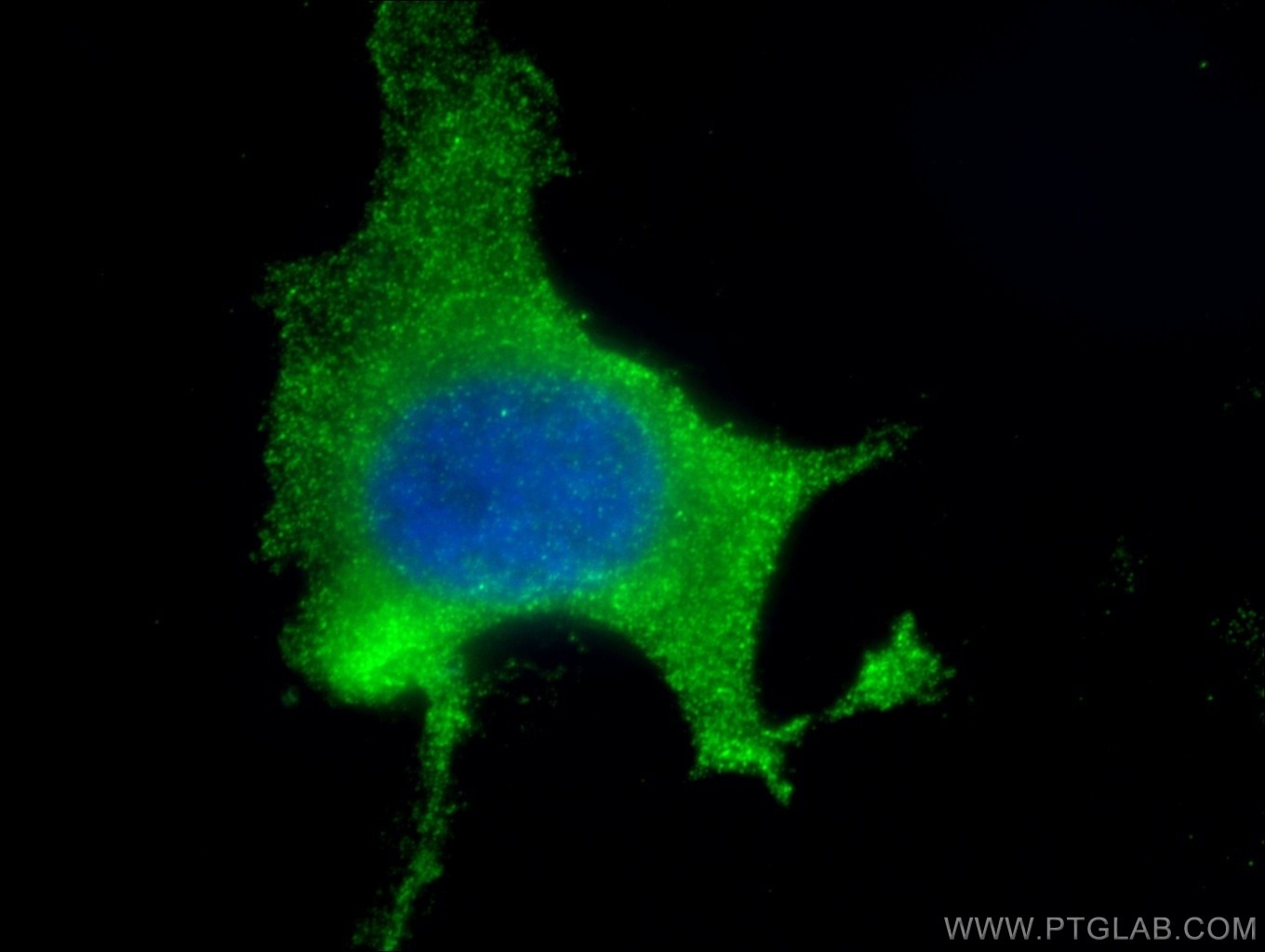
  • Immunofluorescence (IF) / fluorescent staining of HeLa cells using Vinculin Monoclonal antibody (66305-1-Ig)

    IF Staining of HeLa using 66305-1-Ig

    Immunofluorescent analysis of (10% Formaldehyde ) fixed HeLa cells using 66305-1-Ig(Vinculin antibody) at dilution of 1:200 and Alexa Fluor 488-conjugated AffiniPure Goat Anti-Mouse IgG(H+L).

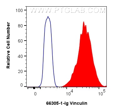
  • Flow cytometry (FC) experiment of HeLa cells using Vinculin Monoclonal antibody (66305-1-Ig)

    FC experiment of HeLa using 66305-1-Ig

    1x10^6 HeLa cells were intracellularly stained with 0.25 ug Vinculin Monoclonal antibody (66305-1-Ig, Clone:2B5A7) and CoraLite®488-Conjugated Goat Anti-Mouse IgG(H+L) (SA00013-1)(red), or 0.25 ug Mouse IgG1 isotype control Mouse McAb (66360-1-Ig, Clone: 1F8D3) (blue). Cells were fixed with 4% PFA and permeabilized with Flow Cytometry Perm Buffer (PF00011-C).

"Vinculin Antibodies" Comparison

View side-by-side comparison of Vinculin antibodies from other vendors to find the one that best suits your research needs.

Geprüfte Anwendungen

Erfolgreiche Detektion in WBHepG2-Zellen, C6-Zellen, HEK-293-Zellen, HeLa-Zellen, HuH-7-Zellen, L02-Zellen, LNCaP-Zellen, NIH/3T3-Zellen, Hausschwein-Herzgewebe, U-251-Zellen, U-87 MG-Zellen
Erfolgreiche Detektion in IHChumanes Mammakarzinomgewebe, humanes Prostatakarzinomgewebe, Mausherzgewebe
Hinweis: Antigendemaskierung mit TE-Puffer pH 9,0 empfohlen. (*) Wahlweise kann die Antigendemaskierung auch mit Citratpuffer pH 6,0 erfolgen.
Erfolgreiche Detektion in IF/ICCHeLa-Zellen
Erfolgreiche Detektion in FC (Intra)HeLa-Zellen

Empfohlene Verdünnung

AnwendungVerdünnung
Western Blot (WB)WB : 1:5000-1:50000
Immunhistochemie (IHC)IHC : 1:50-1:500
Immunfluoreszenz (IF)/ICCIF/ICC : 1:50-1:500
Durchflusszytometrie (FC) (INTRA)FC (INTRA) : 0.25 ug per 10^6 cells in a 100 µl suspension
It is recommended that this reagent should be titrated in each testing system to obtain optimal results.
Sample-dependent, check data in validation data gallery

Produktinformation

66305-1-Ig bindet in WB, IHC, IF/ICC, FC (Intra), CoIP, ELISA Vinculin und zeigt Reaktivität mit Hausschwein, human, Maus, Ratten

Getestete Reaktivität Hausschwein, human, Maus, Ratte
In Publikationen genannte Reaktivitäthuman, hamster, Hausschwein, Kaninchen, Maus, Ratte, Zebrafisch
Wirt / Isotyp Maus / IgG1
Klonalität Monoklonal
Typ Antikörper
Immunogen Vinculin fusion protein Ag24946
Vollständiger Name vinculin
Berechnetes Molekulargewicht 1133 aa, 124 kDa
Beobachtetes Molekulargewicht 117 kDa
GenBank-ZugangsnummerBC039174
Gene symbol Vinculin
Gene ID (NCBI) 7414
Konjugation Unkonjugiert
Form Liquid
Reinigungsmethode Protein-G-Reinigung
Lagerungspuffer PBS with 0.02% sodium azide and 50% glycerol
LagerungsbedingungenBei -20°C lagern. Nach dem Versand ein Jahr lang stabil Aliquotieren ist bei -20oC Lagerung nicht notwendig. 20ul Größen enthalten 0,1% BSA.

Hintergrundinformationen

Vinculin belongs to the vinculin/alpha-catenin family. It is an actin filament (F-actin)-binding protein which involved in cell-matrix adhesion and cell-cell adhesion. Vinculin regulates cell-surface E-cadherin expression and potentiates mechanosensing by the E-cadherin complex. It may also play important roles in cell morphology and locomotion. Vinculin is a 117-kDa, 1,066-amino-acid protein which is ubiquitously expressed. Its splice variant, metavinculin (124 kDa), is muscle-specific.

Protokolle

PRODUKTSPEZIFISCHE PROTOKOLLE
WB protocol for Vinculin antibody 66305-1-IgProtokoll herunterladen
IHC protocol for Vinculin antibody 66305-1-IgProtokoll herunterladenl
IF protocol for Vinculin antibody 66305-1-IgProtokoll herunterladen
STANDARD-PROTOKOLLE
Klicken Sie hier, um unsere Standardprotokolle anzuzeigen

Publikationen

SpeciesApplicationTitle
mouseWB

Crit Care

Recombinant ACE2 protein protects against acute lung injury induced by SARS-CoV-2 spike RBD protein.

Authors - Lingbing Zhang
humanWB

Adv Sci (Weinh)

RBMS1 Coordinates with the m6 A Reader YTHDF1 to Promote NSCLC Metastasis through Stimulating S100P Translation

Authors - Yu Sun
humanWB

Cell Rep Med

Development of an orally bioavailable CDK12/13 degrader and induction of synthetic lethality with AKT pathway inhibition

Authors - Yu Chang
humanWB

Nat Commun

Modular assembly of the principal microtubule nucleator γ-TuRC.

Authors - Martin Würtz
humanWB

Redox Biol

Noscapine derivative 428 suppresses ferroptosis through targeting GPX4

Authors - Youping Zhang
humanWB

Dev Cell

HMOX1-LDHB interaction promotes ferroptosis by inducing mitochondrial dysfunction in foamy macrophages during advanced atherosclerosis

Authors - Xiang Peng

Rezensionen

The reviews below have been submitted by verified Proteintech customers who received an incentive for providing their feedback.


FH

Monifa (Verified Customer) (08-31-2025)

I used vinculin as an alternative loading control to beta actin. After adjusting my transfer buffer to help vinculin transfer to my membrane, I can visualize the protein very well (with chemiluminescence).

  • Applications: Western Blot
  • Primary Antibody Dilution: 1:1000
  • Cell Tissue Type: HeLa
FH

Aditya (Verified Customer) (01-31-2025)

very clean bands, much better than the abcam antibody

  • Applications: Western Blot
  • Primary Antibody Dilution: 1:1000
FH

Morgane (Verified Customer) (01-09-2025)

Very good loading control with larger molecular size

  • Applications: Western Blot
  • Primary Antibody Dilution: 1:1000
  • Cell Tissue Type: HCT116
Vinculin Antibody Western Blot validation (1:1000 dilution) in HCT116 (Cat no:66305-1-Ig)
FH

Daniel (Verified Customer) (10-24-2024)

The antibody works really well and it gives a very clean Western blot.

  • Applications: Western Blot
  • Primary Antibody Dilution: 1:10000
  • Cell Tissue Type: eHAP, RPE1, HCT116 cells
FH

Lisa (Verified Customer) (04-29-2024)

Works super well!

  • Applications: Western Blot
  • Primary Antibody Dilution: 1:5000
FH

Parijat (Verified Customer) (08-21-2023)

Works well as loading control

  • Applications: Western Blot
  • Primary Antibody Dilution: 1:5000
  • Cell Tissue Type: HEK293T
FH

Udesh (Verified Customer) (08-16-2023)

Worked well in WB at 1:3000 and IF at 1:100

  • Applications: Western Blot, Immunofluorescence
  • Primary Antibody Dilution: 1:1000
  • Cell Tissue Type: Bone Marrow MSCs
FH

Mohamad (Verified Customer) (07-03-2023)

Very good antibody

  • Applications: Western Blot
  • Primary Antibody Dilution: 1/1000
FH

Priya (Verified Customer) (01-17-2023)

I have used for human cardiomyocytes, mouse skin and liver tissues

  • Applications: Western Blot
  • Primary Antibody Dilution: 1:1000
  • Cell Tissue Type: Cardiomyoctyes, Keratinocytes, Liver and skin
FH

Macarena (Verified Customer) (10-07-2022)

excellent results.

  • Applications: Western Blot
  • Primary Antibody Dilution: 1:10000
  • Cell Tissue Type: cell lines
FH

Jonas (Verified Customer) (07-29-2022)

This is my go to loading control stain to validate evenly loaded lanes Binding could be stronger but by using 1:500 dilution, a reliable staining can be achieved

  • Applications: Western Blot
  • Primary Antibody Dilution: 1:500
Vinculin Antibody Western Blot validation (1:500 dilution) in (Cat no:66305-1-Ig)
FH

Charlotte (Verified Customer) (07-26-2022)

Very good antibody. Very specific, super fast to reveal. Here we see Gli1 (160 kDa) because it is a mouse antibody too.
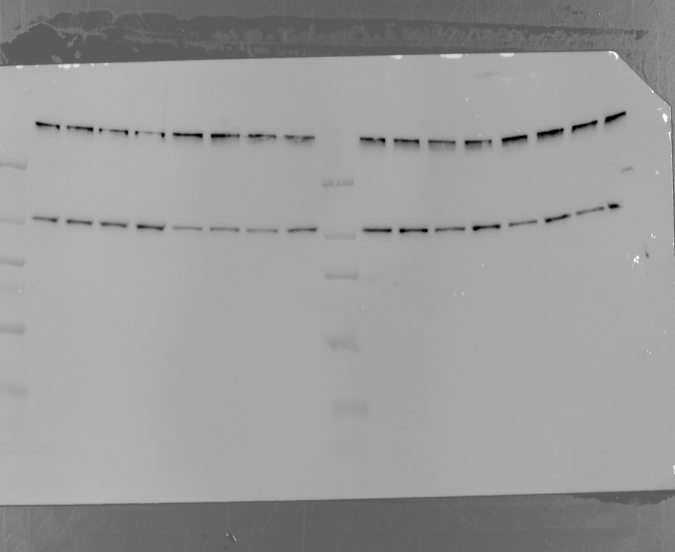
  • Applications: Western Blot
  • Primary Antibody Dilution: 1/5000
  • Cell Tissue Type: NIH-3T3
Vinculin Antibody Western Blot validation (1/5000 dilution) in NIH-3T3 (Cat no:66305-1-Ig)
FH

AKIMASA (Verified Customer) (04-28-2021)

I could get the good quality band!

  • Applications: Western Blot
  • Primary Antibody Dilution: 1:1000
  • Cell Tissue Type: OVCAR8, PEO4, Kuramochi, OVSAHO, A2780, HEY,PEO1
FH

Chun (Verified Customer) (09-07-2020)

This is a fairly good antibody for immunoblotting.

  • Applications: Western Blot
  • Cell Tissue Type: HeLa
FH

Huai-Chin (Verified Customer) (06-08-2019)

Serving as a loading control, this antibody is not that good compare to other. Still work to some extent.

  • Applications: Western Blot,
  • Primary Antibody Dilution: 1:2000
  • Cell Tissue Type: 293T