Phospho-TDP43 (Ser409/410) Polyklonaler Antikörper
Phospho-TDP43 (Ser409/410) Polyklonal Antikörper für WB, ELISA
Wirt / Isotyp
Kaninchen / IgG
Getestete Reaktivität
human, Maus und mehr (2)
Anwendung
WB, IHC, IF, IP, ELISA
Konjugation
Unkonjugiert
Kat-Nr. : 22309-1-AP
Synonyme
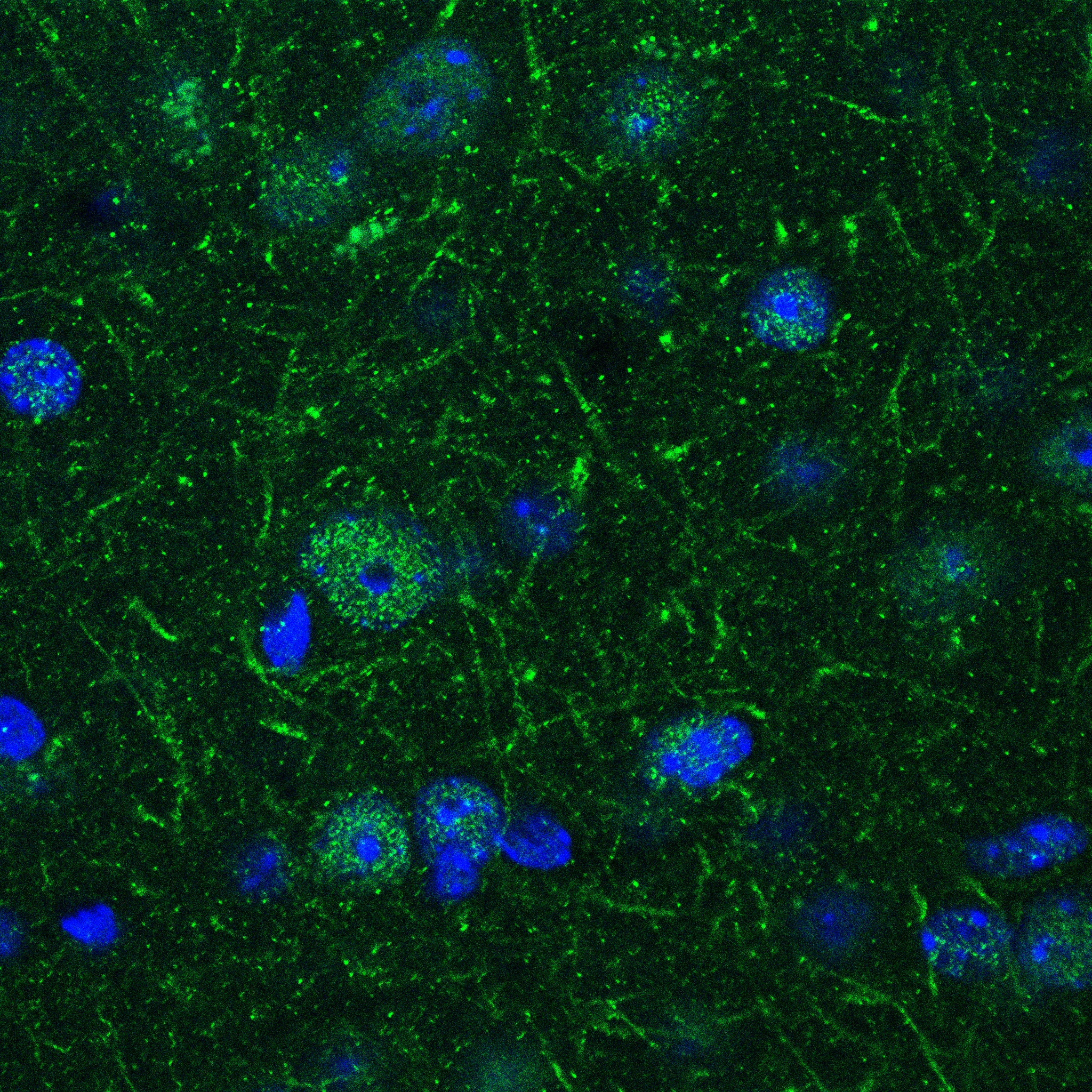
 antibody) at dilution of 1:1000 incubated at room temperature for 1 hours.")
 antibody) at dilution of 1:1500 incubated at room temperature for 1.5 hours.")
Geprüfte Anwendungen
| Erfolgreiche Detektion in WB | Neuro-2a-Zellen, Mit Calyculin A behandelte HeLa-Zellen |
Empfohlene Verdünnung
| Anwendung | Verdünnung |
|---|---|
| Western Blot (WB) | WB : 1:500-1:3000 |
| It is recommended that this reagent should be titrated in each testing system to obtain optimal results. | |
| Sample-dependent, check data in validation data gallery | |
Veröffentlichte Anwendungen
| WB | See 47 publications below |
| IHC | See 43 publications below |
| IF | See 42 publications below |
| IP | See 1 publications below |
Produktinformation
22309-1-AP bindet in WB, IHC, IF, IP, ELISA Phospho-TDP43 (Ser409/410) und zeigt Reaktivität mit human, Maus
| Getestete Reaktivität | human, Maus |
| In Publikationen genannte Reaktivität | human, Affe, Maus, Ratte |
| Wirt / Isotyp | Kaninchen / IgG |
| Klonalität | Polyklonal |
| Typ | Antikörper |
| Immunogen | Peptid |
| Vollständiger Name | TAR DNA binding protein |
| Berechnetes Molekulargewicht | 43 kDa |
| Beobachtetes Molekulargewicht | 40-50 kDa, 25-35 kDa |
| GenBank-Zugangsnummer | NM_007375 |
| Gene symbol | TDP-43 |
| Gene ID (NCBI) | 23435 |
| Konjugation | Unkonjugiert |
| Form | Liquid |
| Reinigungsmethode | Antigen-Affinitätsreinigung |
| Lagerungspuffer | PBS with 0.02% sodium azide and 50% glycerol |
| Lagerungsbedingungen | Bei -20°C lagern. Nach dem Versand ein Jahr lang stabil Aliquotieren ist bei -20oC Lagerung nicht notwendig. 20ul Größen enthalten 0,1% BSA. |
Hintergrundinformationen
Transactivation response (TAR) DNA-binding protein of 43 kDa (also known as TARDBP or TDP-43) was first isolated as a transcriptional inactivator binding to the TAR DNA element of the HIV-1 virus. Neumann et al. (2006) found that a hyperphosphorylated, ubiquitinated, and cleaved form of TARDBP, known as pathologic TDP-43, is the major component of the tau-negative and ubiquitin-positive inclusions that characterize amyotrophic lateral sclerosis (ALS) and the most common pathological subtype of frontotemporal lobar degeneration (FTLD-U). Various forms of TDP-43 exist, including 18-35 kDa of cleaved C-terminal fragments, 45-50 kDa phospho-protein, 55 kDa glycosylated form, 75 kDa hyperphosphorylated form, and 90-300 kDa cross-linked form. (PMID: 17023659,19823856, 21666678, 22193176).22309-1-AP is a rabbit polyclonal antibody recognizing TDP-43 only when phosphorylated at 409/410. Immunohistochemical analyses using this antibody only stain the insoluble inclusions in pathologic tissues without normal diffuse nuclear staining.
Protokolle
| PRODUKTSPEZIFISCHE PROTOKOLLE | |
|---|---|
| WB protocol for Phospho-TDP43 (Ser409/410) antibody 22309-1-AP | Protokoll herunterladen |
| STANDARD-PROTOKOLLE | |
|---|---|
| Klicken Sie hier, um unsere Standardprotokolle anzuzeigen |
Publikationen
| Species | Application | Title |
|---|---|---|
Nat Med The inhibition of TDP-43 mitochondrial localization blocks its neuronal toxicity. | ||
Nat Cell Biol Caspase-2 is a condensate-mediated deubiquitinase in protein quality control | ||
Nat Neurosci TREM2 interacts with TDP-43 and mediates microglial neuroprotection against TDP-43-related neurodegeneration. | ||
Alzheimers Dement Entorhinal vessel density correlates with phosphorylated tau and TDP-43 pathology | ||
JAMA Neurol TDP-43 Accumulation Within Intramuscular Nerve Bundles of Patients With Amyotrophic Lateral Sclerosis. |
Rezensionen
The reviews below have been submitted by verified Proteintech customers who received an incentive for providing their feedback.
FH Rashmi (Verified Customer) (09-25-2024) | Used for WB, Highly Recommended
|
FH Vikas (Verified Customer) (07-11-2024) | Used for western blotting. Highly recommended.
|
FH Madison (Verified Customer) (06-24-2024) | This antibody Stained well in the human tissue there were very clear phospho tangles and there was not a lot of nonspecific staining in the tissue.
|
FH MANOHAR (Verified Customer) (03-06-2024) |
|
FH Shenyi (Verified Customer) (04-27-2022) | Staining with this antibody is very different from with the recombinant one (80007-1-RR) which I received as a free test vial. It showed strong signal in the nucleus while the recombinant one only showed signal in the cytoplasm. Now I'm confused: Could pTDP-43 (Ser409/410) also present in nucleus or it's the antibody being nonspecific?
 |
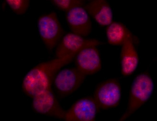
FH Azita (Verified Customer) (03-10-2020) | Immunofluorescent analysis of (4% PFA fixed) NSC-34 cells, using p- (109/110) TDP-43 antibody at 1/500 dilution and Alexa Flour conjugated Donkey anti Rabbit IgG (H+L) (Red). Bisbenzimide (Blue) labels cell nuclei.
 |
FH Laura (Verified Customer) (01-10-2020) | Works well at this concentration on HEK293T lysate.
|
FH Owen (Verified Customer) (10-30-2019) | This antibody was used to image TDP43 in tissue and cell samples. The antibody was useful to image such a difficult target.
|

