- Phare
- Validé par KD/KO
Anticorps Polyclonal de lapin anti-ACOT2
ACOT2 Polyclonal Antibody for WB, IHC, IF/ICC, IP, ELISA
Hôte / Isotype
Lapin / IgG
Réactivité testée
Humain, rat, souris
Applications
WB, IHC, IF/ICC, IP, ELISA
Conjugaison
Non conjugué
N° de cat : 15633-1-AP
Synonymes
Galerie de données de validation
 at dilution of 1:800 incubated at room temperature for 1.5 hours.")
 with sh-Control and sh-ACOT2 transfected HEK-293 cells.")
 at dilution of 1:500 incubated at room temperature for 1.5 hours.")
 at dilution of 1:500 incubated at room temperature for 1.5 hours.")
 at dilution of 1:500 incubated at room temperature for 1.5 hours.")
 at dilution of 1:500 incubated at room temperature for 1.5 hours.")
 with HepG2 cells lysate 1700ug.")
 at dilution of 1:100 (under 10x lens).")
 at dilution of 1:100 (under 40x lens).")
 at dilution of 1:200 (under 10x lens). Heat mediated antigen retrieval with Tris-EDTA buffer (pH 9.0).")
 at dilution of 1:200 (under 40x lens). Heat mediated antigen retrieval with Tris-EDTA buffer (pH 9.0).")
 at dilution of 1:100 (under 10x lens).")
 at dilution of 1:100 (under 40x lens).")
 at dilution of 1:100 (under 10x lens).")
 at dilution of 1:100 (under 40x lens).")
 at dilution of 1:100 (under 10x lens).")
 at dilution of 1:100 (under 40x lens).")
 at dilution of 1:100 (under 10x lens).")
 at dilution of 1:100 (under 40x lens).")
 at dilution of 1:100 (under 10x lens).")
 at dilution of 1:100 (under 40x lens).")
 at dilution of 1:100 (under 40x lens).")
 at dilution of 1:100 (under 10x lens).")
 fixed HepG2 cells using ACOT2 antibody (15633-1-AP) at dilution of 1:200 and CoraLite®488-Conjugated AffiniPure Goat Anti-Rabbit IgG(H+L), CL594-phalloidin (red).")
 fixed HepG2 cells using ACOT2 antibody (15633-1-AP) at dilution of 1:400 and CoraLite®488-Conjugated AffiniPure Goat Anti-Rabbit IgG(H+L), CL594-phalloidin (red).")
Applications testées
| Résultats positifs en WB | cellules HEK-293, cellules HepG2, tissu cérébral humain, tissu rénal de souris, tissu rénal humain, tissu testiculaire humain |
| Résultats positifs en IP | cellules HepG2 |
| Résultats positifs en IHC | tissu rénal humain, tissu cardiaque humain, tissu de muscle squelettique humain, tissu hépatique humain, tissu ovarien humain, tissu splénique humain, tissu testiculaire humain il est suggéré de démasquer l'antigène avec un tampon de TE buffer pH 9.0; (*) À défaut, 'le démasquage de l'antigène peut être 'effectué avec un tampon citrate pH 6,0. |
| Résultats positifs en IF/ICC | cellules HepG2, |
Dilution recommandée
| Application | Dilution |
|---|---|
| Western Blot (WB) | WB : 1:500-1:1000 |
| Immunoprécipitation (IP) | IP : 0.5-4.0 ug for 1.0-3.0 mg of total protein lysate |
| Immunohistochimie (IHC) | IHC : 1:50-1:500 |
| Immunofluorescence (IF)/ICC | IF/ICC : 1:50-1:500 |
| It is recommended that this reagent should be titrated in each testing system to obtain optimal results. | |
| Sample-dependent, check data in validation data gallery | |
Applications publiées
| WB | See 9 publications below |
| IF | See 1 publications below |
Informations sur le produit
15633-1-AP cible ACOT2 dans les applications de WB, IHC, IF/ICC, IP, ELISA et montre une réactivité avec des échantillons Humain, rat, souris
| Réactivité | Humain, rat, souris |
| Réactivité citée | rat, Humain, souris |
| Hôte / Isotype | Lapin / IgG |
| Clonalité | Polyclonal |
| Type | Anticorps |
| Immunogène | ACOT2 Protéine recombinante Ag8093 |
| Nom complet | acyl-CoA thioesterase 2 |
| Masse moléculaire calculée | 483 aa, 53 kDa |
| Poids moléculaire observé | 46-53 kDa |
| Numéro d’acquisition GenBank | BC006335 |
| Symbole du gène | ACOT2 |
| Identification du gène (NCBI) | 10965 |
| Conjugaison | Non conjugué |
| Forme | Liquide |
| Méthode de purification | Purification par affinité contre l'antigène |
| Tampon de stockage | PBS with 0.02% sodium azide and 50% glycerol |
| Conditions de stockage | Stocker à -20°C. Stable pendant un an après l'expédition. L'aliquotage n'est pas nécessaire pour le stockage à -20oC Les 20ul contiennent 0,1% de BSA. |
Informations générales
Acyl-CoA thioesterase (Acot)2 localizes to the mitochondrial matrix and hydrolyses long-chain fatty acyl-CoA into free FA and CoASH. Acot2 is expressed in highly oxidative tissues and is poised to modulate mitochondrial FA oxidation (FAO) (PMID: 25114170). The structure of ACOT2 consists of two domains, N and C domains, and the active site of ACOT2 is located at the interface between the N and C domains (PMID: 19497300).
Protocole
| Product Specific Protocols | |
|---|---|
| WB protocol for ACOT2 antibody 15633-1-AP | Download protocol |
| IHC protocol for ACOT2 antibody 15633-1-AP | Download protocol |
| IF protocol for ACOT2 antibody 15633-1-AP | Download protocol |
| IP protocol for ACOT2 antibody 15633-1-AP | Download protocol |
| Standard Protocols | |
|---|---|
| Click here to view our Standard Protocols |
Publications
| Species | Application | Title |
|---|---|---|
Eur J Immunol YY1 control of mitochondrial-related genes does not account for regulation of immunoglobulin class switch recombination in mice. | ||
Life Sci Alliance High levels of TFAM repress mammalian mitochondrial DNA transcription in vivo | ||
Biomed Res Int Up-Regulated MicroRNA-27b Promotes Adipocyte Differentiation via Induction of Acyl-CoA Thioesterase 2 Expression. | ||
J Biol Chem Requirement of hepatic pyruvate carboxylase during fasting, high fat, and ketogenic diet | ||
EMBO Mol Med A coordinated multiorgan metabolic response contributes to human mitochondrial myopathy |
Avis
The reviews below have been submitted by verified Proteintech customers who received an incentive for providing their feedback.
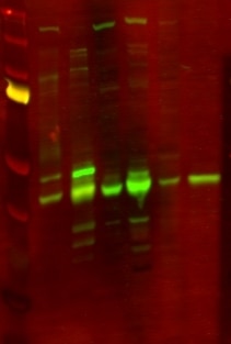
FH Lana (Verified Customer) (06-21-2019) | Lines on WB:1. BioRad Precision Plus Protein standards2. Homogenate of human cortex3. Non-synaptosomal mitochondria of human cortex4. Homogenate of mouse brains5. Non-synaptosomal mitochondria of mouse brains6. Whole cell lysate of rat PC12 cells7. Mitochondria of rat C12 cellsNotes: Specific band 48 kDa in human samples only. Strong non-specific band 40 kDa in all samples types.WB protocol:SDS-PAGE: 15 ug/ul of RIPA lysates, 4-12% Bis-tris gradient gel.Transfer: Immobilon-FL transfer membranes (Millipore), O/N at 30V, 4C.Blocking: SEA Block Blocking Buffer 1hPrimary Ab: O/N incubation at 4C.Secondary Ab: IRDye 800CW Goat anti-Rabbit, 1 h incubation at room temperature.
 |

