- Phare
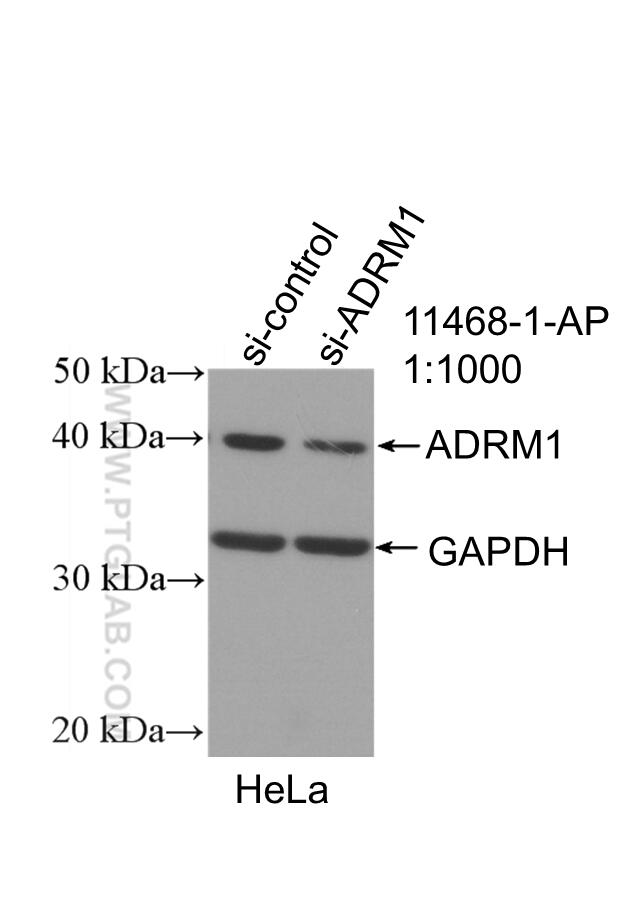
- Validé par KD/KO
Anticorps Polyclonal de lapin anti-ADRM1
ADRM1 Polyclonal Antibody for WB, IHC, IF/ICC, IP, ELISA
Hôte / Isotype
Lapin / IgG
Réactivité testée
Humain, rat, souris
Applications
WB, IHC, IF/ICC, IP, CoIP, ELISA
Conjugaison
Non conjugué
N° de cat : 11468-1-AP
Synonymes
Galerie de données de validation
Applications testées
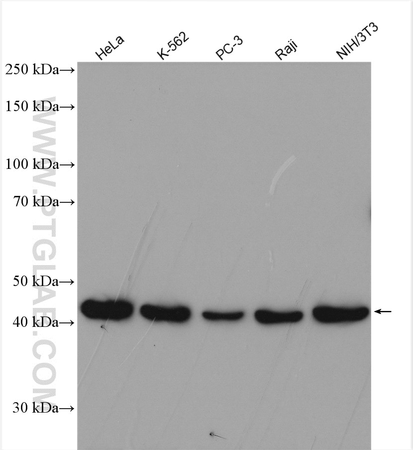
| Résultats positifs en WB | cellules HeLa, cellules K-562, cellules NIH/3T3, cellules PC-3, cellules Raji |
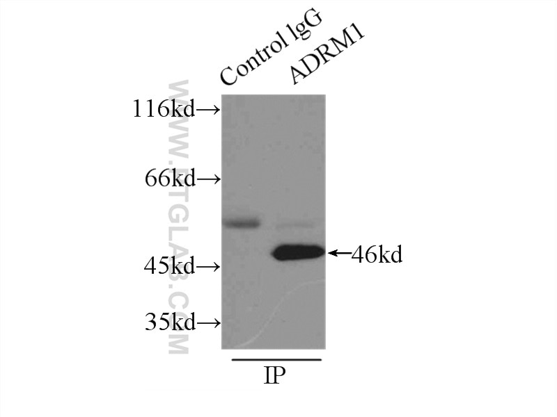
| Résultats positifs en IP | tissu testiculaire de souris |
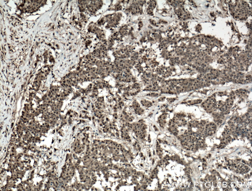
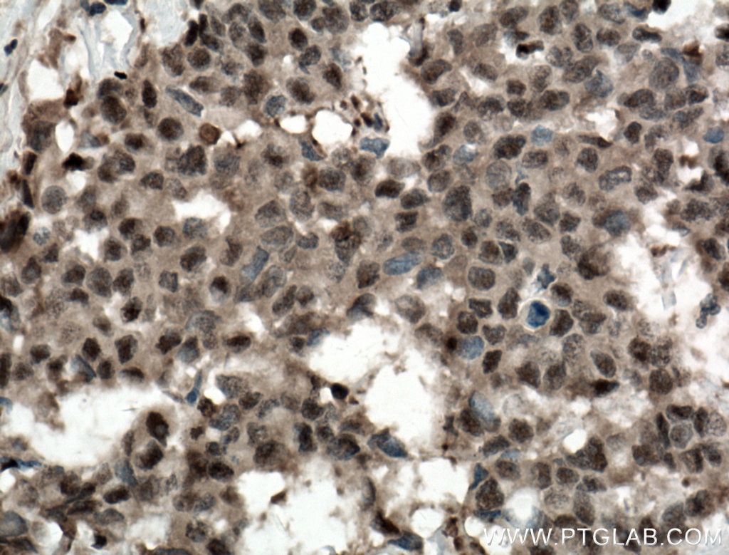
| Résultats positifs en IHC | tissu de cancer du côlon humain, il est suggéré de démasquer l'antigène avec un tampon de TE buffer pH 9.0; (*) À défaut, 'le démasquage de l'antigène peut être 'effectué avec un tampon citrate pH 6,0. |
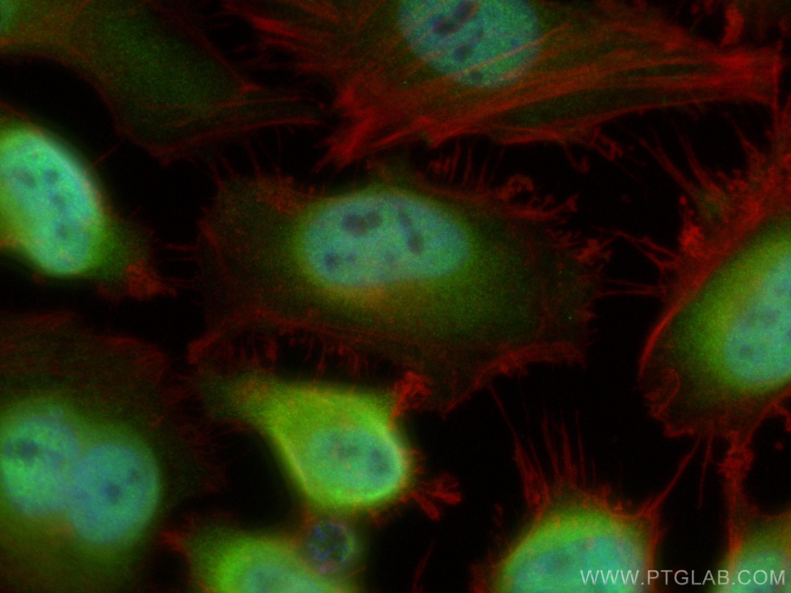
| Résultats positifs en IF/ICC | cellules HeLa, |
Dilution recommandée
| Application | Dilution |
|---|---|
| Western Blot (WB) | WB : 1:1000-1:5000 |
| Immunoprécipitation (IP) | IP : 0.5-4.0 ug for 1.0-3.0 mg of total protein lysate |
| Immunohistochimie (IHC) | IHC : 1:50-1:500 |
| Immunofluorescence (IF)/ICC | IF/ICC : 1:200-1:800 |
| It is recommended that this reagent should be titrated in each testing system to obtain optimal results. | |
| Sample-dependent, check data in validation data gallery | |
Applications publiées
| KD/KO | See 3 publications below |
| WB | See 6 publications below |
| IHC | See 5 publications below |
| IF | See 3 publications below |
| IP | See 1 publications below |
| CoIP | See 1 publications below |
Informations sur le produit
11468-1-AP cible ADRM1 dans les applications de WB, IHC, IF/ICC, IP, CoIP, ELISA et montre une réactivité avec des échantillons Humain, rat, souris
| Réactivité | Humain, rat, souris |
| Réactivité citée | Humain, souris |
| Hôte / Isotype | Lapin / IgG |
| Clonalité | Polyclonal |
| Type | Anticorps |
| Immunogène | ADRM1 Protéine recombinante Ag1997 |
| Nom complet | adhesion regulating molecule 1 |
| Masse moléculaire calculée | 407 aa, 42 kDa |
| Poids moléculaire observé | 46 kDa |
| Numéro d’acquisition GenBank | BC017245 |
| Symbole du gène | ADRM1 |
| Identification du gène (NCBI) | 11047 |
| Conjugaison | Non conjugué |
| Forme | Liquide |
| Méthode de purification | Purification par affinité contre l'antigène |
| Tampon de stockage | PBS with 0.02% sodium azide and 50% glycerol |
| Conditions de stockage | Stocker à -20°C. Stable pendant un an après l'expédition. L'aliquotage n'est pas nécessaire pour le stockage à -20oC Les 20ul contiennent 0,1% de BSA. |
Informations générales
The 26S proteasome is a key component of the ubiquitin-proteasome system, a process responsible for the majority of cellular protein degradation. hRpn13 (also termed ADRM1 or GP110) is a novel 46-kDa subunit of its 19S regulatory complex. hRpn13 binds directly to the proteasome-associated deubiquitinating enzyme, UCH37, and enhances its isopeptidase activity. Overexpression of hRpn13 promotes the activity of the ubiquitin-proteasome system and modulates the influence of osteoblasts on osteoclasts by controlling the stability of regulatory proteins in osteoblasts.
Protocole
| Product Specific Protocols | |
|---|---|
| WB protocol for ADRM1 antibody 11468-1-AP | Download protocol |
| IHC protocol for ADRM1 antibody 11468-1-AP | Download protocol |
| IF protocol for ADRM1 antibody 11468-1-AP | Download protocol |
| IP protocol for ADRM1 antibody 11468-1-AP | Download protocol |
| Standard Protocols | |
|---|---|
| Click here to view our Standard Protocols |
Publications
| Species | Application | Title |
|---|---|---|
Exp Hematol Oncol Downregulation of pro-surfactant protein B contributes to the recurrence of early-stage non-small cell lung cancer by activating PGK1-mediated Akt signaling
| ||
Proc Natl Acad Sci U S A Dendritic spinopathy in transgenic mice expressing ALS/dementia-linked mutant UBQLN2. | ||
Oncogene The degradation of p53 and its major E3 ligase Mdm2 is differentially dependent on the proteasomal ubiquitin receptor S5a. | ||
Genes Chromosomes Cancer Comprehensive analysis of 20q13 genes in ovarian cancer identifies ADRM1 as amplification target. |
