- Phare
- Validé par KD/KO
Anticorps Polyclonal de lapin anti-AIF
AIF Polyclonal Antibody for WB, IHC, IF/ICC, IP, ELISA
Hôte / Isotype
Lapin / IgG
Réactivité testée
Humain, rat, souris et plus (2)
Applications
WB, IHC, IF/ICC, IP, ELISA
Conjugaison
Non conjugué
N° de cat : 17984-1-AP
Synonymes
Galerie de données de validation
 at dilution of 1:4000 incubated at room temperature for 1.5 hours.")
 at dilution of 1:4000 incubated at room temperature for 1.5 hours.")
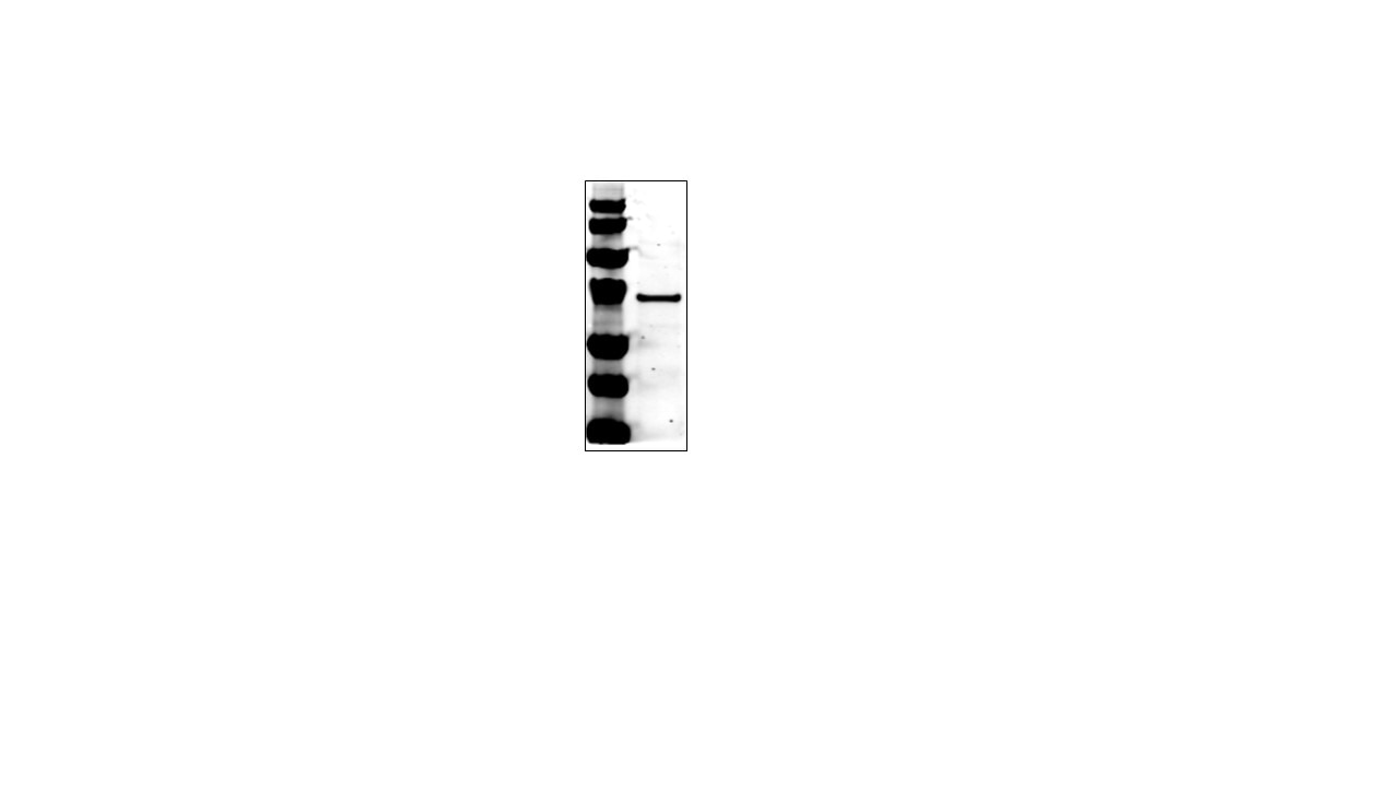
 with sh-Control and sh-AIF transfected HeLa cells.")
 with HeLa cells lysate 1320ug.")
 at dilution of 1:200 (under 40x lens).")
 at dilution of 1:200 (under 10x lens).")
 at dilution of 1:1000 (under 40x lens). Heat mediated antigen retrieval with Tris-EDTA buffer (pH 9.0).")
 at dilution of 1:1000 (under 40x lens). Heat mediated antigen retrieval with Tris-EDTA buffer (pH 9.0).")
 at dilution of 1:1000 (under 40x lens). Heat mediated antigen retrieval with Tris-EDTA buffer (pH 9.0).")
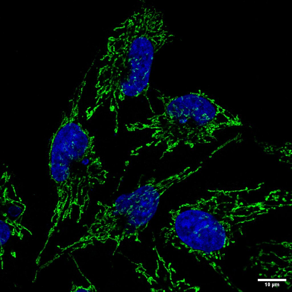
 fixed HeLa cells using AIF antibody (17984-1-AP) at dilution of 1:200 and CoraLite®488-Conjugated Goat Anti-Rabbit IgG(H+L), CL594-phalloidin (red).")
Applications testées
| Résultats positifs en WB | cellules HeLa, cellules NIH/3T3 |
| Résultats positifs en IP | cellules HeLa |
| Résultats positifs en IHC | tissu rénal humain, tissu d'estomac de souris, tissu rénal de rat, tissu rénal de souris il est suggéré de démasquer l'antigène avec un tampon de TE buffer pH 9.0; (*) À défaut, 'le démasquage de l'antigène peut être 'effectué avec un tampon citrate pH 6,0. |
| Résultats positifs en IF/ICC | cellules HeLa, |
Dilution recommandée
| Application | Dilution |
|---|---|
| Western Blot (WB) | WB : 1:1000-1:8000 |
| Immunoprécipitation (IP) | IP : 0.5-4.0 ug for 1.0-3.0 mg of total protein lysate |
| Immunohistochimie (IHC) | IHC : 1:100-1:400 |
| Immunofluorescence (IF)/ICC | IF/ICC : 1:50-1:500 |
| It is recommended that this reagent should be titrated in each testing system to obtain optimal results. | |
| Sample-dependent, check data in validation data gallery | |
Applications publiées
| KD/KO | See 4 publications below |
| WB | See 78 publications below |
| IHC | See 7 publications below |
| IF | See 21 publications below |
Informations sur le produit
17984-1-AP cible AIF dans les applications de WB, IHC, IF/ICC, IP, ELISA et montre une réactivité avec des échantillons Humain, rat, souris
| Réactivité | Humain, rat, souris |
| Réactivité citée | rat, canin, Humain, porc, souris |
| Hôte / Isotype | Lapin / IgG |
| Clonalité | Polyclonal |
| Type | Anticorps |
| Immunogène | AIF Protéine recombinante Ag12400 |
| Nom complet | apoptosis-inducing factor, mitochondrion-associated, 1 |
| Masse moléculaire calculée | 609 aa, 66 kDa |
| Poids moléculaire observé | 67 kDa, 57 kDa |
| Numéro d’acquisition GenBank | BC111065 |
| Symbole du gène | AIF |
| Identification du gène (NCBI) | 9131 |
| Conjugaison | Non conjugué |
| Forme | Liquide |
| Méthode de purification | Purification par affinité contre l'antigène |
| Tampon de stockage | PBS with 0.02% sodium azide and 50% glycerol |
| Conditions de stockage | Stocker à -20°C. Stable pendant un an après l'expédition. L'aliquotage n'est pas nécessaire pour le stockage à -20oC Les 20ul contiennent 0,1% de BSA. |
Informations générales
Apoptosis-inducing factor (AIF) is one of the mitochondrial proteins to be released into the cytosol during apoptosis, and it is discovered as the first protein that regulates caspase-independent apoptosis(PMID:20494118). AIF is encoded as a 67 kDa protein that contains a mitochondrial localization signal (MLS) in the N-terminus.It is cleaved from the 62 kDa to the 57 kDa form following ischemic injury and translocated from the mitochondria to the nucleus in a calpain-dependent manner(PMID: 25101006).
Protocole
| Product Specific Protocols | |
|---|---|
| WB protocol for AIF antibody 17984-1-AP | Download protocol |
| IHC protocol for AIF antibody 17984-1-AP | Download protocol |
| IF protocol for AIF antibody 17984-1-AP | Download protocol |
| IP protocol for AIF antibody 17984-1-AP | Download protocol |
| Standard Protocols | |
|---|---|
| Click here to view our Standard Protocols |
Publications
| Species | Application | Title |
|---|---|---|
Cell Rep Newly synthesized AIFM1 determines the hypersensitivity of T lymphocytes to STING activation-induced cell apoptosis
| ||
Phytomedicine LC-MS based metabonomics study on protective mechanism of ESWW in cerebral ischemia via CYTC/Apaf-1/NDRG4 pathway | ||
Oxid Med Cell Longev miRNA-27a Transcription Activated by c-Fos Regulates Myocardial Ischemia-Reperfusion Injury by Targeting ATAD3a. | ||
Elife Genome-wide CRISPRi screening identifies OCIAD1 as a prohibitin client and regulatory determinant of mitochondrial Complex III assembly in human cells. | ||
PLoS Pathog GP73 represses host innate immune response to promote virus replication by facilitating MAVS and TRAF6 degradation. | ||
J Med Chem Anticancer Effects of Honokiol via Mitochondrial Dysfunction Are Strongly Enhanced by the Mitochondria-Targeting Carrier Berberine. |
Avis
The reviews below have been submitted by verified Proteintech customers who received an incentive for providing their feedback.
FH Tom (Verified Customer) (11-13-2020) | HeLa cells permeablisied (0.1% Triton X-100 in PBS) then blocked (1% BSA in PBS). Cells incubated with primary antibody (1:100) for 1 hour, 3xPBS prior to 1 hour incubation with goat anti-rabbit 488 Alexa Fluor secondary antibody.
 |
FH Praveen (Verified Customer) (05-17-2019) | Excellent Antibody
 |

