- Phare
- Validé par KD/KO
Anticorps Polyclonal de lapin anti-AMOT
AMOT Polyclonal Antibody for WB, IHC, IF/ICC, IP, ELISA
Hôte / Isotype
Lapin / IgG
Réactivité testée
Humain
Applications
WB, IHC, IF/ICC, IP, ELISA
Conjugaison
Non conjugué
N° de cat : 24550-1-AP
Synonymes
Galerie de données de validation
 with si-Control and si-AMOT transfected HEK293 cells.")
 at dilution of 1:4500 incubated at room temperature for 1.5 hours.")
 with HEK-293 cells lysate 1280 ug.")
 at dilution of 1:200 (under 10x lens. Heat mediated antigen retrieval with Tris-EDTA buffer (pH 9.0).")
 at dilution of 1:200 (under 40x lens. Heat mediated antigen retrieval with Tris-EDTA buffer (pH 9.0).")
 fixed HEK-293 cells using AMOT antibody (24550-1-AP) at dilution of 1:200 and CoraLite®488-Conjugated AffiniPure Goat Anti-Rabbit IgG(H+L) (SA00013-2).")
Applications testées
| Résultats positifs en WB | cellules HEK-293, HEK-293T |
| Résultats positifs en IP | cellules HEK-293, |
| Résultats positifs en IHC | tissu de cancer du côlon humain, il est suggéré de démasquer l'antigène avec un tampon de TE buffer pH 9.0; (*) À défaut, 'le démasquage de l'antigène peut être 'effectué avec un tampon citrate pH 6,0. |
| Résultats positifs en IF/ICC | cellules HEK-293, |
Dilution recommandée
| Application | Dilution |
|---|---|
| Western Blot (WB) | WB : 1:1000-1:9000 |
| Immunoprécipitation (IP) | IP : 0.5-4.0 ug for 1.0-3.0 mg of total protein lysate |
| Immunohistochimie (IHC) | IHC : 1:50-1:500 |
| Immunofluorescence (IF)/ICC | IF/ICC : 1:50-1:500 |
| It is recommended that this reagent should be titrated in each testing system to obtain optimal results. | |
| Sample-dependent, check data in validation data gallery | |
Applications publiées
| WB | See 2 publications below |
| IHC | See 1 publications below |
| IF | See 1 publications below |
| IP | See 1 publications below |
Informations sur le produit
24550-1-AP cible AMOT dans les applications de WB, IHC, IF/ICC, IP, ELISA et montre une réactivité avec des échantillons Humain
| Réactivité | Humain |
| Réactivité citée | Humain |
| Hôte / Isotype | Lapin / IgG |
| Clonalité | Polyclonal |
| Type | Anticorps |
| Immunogène | AMOT Protéine recombinante Ag19784 |
| Nom complet | angiomotin |
| Masse moléculaire calculée | 1084 aa, 118 kDa |
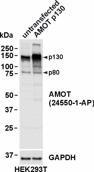
| Poids moléculaire observé | 80 kDa, 130 kDa |
| Numéro d’acquisition GenBank | BC130294 |
| Symbole du gène | AMOT |
| Identification du gène (NCBI) | 154796 |
| Conjugaison | Non conjugué |
| Forme | Liquide |
| Méthode de purification | Purification par affinité contre l'antigène |
| Tampon de stockage | PBS with 0.02% sodium azide and 50% glycerol |
| Conditions de stockage | Stocker à -20°C. Stable pendant un an après l'expédition. L'aliquotage n'est pas nécessaire pour le stockage à -20oC Les 20ul contiennent 0,1% de BSA. |
Informations générales
Angiomotin belongs to the motion family of angiostatin-binding proteins. The encoded protein is expressed predominantly in endothelial cells of capillaries as well as larger vessels of the placenta where it may mediate the inhibitory effect of angiostatin on tube formation and the migration of endothelial cells during the formation of new blood vessels. The most abundant expression was found in the placenta and skeletal muscle. AMOT has two isoforms with MW 130 kDa (p130) and 80 kDa (p80). The p130 isoform can interact with F-actin. This antibody recognizes both p130 and p80.
Protocole
| Product Specific Protocols | |
|---|---|
| WB protocol for AMOT antibody 24550-1-AP | Download protocol |
| IHC protocol for AMOT antibody 24550-1-AP | Download protocol |
| IF protocol for AMOT antibody 24550-1-AP | Download protocol |
| IP protocol for AMOT antibody 24550-1-AP | Download protocol |
| Standard Protocols | |
|---|---|
| Click here to view our Standard Protocols |
Publications
| Species | Application | Title |
|---|---|---|
Theranostics The WW domains dictate isoform-specific regulation of YAP1 stability and pancreatic cancer cell malignancy. | ||
Sci Rep MAGI1 inhibits the AMOTL2/p38 stress pathway and prevents luminal breast tumorigenesis. | ||
Mol Cell Biochem PSAT1 promotes the progression of colorectal cancer by regulating Hippo-YAP/TAZ-ID1 axis via AMOT |
Avis
The reviews below have been submitted by verified Proteintech customers who received an incentive for providing their feedback.
FH Jonasz Jeremiasz (Verified Customer) (07-30-2024) | The antibody works perfectly in western blot analysis of human cell lines, detecting both isoforms of the AMOT protein at endogenous expression levels and when overexpressed.
 |

