- Phare
- Validé par KD/KO
Anticorps Monoclonal anti-Arginase-1
Arginase-1 Monoclonal Antibody for WB, IHC, IF/ICC, IF-P, IP, ELISA
Hôte / Isotype
Mouse / IgG1
Réactivité testée
Humain, Lapin, porc, rat, souris
Applications
WB, IHC, IF/ICC, IF-P, IP, ELISA
Conjugaison
Non conjugué
CloneNo.
5D6D12
N° de cat : 66129-1-Ig
Synonymes
Galerie de données de validation
 at dilution of 1:20000 incubated at room temperature for 1.5 hours.")
 with mouse liver tissue lysate 2240 ug.")
 at dilution of 1:4000 (under 10x lens). Heat mediated antigen retrieval with Tris-EDTA buffer (pH 9.0).")
 at dilution of 1:4800 (under 10x lens). Heat mediated antigen retrieval with Tris-EDTA buffer (pH 9.0).")
 at dilution of 1:4800 (under 40x lens). Heat mediated antigen retrieval with Tris-EDTA buffer (pH 9.0).")
 fixed paraffin-embedded mouse liver tissue using Arginase-1 antibody (66129-1-Ig, Clone: 5D6D12 ) at dilution of 1:400 and Multi-rAb CoraLite ® Plus 488-Goat Anti-Mouse Recombinant Secondary Antibody (H+L) (RGAM002). Heat mediated antigen retrieval with Tris-EDTA buffer (pH 9.0).")
 fixed paraffin-embedded mouse liver tissue using Arginase-1 antibody (66129-1-Ig, Clone: 5D6D12 ) at dilution of 1:400 and Multi-rAb CoraLite ® Plus 488-Goat Anti-Mouse Recombinant Secondary Antibody (H+L) (RGAM002). Heat mediated antigen retrieval with Tris-EDTA buffer (pH 9.0).")
 fixed paraffin-embedded mouse liver tissue using Arginase-1 antibody (66129-1-Ig, Clone: 5D6D12 ) at dilution of 1:400 and CoraLite®488-Conjugated AffiniPure Goat Anti-Mouse IgG(H+L). Heat mediated antigen retrieval with Tris-EDTA buffer (pH 9.0).")
 fixed paraffin-embedded mouse liver tissue using Arginase-1 antibody (66129-1-Ig, Clone: 5D6D12 ) at dilution of 1:400 and CoraLite®488-Conjugated AffiniPure Goat Anti-Mouse IgG(H+L). Heat mediated antigen retrieval with Tris-EDTA buffer (pH 9.0).")
 fixed HepG2 cells using Arginase-1 antibody (66129-1-Ig, Clone: 5D6D12 ) at dilution of 1:800 and CoraLite®488-Conjugated AffiniPure Goat Anti-Mouse IgG(H+L).")
Applications testées
| Résultats positifs en WB | tissu hépatique de porc, tissu hépatique de lapin, tissu hépatique de rat, tissu hépatique de souris |
| Résultats positifs en IP | tissu hépatique de souris, |
| Résultats positifs en IHC | tissu hépatique de souris, tissu hépatique humain il est suggéré de démasquer l'antigène avec un tampon de TE buffer pH 9.0; (*) À défaut, 'le démasquage de l'antigène peut être 'effectué avec un tampon citrate pH 6,0. |
| Résultats positifs en IF-P | tissu hépatique de souris, |
| Résultats positifs en IF/ICC | cellules HepG2, tissu hépatique de souris |
Dilution recommandée
| Application | Dilution |
|---|---|
| Western Blot (WB) | WB : 1:5000-1:50000 |
| Immunoprécipitation (IP) | IP : 0.5-4.0 ug for 1.0-3.0 mg of total protein lysate |
| Immunohistochimie (IHC) | IHC : 1:2000-1:5000 |
| Immunofluorescence (IF)-P | IF-P : 1:200-1:800 |
| Immunofluorescence (IF)/ICC | IF/ICC : 1:400-1:1600 |
| It is recommended that this reagent should be titrated in each testing system to obtain optimal results. | |
| Sample-dependent, check data in validation data gallery | |
Applications publiées
| KD/KO | See 1 publications below |
| WB | See 86 publications below |
| IHC | See 8 publications below |
| IF | See 53 publications below |
Informations sur le produit
66129-1-Ig cible Arginase-1 dans les applications de WB, IHC, IF/ICC, IF-P, IP, ELISA et montre une réactivité avec des échantillons Humain, Lapin, porc, rat, souris
| Réactivité | Humain, Lapin, porc, rat, souris |
| Réactivité citée | rat, Humain, souris |
| Hôte / Isotype | Mouse / IgG1 |
| Clonalité | Monoclonal |
| Type | Anticorps |
| Immunogène | Arginase-1 Protéine recombinante Ag8810 |
| Nom complet | arginase, liver |
| Masse moléculaire calculée | 236aa,25 kDa; 322aa,35 kDa |
| Poids moléculaire observé | 36 kDa |
| Numéro d’acquisition GenBank | BC005321 |
| Symbole du gène | Arginase-1 |
| Identification du gène (NCBI) | 383 |
| Conjugaison | Non conjugué |
| Forme | Liquide |
| Méthode de purification | Purification par protéine G |
| Tampon de stockage | PBS with 0.02% sodium azide and 50% glycerol |
| Conditions de stockage | Stocker à -20°C. Stable pendant un an après l'expédition. L'aliquotage n'est pas nécessaire pour le stockage à -20oC Les 20ul contiennent 0,1% de BSA. |
Informations générales
Arginase-1 (Liver arginase) belongs to the arginase family. ARG1 is a novel immunohistochemical marker of hepatocellular differentiation in fine needle aspiration cytology and a marker of hepatocytes and hepatocellular neoplasms. ARG1 is closely associated with alternative macrophage activation and ARG1 has been shown to protectmotor neurons from trophic factor deprivation and allow sensory neurons to overcome neurite outgrowth inhibition by myelin proteins (PMID: 20071539, PMID:12098359). It can exsit as a homotrimer and it has 3 isoforms produced by alternative splicing (PMID:16141327). Defects in ARG1 are the cause of argininemia (ARGIN). Deletion or TNF-mediated restriction of ARG1 unleashes the production of NO by NOS2, which is critical for pathogen control (PMID:27117406). Before stroke, ARG1 mainly expressed in neurons in a normal brain (PMID: 23311438). The expression of ARG1 increases in microglia/macrophages and astrocytes early after CNS injuries. ARG1 has been regarded as a marker for beneficial microglia/macrophages and possesses antiinflammatory and tissue repair properties under various pathological conditions (PMID: 26538310, PMID: 31619589).
Protocole
| Product Specific Protocols | |
|---|---|
| WB protocol for Arginase-1 antibody 66129-1-Ig | Download protocol |
| IHC protocol for Arginase-1 antibody 66129-1-Ig | Download protocol |
| IF protocol for Arginase-1 antibody 66129-1-Ig | Download protocol |
| IP protocol for Arginase-1 antibody 66129-1-Ig | Download protocol |
| Standard Protocols | |
|---|---|
| Click here to view our Standard Protocols |
Publications
| Species | Application | Title |
|---|---|---|
Immunity Excessive Polyamine Generation in Keratinocytes Promotes Self-RNA Sensing by Dendritic Cells in Psoriasis. | ||
Bioact Mater 3D printing of interferon γ-preconditioned NSC-derived exosomes/collagen/chitosan biological scaffolds for neurological recovery after TBI | ||
J Control Release Gut commensal bacteria Parabacteroides goldsteinii-derived outer membrane vesicles suppress skin inflammation in psoriasis | ||
J Nanobiotechnology Inflammatory memory-activated biomimetic nanovesicles regulate neutrophil plasticity and metabolic reprogramming for rapid diabetic wound healing via targeting miR-193a-5p/TLR4/JNK/P38 MAPK pathways | ||
Adv Healthc Mater A Smart MMP-9-responsive Hydrogel Releasing M2 Macrophage-derived Exosomes for Diabetic Wound Healing | ||
J Neuroinflammation Succinate-induced macrophage polarization and RBP4 secretion promote vascular sprouting in ocular neovascularization |
Avis
The reviews below have been submitted by verified Proteintech customers who received an incentive for providing their feedback.
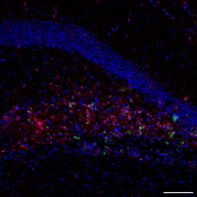
FH Marion (Verified Customer) (11-24-2024) | DAPI (blue) + Iba1 (Red) + Arg1 (green) Arg1 colocalizes with Iba1 staining M2-type microglia in this model of AD. We are satisfied with the results.
 |

