- Phare
- Validé par KD/KO
Anticorps Monoclonal anti-ATP5A1
ATP5A1 Monoclonal Antibody for WB, IHC, IF/ICC, IP, ELISA
Hôte / Isotype
Mouse / IgG2b
Réactivité testée
Humain, rat, singe, souris et plus (1)
Applications
WB, IHC, IF/ICC, IP, ELISA
Conjugaison
Non conjugué
CloneNo.
1B10H3
N° de cat : 66037-1-Ig
Synonymes
Galerie de données de validation
 at dilution of 1:25000 incubated at room temperature for 1.5 hours.")
 with sh-Control and sh-ATP5A1 transfected HepG2 cells.")

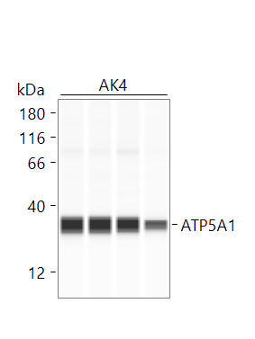
 at dilution of 1:25000 incubated at room temperature for 1.5 hours.")
 at dilution of 1:25000 incubated at room temperature for 1.5 hours.")
 with mouse heart tissue lysate 4000ug.")
 at dilution of 1:50 (under 10x lens).")
 at dilution of 1:50 (under 40x lens).")
 at dilution of 1:2000 (under 40x lens). Heat mediated antigen retrieval with Tris-EDTA buffer (pH 9.0).")
 at dilution of 1:50 (under 10x lens).")
 at dilution of 1:50 (under 40x lens).")
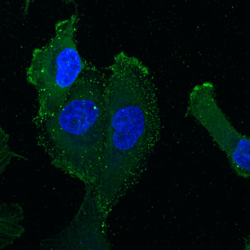
 fixed HepG2 cells using 66037-1-Ig (ATP5A1 antibody) at dilution of 1:300 and CoraLite488-Conjugated AffiniPure Goat Anti-Mouse IgG(H+L). Red: CL555-phalloidin staining of F-actin. .")
 fixed HeLa cells using ATP5A1 antibody (66037-1-Ig, Clone: 1B10H3 ) at dilution of 1:800 and CoraLite®594-Conjugated AffiniPure Goat Anti-Mouse IgG(H+L).")
 fixed HeLa cells using ATP5A1 antibody (66037-1-Ig, Clone: 1B10H3 ) at dilution of 1:800 and CoraLite®488-Conjugated Goat Anti-Mouse IgG(H+L) (SA00013-1), CL594-phalloidin (red).")
 fixed HeLa cells using 66037-1-Ig (ATP5A1 antibody) at dilution of 1:100 and CoraLite488-Conjugated AffiniPure Goat Anti-Mouse IgG(H+L).")
Applications testées
| Résultats positifs en WB | cellules MCF-7, cellules HEK-293, cellules HeLa, cellules HepG2, cellules HSC-T6, cellules NIH/3T3, cellules RAW 264.7 |
| Résultats positifs en IP | tissu cardiaque de souris |
| Résultats positifs en IHC | tissu de cancer du foie humain, tissu cardiaque humain, tissu hépatique humain il est suggéré de démasquer l'antigène avec un tampon de TE buffer pH 9.0; (*) À défaut, 'le démasquage de l'antigène peut être 'effectué avec un tampon citrate pH 6,0. |
| Résultats positifs en IF/ICC | cellules HepG2, cellules HeLa |
Dilution recommandée
| Application | Dilution |
|---|---|
| Western Blot (WB) | WB : 1:5000-1:50000 |
| Immunoprécipitation (IP) | IP : 0.5-4.0 ug for 1.0-3.0 mg of total protein lysate |
| Immunohistochimie (IHC) | IHC : 1:1000-1:4000 |
| Immunofluorescence (IF)/ICC | IF/ICC : 1:150-1:600 |
| It is recommended that this reagent should be titrated in each testing system to obtain optimal results. | |
| Sample-dependent, check data in validation data gallery | |
Applications publiées
| WB | See 23 publications below |
| IHC | See 1 publications below |
| IF | See 10 publications below |
| IP | See 1 publications below |
Informations sur le produit
66037-1-Ig cible ATP5A1 dans les applications de WB, IHC, IF/ICC, IP, ELISA et montre une réactivité avec des échantillons Humain, rat, singe, souris
| Réactivité | Humain, rat, singe, souris |
| Réactivité citée | rat, Humain, souris, Hamster |
| Hôte / Isotype | Mouse / IgG2b |
| Clonalité | Monoclonal |
| Type | Anticorps |
| Immunogène | ATP5A1 Protéine recombinante Ag8119 |
| Nom complet | ATP synthase, H+ transporting, mitochondrial F1 complex, alpha subunit 1, cardiac muscle |
| Masse moléculaire calculée | 60 kDa |
| Poids moléculaire observé | 50 kDa |
| Numéro d’acquisition GenBank | BC064562 |
| Symbole du gène | ATP5A1 |
| Identification du gène (NCBI) | 498 |
| Conjugaison | Non conjugué |
| Forme | Liquide |
| Méthode de purification | Purification par protéine A |
| Tampon de stockage | PBS with 0.02% sodium azide and 50% glycerol |
| Conditions de stockage | Stocker à -20°C. Stable pendant un an après l'expédition. L'aliquotage n'est pas nécessaire pour le stockage à -20oC Les 20ul contiennent 0,1% de BSA. |
Informations générales
The ATP5A1 gene encodes the α subunit of mitochondrial ATP synthase which produces ATP from ADP in the presence of a proton gradient across the membrane. The mitochondrial ATP synthase, also known as Complex V or F1F0 ATP synthase, is a multi-subunit enzyme complex consisting of two functional domains, the F1-containing the catalytic core and the Fo- containing the membrane proton channel. F0 domain has 10 subunits: a, b, c, d, e, f, g, OSCP, A6L, and F6. F1 is composed of subunits α, β, γ, δ, ε, and a loosely attached inhibitor protein IF1. Recently defect in ATP5A1 has been linked to the fatal neonatal mitochondrial encephalopathy. ATP5A1 is localized in the mitochondria and anti-ATP5A1 can be used as the loading control for mitochondrial or Complex V proteins. This antibody recognizes the endogenous ATP5A1 protein in lysates from various cell lines and tissues. The predicted MW of ATP5A1 is 60 kDa, while it undergoes the transit peptide cleavage to become a mature form around 50-55 kDa. Several isoforms of ATP5A1 exist due to the alternative splicing.
Protocole
| Product Specific Protocols | |
|---|---|
| WB protocol for ATP5A1 antibody 66037-1-Ig | Download protocol |
| IHC protocol for ATP5A1 antibody 66037-1-Ig | Download protocol |
| IF protocol for ATP5A1 antibody 66037-1-Ig | Download protocol |
| IP protocol for ATP5A1 antibody 66037-1-Ig | Download protocol |
| Standard Protocols | |
|---|---|
| Click here to view our Standard Protocols |
Publications
| Species | Application | Title |
|---|---|---|
Nat Cell Biol AIDA directly connects sympathetic innervation to adaptive thermogenesis by UCP1. | ||
Nat Commun The non-redundant functions of PIWI family proteins in gametogenesis in golden hamsters | ||
Dev Cell PDGF signaling inhibits mitophagy in glioblastoma stem cells through N6-methyladenosine. | ||
Autophagy AKT-mediated phosphorylation of ATG4B impairs mitochondrial activity and enhances the Warburg effect in hepatocellular carcinoma cells. |
Avis
The reviews below have been submitted by verified Proteintech customers who received an incentive for providing their feedback.
FH Anis (Verified Customer) (06-26-2025) | Jess - SimpleWestern, 1:40 in antibody diluent 2 Used as a marker for mitochondria isolation purity
 |
FH Paulina (Verified Customer) (03-26-2025) | Fixation: 2% PFA for 20min. Permeabilization/antibody dilution solution: 0.05% saponin, 5% horse serum in PBS. Primary antibody diluted 1:100 in saponin solution for 1hr at room temperature, followed by anti-mouse conjugated to AlexaFluor 568 (1:200 in saponin solution) for 1 hr at room temperature. I got very low signal and a lot of background specks, so this antibody does not work well with the saponin permeabilization.
 |
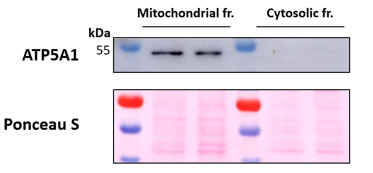
FH Marina (Verified Customer) (09-12-2022) | Mitochondrial fractions were obtained from whole cell lysates of 2 immortalized human breast cancer cell lines using a commercial kit, leading to pure mitochondrial fractions (ATP5A1 positive) and fractions comprising the cytosols minus the mitochondria (ATP5A1 negative). Primary antibody incubation was performed overnight at 4ºC. Ponceau S is used as total protein loading control.
 |
FH Cassandr (Verified Customer) (02-10-2022) | Consistently works antibody
 |

