- Phare
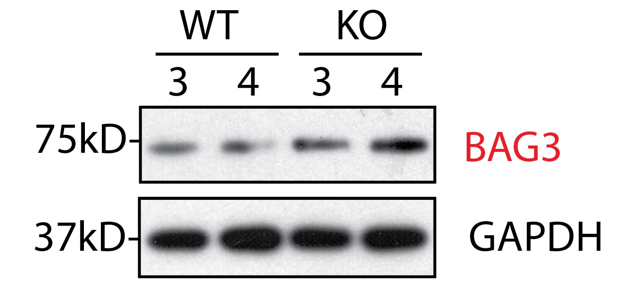
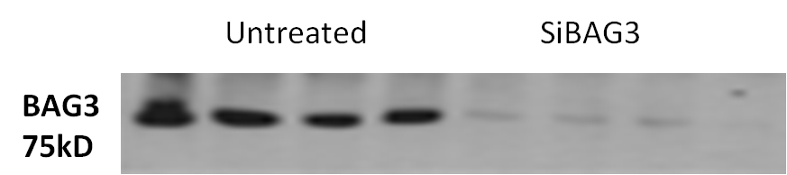
- Validé par KD/KO
Anticorps Polyclonal de lapin anti-BAG3
BAG3 Polyclonal Antibody for WB, IHC, IF/ICC, IP, ELISA
Hôte / Isotype
Lapin / IgG
Réactivité testée
Humain, rat, souris et plus (2)
Applications
WB, IHC, IF/ICC, IP, CoIP, ELISA
Conjugaison
Non conjugué
N° de cat : 10599-1-AP
Synonymes
Galerie de données de validation
 at dilution of 1:3000 incubated at room temperature for 1.5 hours.")
 at dilution of 1:8000 incubated at room temperature for 1.5 hours.")
 with K-562 cells lysate 11000ug.")
 at dilution of 1:1000 (under 40x lens). Heat mediated antigen retrieval with Tris-EDTA buffer (pH 9.0).")
 at dilution of 1:1000 (under 10x lens). Heat mediated antigen retrieval with Tris-EDTA buffer (pH 9.0).")
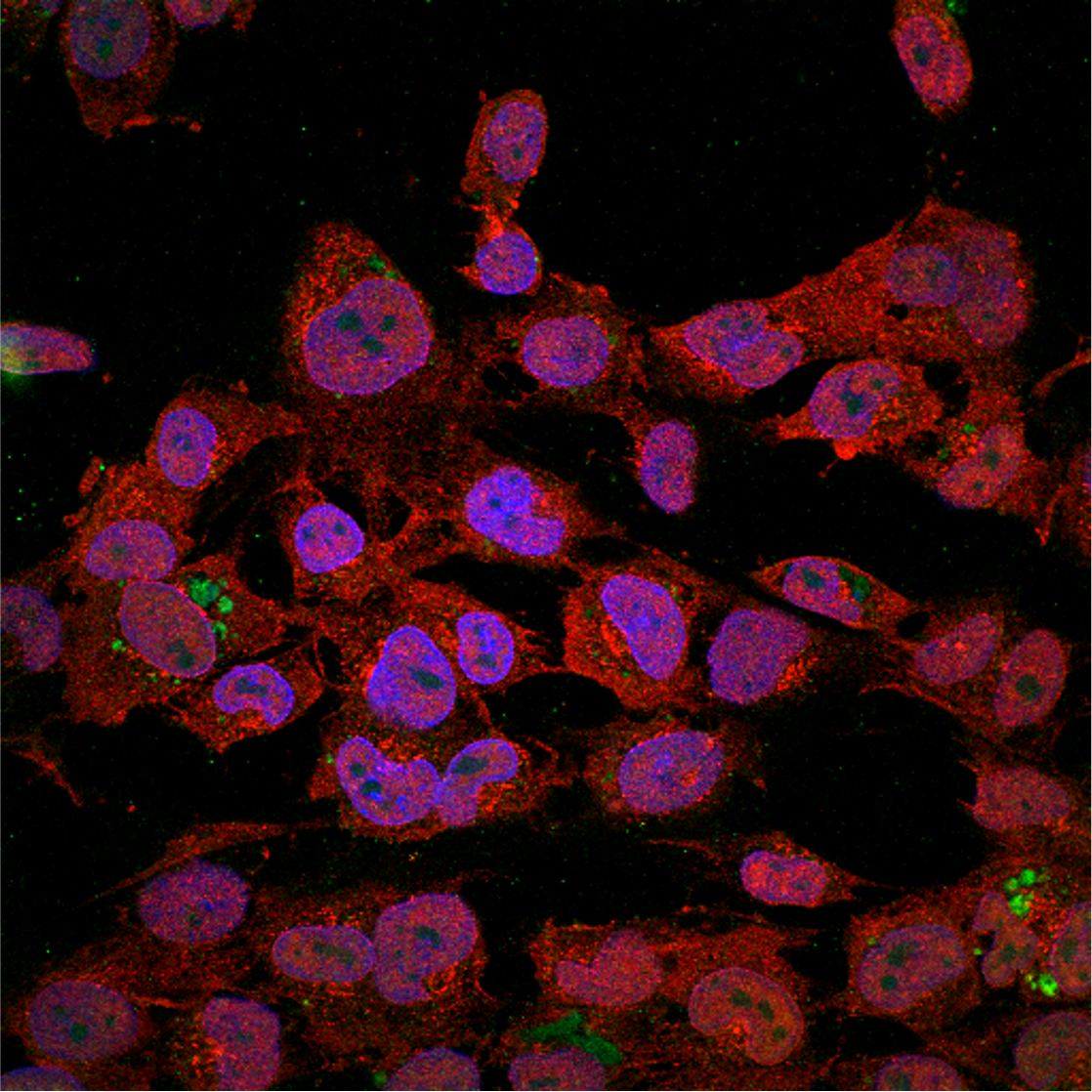
 fixed A549 cells using BAG3 antibody (10599-1-AP) at dilution of 1:200 and CoraLite®488-Conjugated AffiniPure Goat Anti-Rabbit IgG(H+L), CL594-Phalloidin (red).")
 fixed HepG2 cells using BAG3 antibody (10599-1-AP) at dilution of 1:200 and CoraLite®488-Conjugated AffiniPure Goat Anti-Rabbit IgG(H+L), CL594-Phalloidin (red).")
.")
Applications testées
| Résultats positifs en WB | cellules HEK-293, cellules HeLa, cellules K-562, cœur de rat, cœur de souris |
| Résultats positifs en IP | cellules K-562 |
| Résultats positifs en IHC | tissu de cancer du poumon humain, tissu de gliome humain il est suggéré de démasquer l'antigène avec un tampon de TE buffer pH 9.0; (*) À défaut, 'le démasquage de l'antigène peut être 'effectué avec un tampon citrate pH 6,0. |
| Résultats positifs en IF/ICC | cellules A549, cellules HeLa, cellules HepG2 |
Dilution recommandée
| Application | Dilution |
|---|---|
| Western Blot (WB) | WB : 1:30000-1:60000 |
| Immunoprécipitation (IP) | IP : 0.5-4.0 ug for 1.0-3.0 mg of total protein lysate |
| Immunohistochimie (IHC) | IHC : 1:500-1:2000 |
| Immunofluorescence (IF)/ICC | IF/ICC : 1:50-1:500 |
| It is recommended that this reagent should be titrated in each testing system to obtain optimal results. | |
| Sample-dependent, check data in validation data gallery | |
Applications publiées
| KD/KO | See 28 publications below |
| WB | See 111 publications below |
| IHC | See 6 publications below |
| IF | See 48 publications below |
| IP | See 6 publications below |
| CoIP | See 3 publications below |
Informations sur le produit
10599-1-AP cible BAG3 dans les applications de WB, IHC, IF/ICC, IP, CoIP, ELISA et montre une réactivité avec des échantillons Humain, rat, souris
| Réactivité | Humain, rat, souris |
| Réactivité citée | rat, Humain, singe, souris, Hamster |
| Hôte / Isotype | Lapin / IgG |
| Clonalité | Polyclonal |
| Type | Anticorps |
| Immunogène | BAG3 Protéine recombinante Ag0956 |
| Nom complet | BCL2-associated athanogene 3 |
| Masse moléculaire calculée | 61 kDa |
| Poids moléculaire observé | 74-80 kDa |
| Numéro d’acquisition GenBank | BC006418 |
| Symbole du gène | BAG3 |
| Identification du gène (NCBI) | 9531 |
| Conjugaison | Non conjugué |
| Forme | Liquide |
| Méthode de purification | Purification par affinité contre l'antigène |
| Tampon de stockage | PBS with 0.02% sodium azide and 50% glycerol |
| Conditions de stockage | Stocker à -20°C. Stable pendant un an après l'expédition. L'aliquotage n'est pas nécessaire pour le stockage à -20oC Les 20ul contiennent 0,1% de BSA. |
Informations générales
BAG3 (Bcl2-associated athanogene 3) belongs to the BAG protein family, the co-chaperone that binds to Hsc70/Hsp70 through the BAG domain and modulates their activity in polypeptide folding. BAG3 contains also a WW domain and a proline-rich (PXXP) repeat, that mediate binding to partners different from Hsp70. Through interacting with different molecular partner, BAG3 influences several cell processes, such as apoptosis, autophagy and cell motility. BAG3 protein has been reported to sustain cell survival, resistance to therapy, and/or motility and metastatization in several tumor types, thus being identified as a potential target for anticancer therapies. In addition, defects in BAG3 are the cause of some myopathy. BAG3 normally migrates around 74-80 kDa; a slightly different molecular weight or a doublet form can be observed in some cell types and/or following cell exposure to stressors. A synaptosome associated form of 40 kDa has recently been described.
Protocole
| Product Specific Protocols | |
|---|---|
| WB protocol for BAG3 antibody 10599-1-AP | Download protocol |
| IHC protocol for BAG3 antibody 10599-1-AP | Download protocol |
| IF protocol for BAG3 antibody 10599-1-AP | Download protocol |
| IP protocol for BAG3 antibody 10599-1-AP | Download protocol |
| Standard Protocols | |
|---|---|
| Click here to view our Standard Protocols |
Publications
| Species | Application | Title |
|---|---|---|
Nat Genet Mutations affecting the cytoplasmic functions of the co-chaperone DNAJB6 cause limb-girdle muscular dystrophy. | ||
Nat Neurosci A tau homeostasis signature is linked with the cellular and regional vulnerability of excitatory neurons to tau pathology.
| ||
Acta Neuropathol Missense mutations in small muscle protein X-linked (SMPX) cause distal myopathy with protein inclusions. | ||
J Intern Med Transglutaminase type 2 plays a key role in the pathogenesis of Mycobacterium tuberculosis infection. | ||
Nat Commun Misfolded polypeptides are selectively recognized and transported toward aggresomes by a CED complex. | ||
Nat Commun Cardiomyocyte contractile impairment in heart failure results from reduced BAG3-mediated sarcomeric protein turnover. |
Avis
The reviews below have been submitted by verified Proteintech customers who received an incentive for providing their feedback.
FH Andrea (Verified Customer) (01-03-2023) | Strong, clear band on the membrane using a 1:500 ratio.
|
FH WEI (Verified Customer) (03-03-2022) | Very strong antibody, confirmed by KO tissue
|
FH Jane (Verified Customer) (02-03-2022) | One specific band at 80kDa in mouse heart tissue lysate
|
FH Eric (Verified Customer) (01-25-2021) | The red stain is 1:100 BAG3 under laser confocal microscopy in human microglia cells.
 |
FH Tongbin (Verified Customer) (08-25-2020) | This bag3 antibody is very specific without any non-specific bands. It is the best Bag3 antibody out there for western blots.
 |
FH Afaque (Verified Customer) (10-17-2019) | I have used this antibody to study autophagy. It has worked very well for my Western blots. I also tried for IF and got very good results.
 |
FH SHUBHAM (Verified Customer) (02-25-2019) | excellent Antibody
 |
FH Praveen (Verified Customer) (12-03-2018) |
|

