- Phare
- Validé par KD/KO
Anticorps Polyclonal de lapin anti-TGFBI/BIGH3
TGFBI/BIGH3 Polyclonal Antibody for WB, IHC, IF/ICC, FC (Intra), IP, ELISA
Hôte / Isotype
Lapin / IgG
Réactivité testée
Humain, souris et plus (1)
Applications
WB, IHC, IF/ICC, FC (Intra), IP, Neutralization, ELISA, Cell treatment
Conjugaison
Non conjugué
N° de cat : 10188-1-AP
Synonymes
Galerie de données de validation
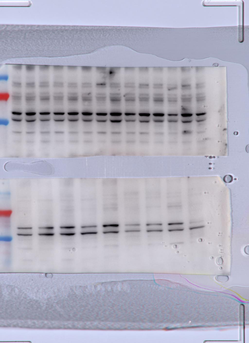
 at dilution of 1:2000 incubated at room temperature for 1.5 hours.")
 with sh-Control and sh-TGFBI / BIGH3 transfected HeLa cells.")
 at dilution of 1:5000 incubated at room temperature for 1.5 hours.")
 at dilution of 1:8000 incubated at room temperature for 1.5 hours.")

 at dilution of 1:300 incubated at room temperature for 1.5 hours.")
 at dilution of 1:400 incubated at room temperature for 1.5 hours.")
 with HeLa cells lysate 1000ug.")
 at dilution of 1:100 (under 10x lens).")
 at dilution of 1:100 (under 40x lens).")
 at dilution of 1:50 (under 10x lens).")
 at dilution of 1:50 (under 40x lens).")
 at dilution of 1:50 (under 10x lens).")
 at dilution of 1:50 (under 40x lens).")
 at dilution of 1:50 (under 10x lens).")
 at dilution of 1:50 (under 40x lens).")
 at dilution of 1:200 (under 10x lens). Heat mediated antigen retrieval with Tris-EDTA buffer (pH 9.0).")
 at dilution of 1:200 (under 40x lens). Heat mediated antigen retrieval with Tris-EDTA buffer (pH 9.0).")
 fixed A549 cells, untreated (left) or TGF-β-treated (right), using TGFBI / BIGH3 antibody (10188-1-AP) at dilution of 1:400 and CoraLite®488-Conjugated AffiniPure Goat Anti-Rabbit IgG(H+L).")
 fixed Y79 cells using TGFBI / BIGH3 antibody (10188-1-AP) at dilution of 1:200 and CoraLite®488-Conjugated AffiniPure Goat Anti-Rabbit IgG(H+L).")
 and CoraLite®488-Conjugated AffiniPure Goat Anti-Rabbit IgG(H+L) at dilution 1:1000 (red), or 0.4 ug Control Antibody. Cells were fixed with 4% PFA and permeabilized with Flow Cytometry Perm Buffer (PF00011-C).")
Applications testées
| Résultats positifs en WB | tissu oculaire de souris, cellules HeLa, cellules Y79, tissu hépatique de souris, tissu rénal humain |
| Résultats positifs en IP | cellules HeLa |
| Résultats positifs en IHC | tissu rénal humain, tissu de cancer du foie humain, tissu oculaire de souris il est suggéré de démasquer l'antigène avec un tampon de TE buffer pH 9.0; (*) À défaut, 'le démasquage de l'antigène peut être 'effectué avec un tampon citrate pH 6,0. |
| Résultats positifs en IF/ICC | cellules A549 traitées au TGF bêta 1, cellules Y79 |
| Résultats positifs en FC (Intra) | cellules Y79 |
Dilution recommandée
| Application | Dilution |
|---|---|
| Western Blot (WB) | WB : 1:1000-1:4000 |
| Immunoprécipitation (IP) | IP : 0.5-4.0 ug for 1.0-3.0 mg of total protein lysate |
| Immunohistochimie (IHC) | IHC : 1:50-1:500 |
| Immunofluorescence (IF)/ICC | IF/ICC : 1:200-1:800 |
| Flow Cytometry (FC) (INTRA) | FC (INTRA) : 0.40 ug per 10^6 cells in a 100 µl suspension |
| It is recommended that this reagent should be titrated in each testing system to obtain optimal results. | |
| Sample-dependent, check data in validation data gallery | |
Applications publiées
| KD/KO | See 9 publications below |
| WB | See 41 publications below |
| IHC | See 36 publications below |
| IF | See 16 publications below |
| IP | See 4 publications below |
| FC | See 1 publications below |
Informations sur le produit
10188-1-AP cible TGFBI/BIGH3 dans les applications de WB, IHC, IF/ICC, FC (Intra), IP, Neutralization, ELISA, Cell treatment et montre une réactivité avec des échantillons Humain, souris
| Réactivité | Humain, souris |
| Réactivité citée | rat, Humain, souris |
| Hôte / Isotype | Lapin / IgG |
| Clonalité | Polyclonal |
| Type | Anticorps |
| Immunogène | TGFBI/BIGH3 Protéine recombinante Ag0241 |
| Nom complet | transforming growth factor, beta-induced, 68kDa |
| Masse moléculaire calculée | 683 aa, 75 kDa |
| Poids moléculaire observé | 64 kDa |
| Numéro d’acquisition GenBank | BC000097 |
| Symbole du gène | TGFBI |
| Identification du gène (NCBI) | 7045 |
| Conjugaison | Non conjugué |
| Forme | Liquide |
| Méthode de purification | Purification par affinité contre l'antigène |
| Tampon de stockage | PBS with 0.02% sodium azide and 50% glycerol |
| Conditions de stockage | Stocker à -20°C. Stable pendant un an après l'expédition. L'aliquotage n'est pas nécessaire pour le stockage à -20oC Les 20ul contiennent 0,1% de BSA. |
Informations générales
TGFBI, also named as BIGH3, Kerato-epithelin and RGD-CAP, binds to type I, II, and IV collagens. TGFBI is an adhesion protein which may play an important role in cell-collagen interactions. In cartilage, it may be involved in endochondral bone formation. TGFBI is an extracellular matrix adaptor protein, it has been reported to be differentially expressed in transformed tissues. TGFBI is a predictive factor of the response to chemotherapy, and suggest the use of TGFBI-derived peptides as possible therapeutic adjuvants for the enhancement of responses to chemotherapy.(PMID:20509890) Defects in TGFBI are the cause of epithelial basement membrane corneal dystrophy (EBMD). Defects in TGFBI are the cause of corneal dystrophy Groenouw type 1 (CDGG1). Defects in TGFBI are the cause of corneal dystrophy lattice type 1 (CDL1). Defects in TGFBI are a cause of corneal dystrophy Thiel-Behnke type (CDTB). Defects in TGFBI are the cause of Reis-Buecklers corneal dystrophy (CDRB). Defects in TGFBI are the cause of lattice corneal dystrophy type 3A (CDL3A). Defects in TGFBI are the cause of Avellino corneal dystrophy (ACD).
Protocole
| Product Specific Protocols | |
|---|---|
| WB protocol for TGFBI/BIGH3 antibody 10188-1-AP | Download protocol |
| IHC protocol for TGFBI/BIGH3 antibody 10188-1-AP | Download protocol |
| IF protocol for TGFBI/BIGH3 antibody 10188-1-AP | Download protocol |
| IP protocol for TGFBI/BIGH3 antibody 10188-1-AP | Download protocol |
| Standard Protocols | |
|---|---|
| Click here to view our Standard Protocols |
Publications
| Species | Application | Title |
|---|---|---|
Nat Cell Biol Supermeres are functional extracellular nanoparticles replete with disease biomarkers and therapeutic targets. | ||
Sci Adv Acetylated K676 TGFBIp as a severity diagnostic blood biomarker for SARS-CoV-2 pneumonia. | ||
Genes Dev Extracellular matrix protein betaig-h3/TGFBI promotes metastasis of colon cancer by enhancing cell extravasation.
| ||
Biomaterials Dual peptide-dendrimer conjugate inhibits acetylation of transforming growth factor β-induced protein and improves survival in sepsis. |
Avis
The reviews below have been submitted by verified Proteintech customers who received an incentive for providing their feedback.
FH Carmelo (Verified Customer) (04-25-2022) | I tested the abs 1:1000 ON at 4C on 40ug spinal cord protein lysates. unfortunately I was not able to distinguish a proper band at the expected size. I will run further optimisation in the incoming weeks.
 |

