- Phare
- Validé par KD/KO
Anticorps Polyclonal de lapin anti-BIN1
BIN1 Polyclonal Antibody for WB, IHC, IF-P, IP, ELISA
Hôte / Isotype
Lapin / IgG
Réactivité testée
Humain, rat, souris et plus (1)
Applications
WB, IHC, IF-P, IP, ELISA
Conjugaison
Non conjugué
N° de cat : 14647-1-AP
Synonymes
Galerie de données de validation
 at dilution of 1:3000 incubated at room temperature for 1.5 hours.")
 at dilution of 1:500 incubated at room temperature for 1.5 hours.")
 at dilution of 1:500 incubated at room temperature for 1.5 hours.")
 at dilution of 1:1000 incubated at room temperature for 1.5 hours.")
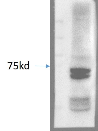
 with mouse brain tissue lysate 3440ug.")
 at dilution of 1:200 (under 10x lens). Heat mediated antigen retrieval with Tris-EDTA buffer (pH 9.0).")
 at dilution of 1:200 (under 40x lens). Heat mediated antigen retrieval with Tris-EDTA buffer (pH 9.0).")
 at dilution of 1:100 (under 10x lens).")
 at dilution of 1:100 (under 40x lens).")
 at dilution of 1:200 (under 10x lens). Heat mediated antigen retrieval with Tris-EDTA buffer (pH 9.0).")
 at dilution of 1:200 (under 40x lens). Heat mediated antigen retrieval with Tris-EDTA buffer (pH 9.0).")
 fixed mouse brain tissue using BIN1 antibody (14647-1-AP) at dilution of 1:200 and CoraLite®488-Conjugated Goat Anti-Rabbit IgG(H+L).")
 fixed mouse brain tissue using BIN1 antibody (14647-1-AP) at dilution of 1:200 and CoraLite®488-Conjugated Goat Anti-Rabbit IgG(H+L).")
Applications testées
| Résultats positifs en WB | cellules Jurkat, tissu cérébral de souris, tissu de muscle squelettique de rat, tissu de muscle squelettique de souris |
| Résultats positifs en IP | tissu cérébral de souris |
| Résultats positifs en IHC | tissu de muscle squelettique de souris, tissu cérébral de souris, tissu d'ostéosarcome humain il est suggéré de démasquer l'antigène avec un tampon de TE buffer pH 9.0; (*) À défaut, 'le démasquage de l'antigène peut être 'effectué avec un tampon citrate pH 6,0. |
| Résultats positifs en IF-P | tissu cérébral de souris, |
Dilution recommandée
| Application | Dilution |
|---|---|
| Western Blot (WB) | WB : 1:1000-1:6000 |
| Immunoprécipitation (IP) | IP : 0.5-4.0 ug for 1.0-3.0 mg of total protein lysate |
| Immunohistochimie (IHC) | IHC : 1:50-1:500 |
| Immunofluorescence (IF)-P | IF-P : 1:50-1:500 |
| It is recommended that this reagent should be titrated in each testing system to obtain optimal results. | |
| Sample-dependent, check data in validation data gallery | |
Applications publiées
| KD/KO | See 4 publications below |
| WB | See 7 publications below |
| IHC | See 4 publications below |
| IF | See 5 publications below |
Informations sur le produit
14647-1-AP cible BIN1 dans les applications de WB, IHC, IF-P, IP, ELISA et montre une réactivité avec des échantillons Humain, rat, souris
| Réactivité | Humain, rat, souris |
| Réactivité citée | Humain, porc, souris |
| Hôte / Isotype | Lapin / IgG |
| Clonalité | Polyclonal |
| Type | Anticorps |
| Immunogène | BIN1 Protéine recombinante Ag6240 |
| Nom complet | bridging integrator 1 |
| Masse moléculaire calculée | 65 kDa |
| Poids moléculaire observé | 50-65 kDa |
| Numéro d’acquisition GenBank | BC004101 |
| Symbole du gène | BIN1 |
| Identification du gène (NCBI) | 274 |
| Conjugaison | Non conjugué |
| Forme | Liquide |
| Méthode de purification | Purification par affinité contre l'antigène |
| Tampon de stockage | PBS with 0.02% sodium azide and 50% glycerol |
| Conditions de stockage | Stocker à -20°C. Stable pendant un an après l'expédition. L'aliquotage n'est pas nécessaire pour le stockage à -20oC Les 20ul contiennent 0,1% de BSA. |
Informations générales
BIN1 (Bridging integrator 1), also known as amphiphysin II or Myc box-dependent-interacting protein 1, is a ubiquitous nucleocytoplasmic adaptor protein that was identified initially as an MYC-interacting proapoptotic tumor suppressor. Alternative splicing of the gene results in multiple transcript variants encoding different isoforms. BIN1 is a key regulator of different cellular functions, including endocytosis and membrane recycling, cytoskeleton regulation, DNA repair, cell cycle progression, and apoptosis (PMID: 24590001).
Protocole
| Product Specific Protocols | |
|---|---|
| WB protocol for BIN1 antibody 14647-1-AP | Download protocol |
| IHC protocol for BIN1 antibody 14647-1-AP | Download protocol |
| IF protocol for BIN1 antibody 14647-1-AP | Download protocol |
| IP protocol for BIN1 antibody 14647-1-AP | Download protocol |
| Standard Protocols | |
|---|---|
| Click here to view our Standard Protocols |
Publications
| Species | Application | Title |
|---|---|---|
Mol Neurodegener BIN1 is a key regulator of proinflammatory and neurodegeneration-related activation in microglia. | ||
Int J Cancer Low expression of Bin1, along with high expression of IDO in tumor tissue and draining lymph nodes, are predictors of poor prognosis for esophageal squamous cell cancer patients. | ||
Mol Biol Cell Cooperation of MICAL-L1, syndapin2, and phosphatidic acid in tubular recycling endosome biogenesis.
| ||
Toxicol Appl Pharmacol Salinomycin promotes T-cell proliferation by inhibiting the expression and enzymatic activity of immunosuppressive indoleamine-2,3-dioxygenase in human breast cancer cells. | ||
J Neuropathol Exp Neurol Combining Hypothermia and Oleuropein Subacutely Protects Subcortical White Matter in a Swine Model of Neonatal Hypoxic-Ischemic Encephalopathy. | ||
J Comp Neurol Fractional anisotropy from diffusion tensor imaging correlates with acute astrocyte and myelin swelling in neonatal swine models of excitotoxic and hypoxic-ischemic brain injury. |
Avis
The reviews below have been submitted by verified Proteintech customers who received an incentive for providing their feedback.
FH CaX (Verified Customer) (10-24-2023) | An excellent Ab to probe Bin1 at the right MW.
 |

