- Phare
- Validé par KD/KO
Anticorps Polyclonal de lapin anti-Carbonic anhydrase 1/CA1
Carbonic anhydrase 1/CA1 Polyclonal Antibody for WB, IHC, IP, ELISA
Hôte / Isotype
Lapin / IgG
Réactivité testée
Humain, rat, souris
Applications
WB, IHC, IF, IP, ELISA
Conjugaison
Non conjugué
N° de cat : 13198-2-AP
Synonymes
Galerie de données de validation
 at dilution of 1:1500 incubated at room temperature for 1.5 hours.")
 at dilution of 1:300 incubated at room temperature for 1.5 hours.")
 at dilution of 1:300 incubated at room temperature for 1.5 hours.")
 at dilution of 1:500 incubated at room temperature for 1.5 hours.")
 at dilution of 1:800 incubated at room temperature for 1.5 hours.")
 with K-562 cells lysate 1200ug.")
 at dilution of 1:200 (under 10x lens. Heat mediated antigen retrieval with Tris-EDTA buffer (pH 9.0).")
 at dilution of 1:200 (under 40x lens. Heat mediated antigen retrieval with Tris-EDTA buffer (pH 9.0).")
 at dilution of 1:300 (under 40x lens). Heat mediated antigen retrieval with Tris-EDTA buffer (pH 9.0).")
Applications testées
| Résultats positifs en WB | tissu splénique de souris, cellules Jurkat, tissu splénique de rat |
| Résultats positifs en IP | cellules K-562 |
| Résultats positifs en IHC | tissu de cancer du sein humain, tissu de cancer du côlon humain il est suggéré de démasquer l'antigène avec un tampon de TE buffer pH 9.0; (*) À défaut, 'le démasquage de l'antigène peut être 'effectué avec un tampon citrate pH 6,0. |
Dilution recommandée
| Application | Dilution |
|---|---|
| Western Blot (WB) | WB : 1:500-1:3000 |
| Immunoprécipitation (IP) | IP : 0.5-4.0 ug for 1.0-3.0 mg of total protein lysate |
| Immunohistochimie (IHC) | IHC : 1:50-1:500 |
| It is recommended that this reagent should be titrated in each testing system to obtain optimal results. | |
| Sample-dependent, check data in validation data gallery | |
Applications publiées
| KD/KO | See 1 publications below |
| WB | See 7 publications below |
| IHC | See 1 publications below |
| IF | See 4 publications below |
| IP | See 1 publications below |
Informations sur le produit
13198-2-AP cible Carbonic anhydrase 1/CA1 dans les applications de WB, IHC, IF, IP, ELISA et montre une réactivité avec des échantillons Humain, rat, souris
| Réactivité | Humain, rat, souris |
| Réactivité citée | rat, Humain, souris |
| Hôte / Isotype | Lapin / IgG |
| Clonalité | Polyclonal |
| Type | Anticorps |
| Immunogène | Carbonic anhydrase 1/CA1 Protéine recombinante Ag3932 |
| Nom complet | carbonic anhydrase I |
| Masse moléculaire calculée | 261 aa, 29 kDa |
| Poids moléculaire observé | 29 kDa |
| Numéro d’acquisition GenBank | BC027890 |
| Symbole du gène | CA1 |
| Identification du gène (NCBI) | 759 |
| Conjugaison | Non conjugué |
| Forme | Liquide |
| Méthode de purification | Purification par affinité contre l'antigène |
| Tampon de stockage | PBS with 0.02% sodium azide and 50% glycerol |
| Conditions de stockage | Stocker à -20°C. Stable pendant un an après l'expédition. L'aliquotage n'est pas nécessaire pour le stockage à -20oC Les 20ul contiennent 0,1% de BSA. |
Informations générales
CA1(Carbonic anhydrase 1) is also named as CAB and belongs to the alpha-carbonic anhydrase family, which may reside in cytoplasm, in mitochondria, or in secretory granules, or associate with membranes in cell. It forms a large family of genes encoding zinc metalloenzymes of great physiologic importance. Extracellular CA1 mediates hemorrhagic retinal and cerebral vascular permeability through prekallikrein activation(PMID:17259996).
Protocole
| Product Specific Protocols | |
|---|---|
| WB protocol for Carbonic anhydrase 1/CA1 antibody 13198-2-AP | Download protocol |
| IHC protocol for Carbonic anhydrase 1/CA1 antibody 13198-2-AP | Download protocol |
| IP protocol for Carbonic anhydrase 1/CA1 antibody 13198-2-AP | Download protocol |
| Standard Protocols | |
|---|---|
| Click here to view our Standard Protocols |
Publications
| Species | Application | Title |
|---|---|---|
Acta Neuropathol Commun Upregulation of carbonic anhydrase 1 beneficial for depressive disorder
| ||
J Ginseng Res Ginsenoside Rg1 attenuates mechanical stress-induced cardiac injury via calcium sensing receptor-related pathway | ||
FASEB J Loss of interleukin-10 receptor disrupts intestinal epithelial cell proliferation and skews differentiation towards the goblet cell fate. | ||
J Inflamm Res M1-Type Macrophages Secrete TNF-α to Stimulate Vascular Calcification by Upregulating CA1 and CA2 Expression in VSMCs | ||
Am J Physiol Lung Cell Mol Physiol Carbonic anhydrase and soluble adenylate cyclase regulation of cystic fibrosis cellular phenotypes. | ||
Front Pharmacol Calcium Sensing Receptor-Related Pathway Contributes to Cardiac Injury and the Mechanism of Astragaloside IV on Cardioprotection. |
Avis
The reviews below have been submitted by verified Proteintech customers who received an incentive for providing their feedback.
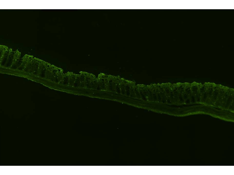
FH Brittany (Verified Customer) (09-27-2019) | Using the CA-1 primary antibody with mouse colon scrapes yielded very clean bands with western blotting. Immunofluorescence also gave a fairly clean, detectable signal, with CA-1 showing up along the brush border in mouse colon tissue. There were some cells that stained more intracellularly, and unsure if that was non-specific binding of the antibody, or another cell type other than colonocytes that expresses CA-1 intracellularly.
 |

