- Phare
- Validé par KD/KO
Anticorps Polyclonal de lapin anti-Carbonic Anhydrase IX/CA9
Carbonic Anhydrase IX/CA9 Polyclonal Antibody for WB, IHC, IP, ELISA
Hôte / Isotype
Lapin / IgG
Réactivité testée
Humain, rat, souris
Applications
WB, IHC, IF, IP, ELISA
Conjugaison
Non conjugué
N° de cat : 11071-1-AP
Synonymes
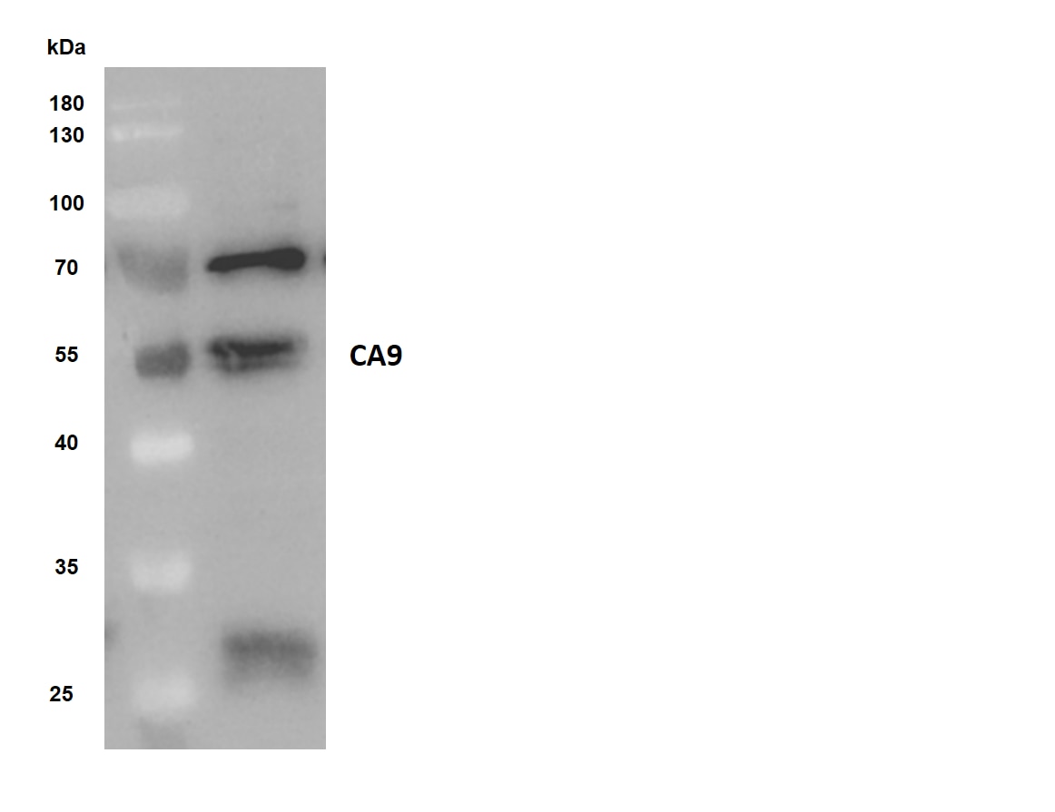
Galerie de données de validation
 at dilution of 1:3000 incubated at room temperature for 1.5 hours.")
 at dilution of 1:8000 incubated at room temperature for 1.5 hours.")
 with sh-Control and sh-Carbonic Anhydrase IX/CA9 transfected A549 cells.")
 with HeLa cells lysate 1200 ug.")
 at dilution of 1:200 (under 40x lens). Heat mediated antigen retrieval with Tris-EDTA buffer (pH 9.0).")
 at dilution of 1:200 (under 10x lens). Heat mediated antigen retrieval with Tris-EDTA buffer (pH 9.0).")
Applications testées
| Résultats positifs en WB | cellules A549, cellules HeLa, cellules HeLa traitées au chlorure de cobalt |
| Résultats positifs en IP | cellules HeLa, |
| Résultats positifs en IHC | tissu de cancer de l'estomac humain, il est suggéré de démasquer l'antigène avec un tampon de TE buffer pH 9.0; (*) À défaut, 'le démasquage de l'antigène peut être 'effectué avec un tampon citrate pH 6,0. |
Dilution recommandée
| Application | Dilution |
|---|---|
| Western Blot (WB) | WB : 1:1000-1:6000 |
| Immunoprécipitation (IP) | IP : 0.5-4.0 ug for 1.0-3.0 mg of total protein lysate |
| Immunohistochimie (IHC) | IHC : 1:50-1:500 |
| It is recommended that this reagent should be titrated in each testing system to obtain optimal results. | |
| Sample-dependent, check data in validation data gallery | |
Applications publiées
| KD/KO | See 1 publications below |
| WB | See 24 publications below |
| IHC | See 27 publications below |
| IF | See 9 publications below |
Informations sur le produit
11071-1-AP cible Carbonic Anhydrase IX/CA9 dans les applications de WB, IHC, IF, IP, ELISA et montre une réactivité avec des échantillons Humain, rat, souris
| Réactivité | Humain, rat, souris |
| Réactivité citée | Humain, souris |
| Hôte / Isotype | Lapin / IgG |
| Clonalité | Polyclonal |
| Type | Anticorps |
| Immunogène | Carbonic Anhydrase IX/CA9 Protéine recombinante Ag1540 |
| Nom complet | carbonic anhydrase IX |
| Masse moléculaire calculée | 459 aa, 50 kDa |
| Poids moléculaire observé | 50 kDa, 60-70 kDa |
| Numéro d’acquisition GenBank | BC014950 |
| Symbole du gène | CA9 |
| Identification du gène (NCBI) | 768 |
| Conjugaison | Non conjugué |
| Forme | Liquide |
| Méthode de purification | Purification par affinité contre l'antigène |
| Tampon de stockage | PBS with 0.02% sodium azide and 50% glycerol |
| Conditions de stockage | Stocker à -20°C. Stable pendant un an après l'expédition. L'aliquotage n'est pas nécessaire pour le stockage à -20oC Les 20ul contiennent 0,1% de BSA. |
Informations générales
CA9 (Carbonic anhydrase 9) may be involved in the control of cell proliferation and transformation and appears to be a novel specific biomarker for a cervical neoplasia (PMID:18703501). It is a tumor-associated antigen that has been shown to have diagnostic utility in identifying cervical dysplasia and carcinoma. The protein is present in the cytoplasmic membrane and the nucleus, with a molecular weight of 50 kDa. The molecular weight of the glycosylated form is approximately 60-70 kDa. (PMID: 31142270, PMID: 31819036)
Protocole
| Product Specific Protocols | |
|---|---|
| WB protocol for Carbonic Anhydrase IX/CA9 antibody 11071-1-AP | Download protocol |
| IHC protocol for Carbonic Anhydrase IX/CA9 antibody 11071-1-AP | Download protocol |
| IP protocol for Carbonic Anhydrase IX/CA9 antibody 11071-1-AP | Download protocol |
| Standard Protocols | |
|---|---|
| Click here to view our Standard Protocols |
Publications
| Species | Application | Title |
|---|---|---|
Proc Natl Acad Sci U S A CRLX101 nanoparticles localize in human tumors and not in adjacent, nonneoplastic tissue after intravenous dosing. | ||
Cell Commun Signal KCNJ2/HIF1α positive-feedback loop promotes the metastasis of osteosarcoma | ||
Biomed Pharmacother Para-toluenesulfonamide, a novel potent carbonic anhydrase inhibitor, improves hypoxia-induced metastatic breast cancer cell viability and prevents resistance to αPD-1 therapy in triple-negative breast cancer | ||
Oxid Med Cell Longev Carbonic Anhydrase IX Controls Vulnerability to Ferroptosis in Gefitinib-Resistant Lung Cancer | ||
Oxid Med Cell Longev Targeting the Ang2/Tie2 Axis with Tanshinone IIA Elicits Vascular Normalization in Ischemic Injury and Colon Cancer. | ||
Eur J Nucl Med Mol Imaging In vivo three-dimensional evaluation of tumour hypoxia in nasopharyngeal carcinomas using FMT-CT and MSOT. |
Avis
The reviews below have been submitted by verified Proteintech customers who received an incentive for providing their feedback.
FH Aline Seiko (Verified Customer) (05-12-2025) | It was observed a 70kDa and 25kDa isoform.
|
FH Maelle (Verified Customer) (01-03-2025) | Lost efficacy after freeze-defreze even if it is in glycerol
|
FH Josh (Verified Customer) (10-28-2021) | 15 ug of human clear cell renal cell carcinoma cell line RCC4 was resolved on 15% Tris/Glycine and transferred to PVDF membrane. Membrane was blocked in blocking buffer (2% BSA in TBS/0.1% Tween-20) for 1hr at room temperature, followed by overnight incubation with anti-CA9 (1:1000) in blocking buffer at 4oC. After 1hr incubation with anti-Rabbit secondary antibody, membrane was imaged with ECL. Expected band was detected along with several non-specific bands by western blot
 |
FH K (Verified Customer) (08-03-2021) | total cell lysate (50 ug of human HCC cell lines) was resolved on 10% Bis-Tris gel and transferred to nitrocellulose membrane. Membrane was incubated in blocking buffer (5% BSA in TBS/0.1% Tween-20) for 1h. Membrane was incubated with anti-CA9 in blocking buffer (1:500) at 40C overnight. After 1h incubation with appropriate secondary antibody (anti-Rabbit 1:5000) membranes were imaged with ECL and expected band was detected.
 |
FH Paulo (Verified Customer) (03-04-2019) | Total cell lysate (50 ug of human ccRCC cell lines) was resolved on 10% Bis-Tris gel and transferred to PVDF membrane. Membrane was incubated in blocking buffer (5% milk in PBS/0.1% Tween-20) for 1h. Membrane was incubated with anti-CAIX in blocking buffer (1:250) at 4C overnight. After 1h incubation with appropriate secondary antibody (DAKO anti-Rabbit 1:10000) membranes were imaged with ECL and expected band was detected
|

