Anticorps Polyclonal de lapin anti-CACNA1A
CACNA1A Polyclonal Antibody for IHC, ELISA
Hôte / Isotype
Lapin / IgG
Réactivité testée
Humain
Applications
IHC, ELISA
Conjugaison
Non conjugué
N° de cat : 27227-1-AP
Synonymes
Galerie de données de validation
 at dilution of 1:100 (under 10x lens). Heat mediated antigen retrieval with Tris-EDTA buffer (pH 9.0).")
 at dilution of 1:100 (under 40x lens). Heat mediated antigen retrieval with Tris-EDTA buffer (pH 9.0).")
Applications testées
| Résultats positifs en IHC | tissu cérébral humain il est suggéré de démasquer l'antigène avec un tampon de TE buffer pH 9.0; (*) À défaut, 'le démasquage de l'antigène peut être 'effectué avec un tampon citrate pH 6,0. |
Dilution recommandée
| Application | Dilution |
|---|---|
| Immunohistochimie (IHC) | IHC : 1:50-1:500 |
| It is recommended that this reagent should be titrated in each testing system to obtain optimal results. | |
| Sample-dependent, check data in validation data gallery | |
Informations sur le produit
27227-1-AP cible CACNA1A dans les applications de IHC, ELISA et montre une réactivité avec des échantillons Humain
| Réactivité | Humain |
| Hôte / Isotype | Lapin / IgG |
| Clonalité | Polyclonal |
| Type | Anticorps |
| Immunogène | CACNA1A Protéine recombinante Ag25943 |
| Nom complet | calcium channel, voltage-dependent, P/Q type, alpha 1A subunit |
| Masse moléculaire calculée | 282 kDa |
| Numéro d’acquisition GenBank | NM_000068 |
| Symbole du gène | CACNA1A |
| Identification du gène (NCBI) | 773 |
| Conjugaison | Non conjugué |
| Forme | Liquide |
| Méthode de purification | Purification par affinité contre l'antigène |
| Tampon de stockage | PBS with 0.02% sodium azide and 50% glycerol |
| Conditions de stockage | Stocker à -20°C. Stable pendant un an après l'expédition. L'aliquotage n'est pas nécessaire pour le stockage à -20oC Les 20ul contiennent 0,1% de BSA. |
Protocole
| Product Specific Protocols | |
|---|---|
| IHC protocol for CACNA1A antibody 27227-1-AP | Download protocol |
| Standard Protocols | |
|---|---|
| Click here to view our Standard Protocols |
Avis
The reviews below have been submitted by verified Proteintech customers who received an incentive for providing their feedback.
FH Lea (Verified Customer) (10-08-2024) | I transfected HEK 293T cells with 2 different CACNA1A constructs (12Q and 27Q, 2 replicates each) and an empty vector as a negative control. The expected band at ~280 kDa is clearly visible in the western blot and no unspecific bands are visible in the negative control. I was very happy with the results.
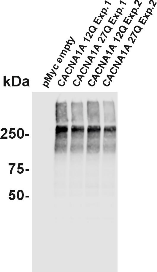 |
FH Ümit (Verified Customer) (08-11-2022) | Cav2.1 (black) staining of two rhesus monkey brainstem sections (5µm &7µm) visualized with immunoperoxidase method (DAB-Nickel).
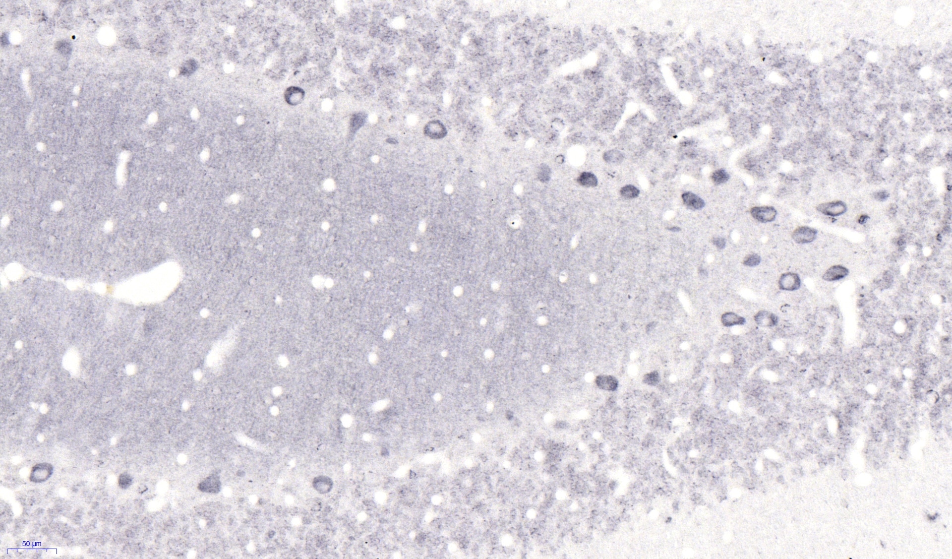 |

