Anticorps Polyclonal de lapin anti-CEP83
CEP83 Polyclonal Antibody for WB, IHC, IF/ICC, ELISA
Hôte / Isotype
Lapin / IgG
Réactivité testée
Humain et plus (1)
Applications
WB, IHC, IF/ICC, ELISA
Conjugaison
Non conjugué
N° de cat : 26013-1-AP
Synonymes
Galerie de données de validation
 at dilution of 1:600 incubated at room temperature for 1.5 hours.")
 at dilution of 1:200 (under 10x lens).")
 at dilution of 1:200 (under 40x lens).")
 fixed hTERT-RPE1 cells using CEP83 antibody (26013-1-AP) at dilution of 1:400 and CoraLite®488-Conjugated Goat Anti-Rabbit IgG(H+L), CoraLite®594 ARL13B antibody (CL594-17711, red).")
 fixed hTERT-RPE1 cells using CEP83 antibody (26013-1-AP) at dilution of 1:800 and CoraLite®594-Conjugated Goat Anti-Rabbit IgG(H+L).")
Applications testées
| Résultats positifs en WB | cellules HEK-293 |
| Résultats positifs en IHC | tissu rénal humain, il est suggéré de démasquer l'antigène avec un tampon de TE buffer pH 9.0; (*) À défaut, 'le démasquage de l'antigène peut être 'effectué avec un tampon citrate pH 6,0. |
| Résultats positifs en IF/ICC | cellules hTERT-RPE1, |
Dilution recommandée
| Application | Dilution |
|---|---|
| Western Blot (WB) | WB : 1:500-1:1000 |
| Immunohistochimie (IHC) | IHC : 1:50-1:500 |
| Immunofluorescence (IF)/ICC | IF/ICC : 1:200-1:800 |
| It is recommended that this reagent should be titrated in each testing system to obtain optimal results. | |
| Sample-dependent, check data in validation data gallery | |
Applications publiées
| WB | See 7 publications below |
| IF | See 6 publications below |
Informations sur le produit
26013-1-AP cible CEP83 dans les applications de WB, IHC, IF/ICC, ELISA et montre une réactivité avec des échantillons Humain
| Réactivité | Humain |
| Réactivité citée | Humain, souris |
| Hôte / Isotype | Lapin / IgG |
| Clonalité | Polyclonal |
| Type | Anticorps |
| Immunogène | CEP83 Protéine recombinante Ag22826 |
| Nom complet | coiled-coil domain containing 41 |
| Masse moléculaire calculée | 693 aa, 82 kDa |
| Poids moléculaire observé | 83 kDa, 68 kDa |
| Numéro d’acquisition GenBank | BC125087 |
| Symbole du gène | CEP83 |
| Identification du gène (NCBI) | 51134 |
| Conjugaison | Non conjugué |
| Forme | Liquide |
| Méthode de purification | Purification par affinité contre l'antigène |
| Tampon de stockage | PBS with 0.02% sodium azide and 50% glycerol |
| Conditions de stockage | Stocker à -20°C. Stable pendant un an après l'expédition. L'aliquotage n'est pas nécessaire pour le stockage à -20oC Les 20ul contiennent 0,1% de BSA. |
Informations générales
CEP83 (centrosomal protein, 83 kDa), also known as CCDC41 (coiled-coil domain containing 41), is required for ciliary vesicle docking to the mother centriole. It is a key component of the centriolar distal appendages. CCDC41 may collaborate with IFT20 in the trafficking of ciliary membrane proteins from the Golgi complex to the cilium during the initiation of primary cilium assembly (PMID: 23530209). Alternative splicing results in transcript variants encoding two isoforms with calculated molecular weights of 83 kDa and 68 kDa, respectively.
Protocole
| Product Specific Protocols | |
|---|---|
| WB protocol for CEP83 antibody 26013-1-AP | Download protocol |
| IHC protocol for CEP83 antibody 26013-1-AP | Download protocol |
| IF protocol for CEP83 antibody 26013-1-AP | Download protocol |
| Standard Protocols | |
|---|---|
| Click here to view our Standard Protocols |
Publications
| Species | Application | Title |
|---|---|---|
Cell Res NudCL2 is an autophagy receptor that mediates selective autophagic degradation of CP110 at mother centrioles to promote ciliogenesis. | ||
J Cell Biol The Cby3/ciBAR1 complex positions the annulus along the sperm flagellum during spermiogenesis | ||
PLoS Biol The evolutionary conserved proteins CEP90, FOPNL, and OFD1 recruit centriolar distal appendage proteins to initiate their assembly | ||
Cell Mol Life Sci Requirement of NPHP5 in the hierarchical assembly of basal feet associated with basal bodies of primary cilia. | ||
J Biol Chem The C7orf43/TRAPPC14 component links the TRAPPII complex to RABIN8 for preciliary vesicle tethering at the mother centriole during ciliogenesis. |
Avis
The reviews below have been submitted by verified Proteintech customers who received an incentive for providing their feedback.
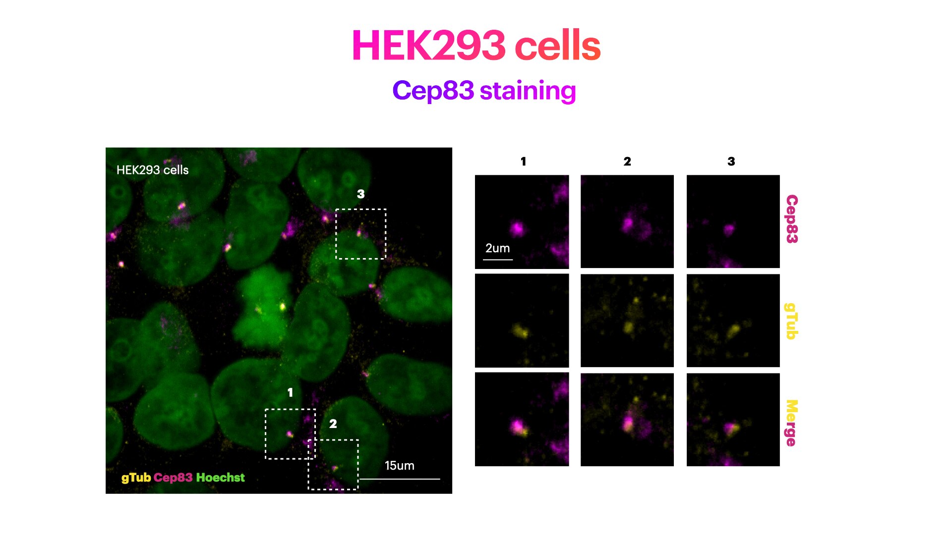
FH Elisa (Verified Customer) (03-01-2022) | HEK293 cells stained for Hoechst (DNA marker, in green), Cep83 (mother centriole distal appendage marker, in magenta) and g-Tubulin (pericentriolar matrix marker, in green). HEK293 cells were plated on Poly-lysine coated coverslips and fixed in cold methanol for 2' at -20C. Cells were then rehydrated with PBS for 5'. Membrane permeabilization was then performed with 0.1% Triton + 0.1% Tween +0.01%SDS in PBS for 5'. Cells were finally incubated with blocking buffer (5% BSA+ 0.1% Tween in PBS) for 30' at RT. Primary antibody was diluted in blocking buffer 1:200 and incubated for 1h at room temperature. Alexa-555-Anti-rabbit was used as secondary antibody (1:600 dilution) (1h at room temperature).
 |
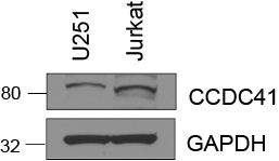
FH Sarah (Verified Customer) (07-03-2019) | Total cell lysate (15 ug) was resolved on a 4-12% Bis-Tris gel and transferred to nitrocellulose membrane. Membrane was incubated in blocking buffer (5% milk/0.1% Tween-20) for 1h. Membrane was incubated with anti-CCDC41 in blocking buffer (1:1000) at 4C overnight. After washing, membrane was incubated in anti-rabbit-HRP in blocking bufffer (1:3000) for 1h at room temperature. Protein was detected using ECL reagent and imaged on a chemiluminescence detection system.
 |

