Tested Applications
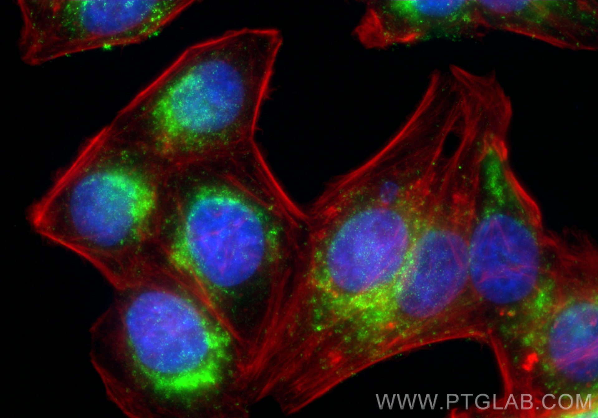
| Positive IF/ICC detected in | HepG2 cells |
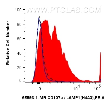
| Positive FC (Intra) detected in | Jurkat cells |
| Positive FC detected in | Thrombin treated human peripheral blood platelets |
Recommended dilution
| Application | Dilution |
|---|---|
| Immunofluorescence (IF)/ICC | IF/ICC : 1:250-1:1000 |
| Flow Cytometry (FC) (INTRA) | FC (INTRA) : 0.25 ug per 10^6 cells in 100 μl suspension |
| Flow Cytometry (FC) | FC : 0.25 ug per 10^6 cells in a 100 µl suspension |
| This reagent has been tested for flow cytometric analysis. It is recommended that this reagent should be titrated in each testing system to obtain optimal results. | |
| Sample-dependent, Check data in validation data gallery. | |
Published Applications
| FC | See 1 publications below |
Product Information
65596-1-MR targets CD107a / LAMP1 in IF/ICC, FC, FC (Intra) applications and shows reactivity with human samples.
| Tested Reactivity | human |
| Cited Reactivity | human |
| Host / Isotype | Mouse / IgG2a |
| Class | Recombinant |
| Type | Antibody |
| Immunogen |
Human adherent peripheral blood cells Predict reactive species |
| Full Name | lysosomal-associated membrane protein 1 |
| Calculated Molecular Weight | 45 kDa |
| GenBank Accession Number | BC006345 |
| Gene Symbol | LAMP1 |
| Gene ID (NCBI) | 3916 |
| ENSEMBL Gene ID | ENSG00000185896 |
| RRID | AB_3670344 |
| Conjugate | Unconjugated |
| Form | Liquid |
| Purification Method | Protein A purification |
| UNIPROT ID | P11279 |
| Storage Buffer | PBS with 0.09% sodium azide, pH 7.3. |
| Storage Conditions | Store at 2-8°C. Stable for one year after shipment. |
Background Information
LAMP1 (CD107a) is a heavily glycosylated membrane protein enriched in the lysosomal membrane. LAMP1 is extensively glycosylated with asparagine-linked oligosaccharides which protect it from intracellular proteolysis (PMID: 10521503). Although LAMP1 is expressed largely in the endosome-lysosomal membrane of cells, it is also found on the plasma membrane (PMID: 16168398). Elevated LAMP1 expression at the cell surface has also been detected during platelet and granulocytic cell activation, as well as in some tumor cells (PMID: 29085473). LAMP1 functions to provide selectins with carbohydrate ligands. This protein has also been shown to be a marker of degranulation on lymphocytes such as CD8+ and NK cells and may also play a role in tumor cell differentiation and metastasis (PMID: 18835598; 29085473; 9426697).
Protocols
| Product Specific Protocols | |
|---|---|
| FC protocol for CD107a / LAMP1 antibody 65596-1-MR | Download protocol |
| IF protocol for CD107a / LAMP1 antibody 65596-1-MR | Download protocol |
| Standard Protocols | |
|---|---|
| Click here to view our Standard Protocols |