- Phare
- Validé par KD/KO
Anticorps Polyclonal de lapin anti-CD3
CD3 Polyclonal Antibody for WB, IHC, IF-P, IF-Fro, IP, ELISA
Hôte / Isotype
Lapin / IgG
Réactivité testée
Humain, porc, rat, souris
Applications
WB, IHC, IF-P, IF-Fro, IP, ELISA, Cell treatment
Conjugaison
Non conjugué
N° de cat : 17617-1-AP
Synonymes
Galerie de données de validation
, MOLT-4 cells (CD3+), U-937 cells (CD3-), Raji cells (CD3-), mouse thymus tissue, and rat thymus tissue were subjected to SDS PAGE followed by western blot with 17617-1-AP (CD3 antibody) at dilution of 1:4000 incubated at room temperature for 1.5 hours.")
 with sh-Control and sh-CD3 transfected Jurkat cells.")
 at dilution of 1:600 incubated at room temperature for 1.5 hours.")
 with Jurkat cells lysate 3600ug.")
 at dilution of 1:1000 (under 10x lens). Heat mediated antigen retrieval with Tris-EDTA buffer (pH 9.0).")
 at dilution of 1:1000 (under 40x lens). Heat mediated antigen retrieval with Tris-EDTA buffer (pH 9.0).")
 at dilution of 1:1000 (under 10x lens). Heat mediated antigen retrieval with Tris-EDTA buffer (pH 9.0).")
 at dilution of 1:1000 (under 40x lens). Heat mediated antigen retrieval with Tris-EDTA buffer (pH 9.0).")
 at dilution of 1:100 (under 10x lens).")
 at dilution of 1:100 (under 40x lens).")
 at dilution of 1:5000 (under 10x lens). Heat mediated antigen retrieval with Tris-EDTA buffer (pH 9.0).")
 at dilution of 1:5000 (under 40x lens). Heat mediated antigen retrieval with Tris-EDTA buffer (pH 9.0).")
 at dilution of 1:1000 (under 10x lens). Heat mediated antigen retrieval with Tris-EDTA buffer (pH 9.0).")
 at dilution of 1:1000 (under 40x lens). Heat mediated antigen retrieval with Tris-EDTA buffer (pH 9.0).")
 at dilution of 1:1000 (under 10x lens. Heat mediated antigen retrieval with Tris-EDTA buffer (pH 9.0).")
 at dilution of 1:1000 (under 40x lens. Heat mediated antigen retrieval with Tris-EDTA buffer (pH 9.0).")
 at dilution of 1:1000 (under 10x lens. Heat mediated antigen retrieval with Tris-EDTA buffer (pH 9.0).")
 at dilution of 1:1000 (under 40x lens. Heat mediated antigen retrieval with Tris-EDTA buffer (pH 9.0).")
 at dilution of 1:1000 (under 10x lens). Heat mediated antigen retrieval with Tris-EDTA buffer (pH 9.0).")
 at dilution of 1:1000 (under 40x lens). Heat mediated antigen retrieval with Tris-EDTA buffer (pH 9.0).")
 at dilution of 1:500 (under 40x lens). Heat mediated antigen retrieval with Tris-EDTA buffer (pH 9.0).")
 fixed human tonsillitis tissue using CD3 antibody (17617-1-AP) at dilution of 1:200 and CoraLite®488-Conjugated AffiniPure Goat Anti-Rabbit IgG(H+L), CD20 antibody (60271-1-Ig, Clone: 4A7G3, red).")
 fixed human tonsillitis tissue using CD3 antibody (17617-1-AP) at dilution of 1:200 and CoraLite®488-Conjugated AffiniPure Goat Anti-Rabbit IgG(H+L), CD20 antibody (60271-1-Ig, Clone: 4A7G3, red).")
 fixed mouse spleen tissue using CD3 antibody (17617-1-AP) at dilution of 1:800 and CoraLite®488-Conjugated AffiniPure Goat Anti-Rabbit IgG(H+L).")
 fixed mouse spleen tissue using CD3 antibody (17617-1-AP) at dilution of 1:800 and CoraLite®488-Conjugated AffiniPure Goat Anti-Rabbit IgG(H+L).")
 fixed human tonsillitis tissue using CD3 antibody (17617-1-AP) at dilution of 1:200 and CoraLite®488-Conjugated AffiniPure Goat Anti-Rabbit IgG(H+L), CD20 antibody (60271-1-Ig, Clone: 4A7G3, red).")
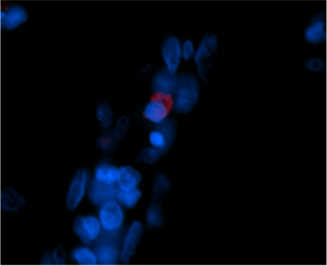
 fixed frozen OCT-embedded mouse colon tissue using CD3 antibody (17617-1-AP) at dilution of 1:400 and CoraLite®594-Conjugated Goat Anti-Rabbit IgG(H+L) (SA00013-4).")
Applications testées
| Résultats positifs en WB | cellules Jurkat, cellules MOLT-4, tissu de thymus de rat, tissu de thymus de souris, tissu splénique de porc |
| Résultats positifs en IP | cellules Jurkat |
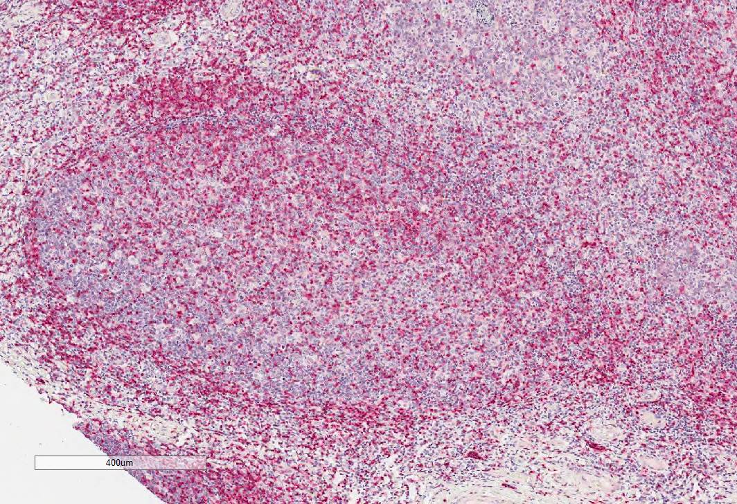
| Résultats positifs en IHC | tissu d'amygdalite humain, tissu de cancer du pancréas humain, tissu de cancer du poumon humain, tissu de cancer du sein humain, tissu de côlon de souris, tissu de lymphome humain, tissu de thymus de rat, tissu splénique de souris il est suggéré de démasquer l'antigène avec un tampon de TE buffer pH 9.0; (*) À défaut, 'le démasquage de l'antigène peut être 'effectué avec un tampon citrate pH 6,0. |
| Résultats positifs en IF-P | tissu d'amygdalite humain, tissu splénique de souris |
| Résultats positifs en IF-Fro | tissu de côlon de souris, |
Dilution recommandée
| Application | Dilution |
|---|---|
| Western Blot (WB) | WB : 1:1000-1:8000 |
| Immunoprécipitation (IP) | IP : 0.5-4.0 ug for 1.0-3.0 mg of total protein lysate |
| Immunohistochimie (IHC) | IHC : 1:500-1:2000 |
| Immunofluorescence (IF)-P | IF-P : 1:50-1:500 |
| Immunofluorescence (IF)-FRO | IF-FRO : 1:200-1:800 |
| It is recommended that this reagent should be titrated in each testing system to obtain optimal results. | |
| Sample-dependent, check data in validation data gallery | |
Applications publiées
| WB | See 9 publications below |
| IHC | See 93 publications below |
| IF | See 62 publications below |
| FC | See 4 publications below |
Informations sur le produit
17617-1-AP cible CD3 dans les applications de WB, IHC, IF-P, IF-Fro, IP, ELISA, Cell treatment et montre une réactivité avec des échantillons Humain, porc, rat, souris
| Réactivité | Humain, porc, rat, souris |
| Réactivité citée | rat, Humain, porc, souris |
| Hôte / Isotype | Lapin / IgG |
| Clonalité | Polyclonal |
| Type | Anticorps |
| Immunogène | CD3 Protéine recombinante Ag11797 |
| Nom complet | CD3e molecule, epsilon (CD3-TCR complex) |
| Masse moléculaire calculée | 207 aa, 23 kDa |
| Poids moléculaire observé | 23-25 kDa |
| Numéro d’acquisition GenBank | BC049847 |
| Symbole du gène | CD3 |
| Identification du gène (NCBI) | 916 |
| Conjugaison | Non conjugué |
| Forme | Liquide |
| Méthode de purification | Purification par affinité contre l'antigène |
| Tampon de stockage | PBS with 0.02% sodium azide and 50% glycerol |
| Conditions de stockage | Stocker à -20°C. Stable pendant un an après l'expédition. L'aliquotage n'est pas nécessaire pour le stockage à -20oC Les 20ul contiennent 0,1% de BSA. |
Informations générales
CD3 is a complex of proteins that directly associates with the T cell receptor (TCR). The TCR/CD3 complex of T-lymphocytes consists of either a TCR alpha/beta or TCR gamma/delta heterodimer coexpressed at the cell surface with the invariant subunits of CD3 labeled gamma, delta, epsilon, zeta, and eta. The TCR recognizes antigens bound to major histocompatibility complex (MHC) molecules. TCR-mediated peptide-MHC recognition is transmitted to the CD3 complex, leading to the intracellular signal transduction. CD3 is considered to be a pan-T cell marker for detection of normal and neoplastic T cells. This anti-CD3 is a polyclonal antibody directed against the epsilon chain of human CD3 molecule.
Protocole
| Product Specific Protocols | |
|---|---|
| WB protocol for CD3 antibody 17617-1-AP | Download protocol |
| IHC protocol for CD3 antibody 17617-1-AP | Download protocol |
| IF protocol for CD3 antibody 17617-1-AP | Download protocol |
| IP protocol for CD3 antibody 17617-1-AP | Download protocol |
| Standard Protocols | |
|---|---|
| Click here to view our Standard Protocols |
Publications
| Species | Application | Title |
|---|---|---|
Blood C1Q labels a highly aggressive macrophage-like leukemia population indicating extramedullary infiltration and relapse | ||
Nat Commun Engineered triple inhibitory receptor resistance improves anti-tumor CAR-T cell performance via CD56. | ||
J Adv Res Zearalenone-14-glucoside specifically promotes dysplasia of Gut-Associated Lymphoid Tissue: A natural product for constructing intestinal nodular lymphatic hyperplasia model | ||
Cancer Res M6A Demethylase ALKBH5 Regulates PD-L1 Expression and Tumor Immunoenvironment in Intrahepatic Cholangiocarcinoma. | ||
Acta Biomater Simultaneous targeting of TGF-β1/PD-L1 via a hydrogel-nanoparticle system to remodel the ECM and immune microenvironment for limiting adhesion formation |
Avis
The reviews below have been submitted by verified Proteintech customers who received an incentive for providing their feedback.
FH Federica (Verified Customer) (12-08-2023) | Leica Bond RXm Red Chromogenic Kit Antigen Retrival ER1 (ph6) for 30 min
 |
FH Niklas (Verified Customer) (07-31-2023) | works well
|
FH Emma (Verified Customer) (11-29-2021) | This works really well by IF on FFPE tissue. We used a 1:50 dilution and Tris-EDTA antigen retrieval.
 |

