Anticorps Monoclonal anti-CD63
CD63 Monoclonal Antibody for WB, IHC, IF-P, ELISA
Hôte / Isotype
Mouse / IgG1
Réactivité testée
Humain et plus (2)
Applications
WB, IHC, IF-P, ELISA
Conjugaison
Non conjugué
CloneNo.
3D4D1
N° de cat : 67605-1-Ig
Synonymes
Galerie de données de validation
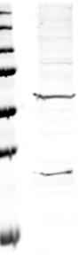
 at dilution of 1:10000 incubated at room temperature for 1.5 hours. The membrane was stripped and reblotted with HRP-conjugated Alpha Tubulin Monoclonal antibody (HRP-66031) as loading control.")
 at dilution of 1:3000 incubated at room temperature for 1.5 hours.")
 at dilution of 1:700 (under 10x lens). Heat mediated antigen retrieval with Tris-EDTA buffer (pH 9.0).")
 at dilution of 1:700 (under 40x lens). Heat mediated antigen retrieval with Tris-EDTA buffer (pH 9.0).")
 at dilution of 1:800 (under 10x lens). Heat mediated antigen retrieval with Tris-EDTA buffer (pH 9.0).")
 at dilution of 1:800 (under 40x lens). Heat mediated antigen retrieval with Tris-EDTA buffer (pH 9.0).")
 at dilution of 1:2000 (under 10x lens). Heat mediated antigen retrieval with Tris-EDTA buffer (pH 9.0).")
 at dilution of 1:2000 (under 40x lens). Heat mediated antigen retrieval with Tris-EDTA buffer (pH 9.0).")
 at dilution of 1:500 (under 10x lens).")
 at dilution of 1:500 (under 40x lens).")
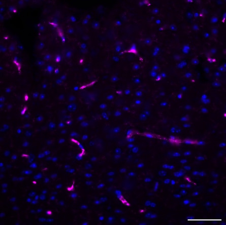
 fixed human tonsillitis tissue using CD63 antibody (67605-1-Ig, Clone: 3D4D1 ) at dilution of 1:400 and CoraLite®488-Conjugated AffiniPure Goat Anti-Mouse IgG(H+L).")
 fixed human tonsillitis tissue using CD63 antibody (67605-1-Ig, Clone: 3D4D1 ) at dilution of 1:400 and CoraLite®488-Conjugated AffiniPure Goat Anti-Mouse IgG(H+L).")
 fixed human lymphoma tissue using CD63 antibody (67605-1-Ig, Clone: 3D4D1 ) at dilution of 1:400 and CoraLite®488-Conjugated AffiniPure Goat Anti-Mouse IgG(H+L).")
 fixed human lymphoma tissue using CD63 antibody (67605-1-Ig, Clone: 3D4D1 ) at dilution of 1:400 and CoraLite®488-Conjugated AffiniPure Goat Anti-Mouse IgG(H+L).")
 fixed human tonsillitis tissue using CD63 antibody (67605-1-Ig, Clone: 3D4D1 ) at dilution of 1:400 and CoraLite®488-Conjugated AffiniPure Goat Anti-Mouse IgG(H+L).")
 fixed human malignant melanoma tissue using CD63 antibody (67605-1-Ig, Clone: 3D4D1 ) at dilution of 1:400 and CoraLite®488-Conjugated AffiniPure Goat Anti-Mouse IgG(H+L).")
 fixed human malignant melanoma tissue using CD63 antibody (67605-1-Ig, Clone: 3D4D1 ) at dilution of 1:400 and CoraLite®488-Conjugated AffiniPure Goat Anti-Mouse IgG(H+L).")
Applications testées
| Résultats positifs en WB | cellules HeLa, cellules A549, cellules HUVEC, cellules K-562, cellules LNCaP, cellules MCF-7, cellules THP-1, cellules U2OS |
| Résultats positifs en IHC | tissu d'amygdalite humain, tissu de cancer du côlon humain, tissu de mélanome malin humain il est suggéré de démasquer l'antigène avec un tampon de TE buffer pH 9.0; (*) À défaut, 'le démasquage de l'antigène peut être 'effectué avec un tampon citrate pH 6,0. |
| Résultats positifs en IF-P | tissu d'amygdalite humain, tissu de lymphome humain, tissu de mélanome malin humain |
Dilution recommandée
| Application | Dilution |
|---|---|
| Western Blot (WB) | WB : 1:5000-1:10000 |
| Immunohistochimie (IHC) | IHC : 1:350-1:1400 |
| Immunofluorescence (IF)-P | IF-P : 1:200-1:800 |
| It is recommended that this reagent should be titrated in each testing system to obtain optimal results. | |
| Sample-dependent, check data in validation data gallery | |
Applications publiées
| WB | See 85 publications below |
| IF | See 5 publications below |
Informations sur le produit
67605-1-Ig cible CD63 dans les applications de WB, IHC, IF-P, ELISA et montre une réactivité avec des échantillons Humain
| Réactivité | Humain |
| Réactivité citée | rat, Chèvre, Humain |
| Hôte / Isotype | Mouse / IgG1 |
| Clonalité | Monoclonal |
| Type | Anticorps |
| Immunogène | CD63 Protéine recombinante Ag19690 |
| Nom complet | CD63 molecule |
| Masse moléculaire calculée | 26 kDa |
| Poids moléculaire observé | 35 kDa |
| Numéro d’acquisition GenBank | BC002349 |
| Symbole du gène | CD63 |
| Identification du gène (NCBI) | 967 |
| Conjugaison | Non conjugué |
| Forme | Liquide |
| Méthode de purification | Purification par protéine G |
| Tampon de stockage | PBS with 0.02% sodium azide and 50% glycerol |
| Conditions de stockage | Stocker à -20°C. Stable pendant un an après l'expédition. L'aliquotage n'est pas nécessaire pour le stockage à -20oC Les 20ul contiennent 0,1% de BSA. |
Informations générales
CD63 is a 30-60 kDa lysosomal membrane protein that belongs to the tetraspanin family. This protein plays many important roles in immuno-physiological functions. It mediate signal transduction events that play a role in the regulation of cell development, activation and motility. CD63 is expressed on activated platelets, thus it may function as a blood platelet activation marker. CD63 is a lysosomal membrane glycoprotein that is translocated to plasma membrane after platelet activation. The CD63 tetraspanin is highly expressed in the early stages of melanoma and decreases in advanced lesions, suggesting it as a possible suppressor of tumor progression. Deficiency of this protein is associated with Hermansky-Pudlak syndrome.
Protocole
| Product Specific Protocols | |
|---|---|
| WB protocol for CD63 antibody 67605-1-Ig | Download protocol |
| IHC protocol for CD63 antibody 67605-1-Ig | Download protocol |
| IF protocol for CD63 antibody 67605-1-Ig | Download protocol |
| Standard Protocols | |
|---|---|
| Click here to view our Standard Protocols |
Publications
| Species | Application | Title |
|---|---|---|
Mol Cancer Exosomal circ_0006896 promotes AML progression via interaction with HDAC1 and restriction of antitumor immunity | ||
Cell Metab Nicotinamide metabolism face-off between macrophages and fibroblasts manipulates the microenvironment in gastric cancer | ||
Nat Commun Lactate dehydrogenase A regulates tumor-macrophage symbiosis to promote glioblastoma progression | ||
Int J Oral Sci Administration of Porphyromonas gingivalis in pregnant mice enhances glycolysis and histone lactylation/ADAM17 leading to cleft palate in offspring | ||
Bone Res Interorgan communication in neurogenic heterotopic ossification: the role of brain-derived extracellular vesicles | ||
Drug Des Devel Ther Keloid Patient Plasma-Derived Exosomal hsa_circ_0020792 Promotes Normal Skin Fibroblasts Proliferation, Migration, and Fibrogenesis via Modulating miR-193a-5p and Activating TGF-β1/Smad2/3 Signaling |
Avis
The reviews below have been submitted by verified Proteintech customers who received an incentive for providing their feedback.
FH Marion (Verified Customer) (11-24-2024) | CD63 stains intracellular endosomal vesiclesd in neuronal extensions. We are satisfied with the results.
 |
FH Guorong (Verified Customer) (03-22-2022) | Two bands at 30 and 60 kDa was detected
 |

