- Phare
- Validé par KD/KO
Anticorps Polyclonal de lapin anti-Cathepsin D
Cathepsin D Polyclonal Antibody for WB, IHC, IF/ICC, ELISA
Hôte / Isotype
Lapin / IgG
Réactivité testée
Humain, souris et plus (3)
Applications
WB, IHC, IF/ICC, ELISA
Conjugaison
Non conjugué
N° de cat : 21327-1-AP
Synonymes
Galerie de données de validation
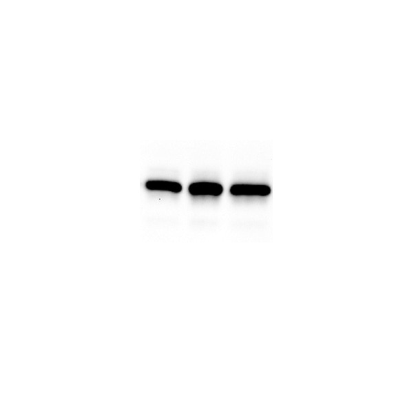
 with wild-type and sg-Cathepsin D transfected HCT-116 cells.")
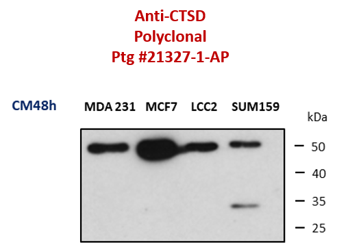
 at dilution of 1:8000 incubated at room temperature for 1.5 hours.")
 at dilution of 1:10000 incubated at room temperature for 1.5 hours.")
 at dilution of 1:1000 (under 10x lens). Heat mediated antigen retrieval with Tris-EDTA buffer (pH 9.0).")
 at dilution of 1:1000 (under 40x lens). Heat mediated antigen retrieval with Tris-EDTA buffer (pH 9.0).")
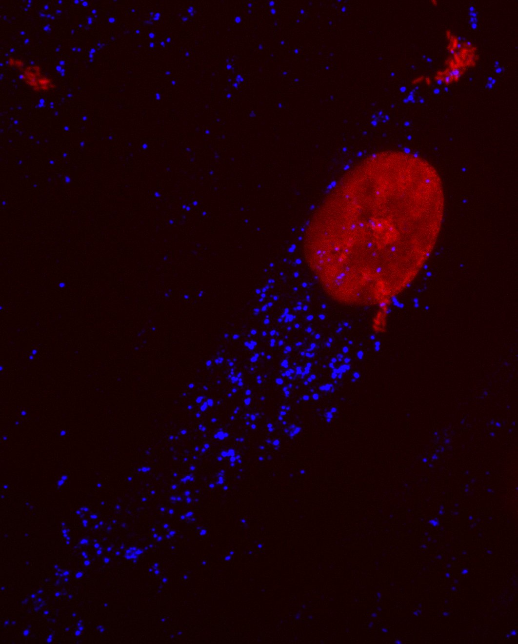
 fixed MCF-7 cells using Cathepsin D antibody (21327-1-AP) at dilution of 1:400 and Multi-rAb CoraLite ® Plus 488-Goat Anti-Rabbit Recombinant Secondary Antibody (H+L) (RGAR002).")
 fixed A431 cells using Cathepsin D antibody (21327-1-AP) at dilution of 1:200 and CoraLite®488-Conjugated Goat Anti-Rabbit IgG(H+L) (SA00013-2).")
Applications testées
| Résultats positifs en WB | cellules A375, cellules HCT 116, cellules HepG2, cellules HuH-7, cellules MCF-7 |
| Résultats positifs en IHC | tissu de cirrhose hépatique humain, il est suggéré de démasquer l'antigène avec un tampon de TE buffer pH 9.0; (*) À défaut, 'le démasquage de l'antigène peut être 'effectué avec un tampon citrate pH 6,0. |
| Résultats positifs en IF/ICC | cellules MCF-7, cellules A431 |
Dilution recommandée
| Application | Dilution |
|---|---|
| Western Blot (WB) | WB : 1:5000-1:50000 |
| Immunohistochimie (IHC) | IHC : 1:500-1:2000 |
| Immunofluorescence (IF)/ICC | IF/ICC : 1:200-1:800 |
| It is recommended that this reagent should be titrated in each testing system to obtain optimal results. | |
| Sample-dependent, check data in validation data gallery | |
Applications publiées
| KD/KO | See 1 publications below |
| WB | See 123 publications below |
| IHC | See 16 publications below |
| IF | See 22 publications below |
Informations sur le produit
21327-1-AP cible Cathepsin D dans les applications de WB, IHC, IF/ICC, ELISA et montre une réactivité avec des échantillons Humain, souris
| Réactivité | Humain, souris |
| Réactivité citée | rat, bovin, Humain, porc, souris |
| Hôte / Isotype | Lapin / IgG |
| Clonalité | Polyclonal |
| Type | Anticorps |
| Immunogène | Cathepsin D Protéine recombinante Ag15254 |
| Nom complet | cathepsin D |
| Masse moléculaire calculée | 412 aa, 45 kDa |
| Poids moléculaire observé | 32 kDa, 48 kDa, 52 kDa |
| Numéro d’acquisition GenBank | BC016320 |
| Symbole du gène | Cathepsin D |
| Identification du gène (NCBI) | 1509 |
| Conjugaison | Non conjugué |
| Forme | Liquide |
| Méthode de purification | Purification par affinité contre l'antigène |
| Tampon de stockage | PBS with 0.02% sodium azide and 50% glycerol |
| Conditions de stockage | Stocker à -20°C. Stable pendant un an après l'expédition. L'aliquotage n'est pas nécessaire pour le stockage à -20oC Les 20ul contiennent 0,1% de BSA. |
Informations générales
CTSD (Cathepsin D) also named CPSD, belongs to the peptidase A1 family. It is ubiquitously expressed and is involved in proteolytic degradation, cell invasion, and apoptosis. Human CTSD is synthesized as a 52-kDa precursor that is converted into an active 48-kDa single-chain intermediate in the endosomes, and then into a fully active mature form, composed of a 34-kDa heavy chain and a 14-kDa light chain, in the lysosomes. It is a lysosomal acid protease found in neutrophils and monocytes and involved in the pathogenesis of several diseases such as breast cancer and possibly Alzheimer disease (PMID: 27114232, PMID: 30717773, PMID: 30051532).
Protocole
| Product Specific Protocols | |
|---|---|
| WB protocol for Cathepsin D antibody 21327-1-AP | Download protocol |
| IHC protocol for Cathepsin D antibody 21327-1-AP | Download protocol |
| IF protocol for Cathepsin D antibody 21327-1-AP | Download protocol |
| Standard Protocols | |
|---|---|
| Click here to view our Standard Protocols |
Publications
| Species | Application | Title |
|---|---|---|
Acta Pharm Sin B Nuciferine protects against high-fat diet-induced hepatic steatosis and insulin resistance via activating TFEB-mediated autophagy-lysosomal pathway. | ||
Autophagy Atractylenolide I inhibits angiogenesis and reverses sunitinib resistance in clear cell renal cell carcinoma through ATP6V0D2-mediated autophagic degradation of EPAS1/HIF2α | ||
Redox Biol Cyclic helix B peptide alleviates proinflammatory cell death and improves functional recovery after traumatic spinal cord injury | ||
Autophagy Restoration of CTSD (cathepsin D) and lysosomal function in stroke is neuroprotective.
|
Avis
The reviews below have been submitted by verified Proteintech customers who received an incentive for providing their feedback.
FH Tae Yeon (Verified Customer) (09-02-2025) | Good puncta staining to detect lysosomal enzyme
|
FH aude (Verified Customer) (05-26-2025) | Sat 5% milk Incubation Primary Ab ON at 4°C
 |
FH Chen (Verified Customer) (06-01-2021) | Very good to detect cathepsin D in postmortem human samples with Western blotting
 |
FH Efil (Verified Customer) (05-29-2019) | cells were fixed in cold MeOH for 8 minutes
 |

