- Phare
- Validé par KD/KO
Anticorps Polyclonal de lapin anti-CUL3
CUL3 Polyclonal Antibody for WB, IHC, IF/ICC, IP, ELISA
Hôte / Isotype
Lapin / IgG
Réactivité testée
Humain, rat, souris
Applications
WB, IHC, IF/ICC, IP, ELISA
Conjugaison
Non conjugué
N° de cat : 11107-1-AP
Synonymes
Galerie de données de validation
Applications testées
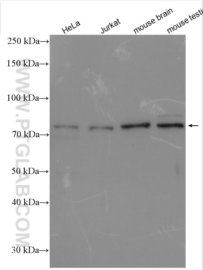
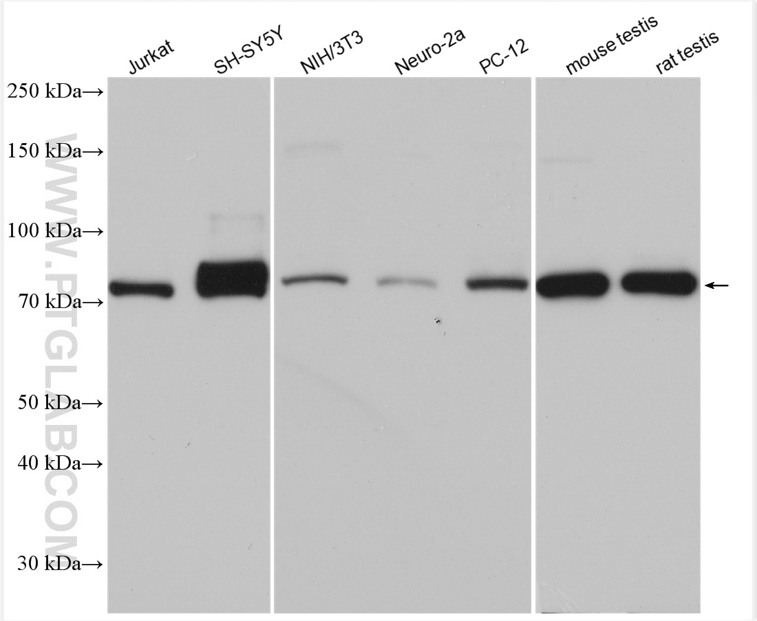
| Résultats positifs en WB | cellules HeLa, cellules Jurkat, cellules NIH/3T3, cellules PC-12, cellules SH-SY5Y, tissu cérébral de souris, tissu testiculaire de rat, tissu testiculaire de souris |
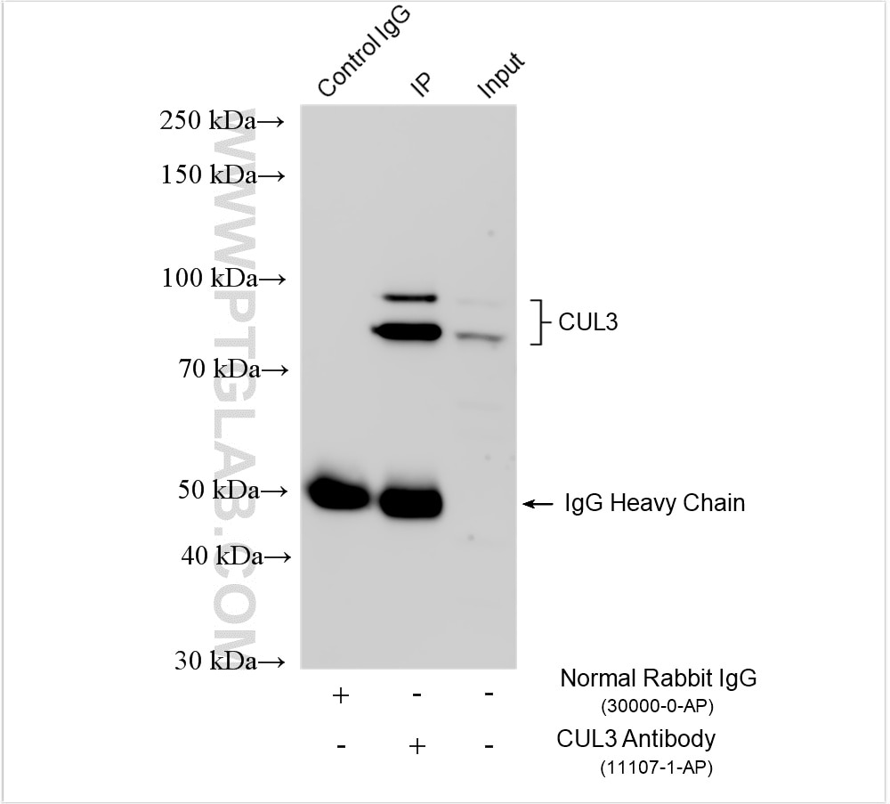
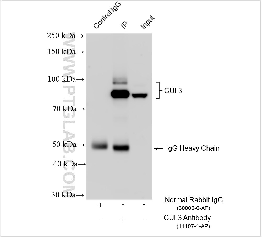
| Résultats positifs en IP | tissu testiculaire de souris, cellules HeLa |
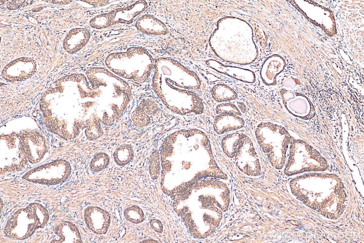
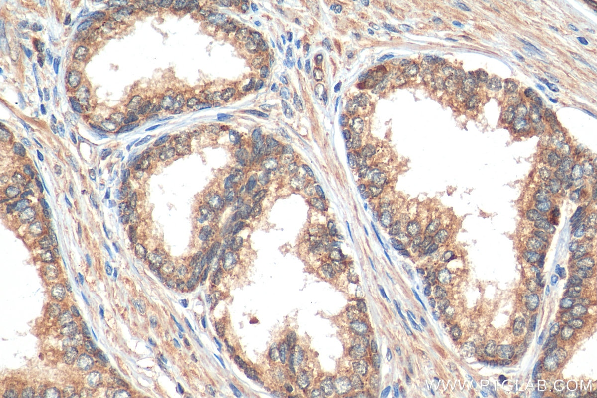
| Résultats positifs en IHC | tissu de cancer de la prostate humain, il est suggéré de démasquer l'antigène avec un tampon de TE buffer pH 9.0; (*) À défaut, 'le démasquage de l'antigène peut être 'effectué avec un tampon citrate pH 6,0. |
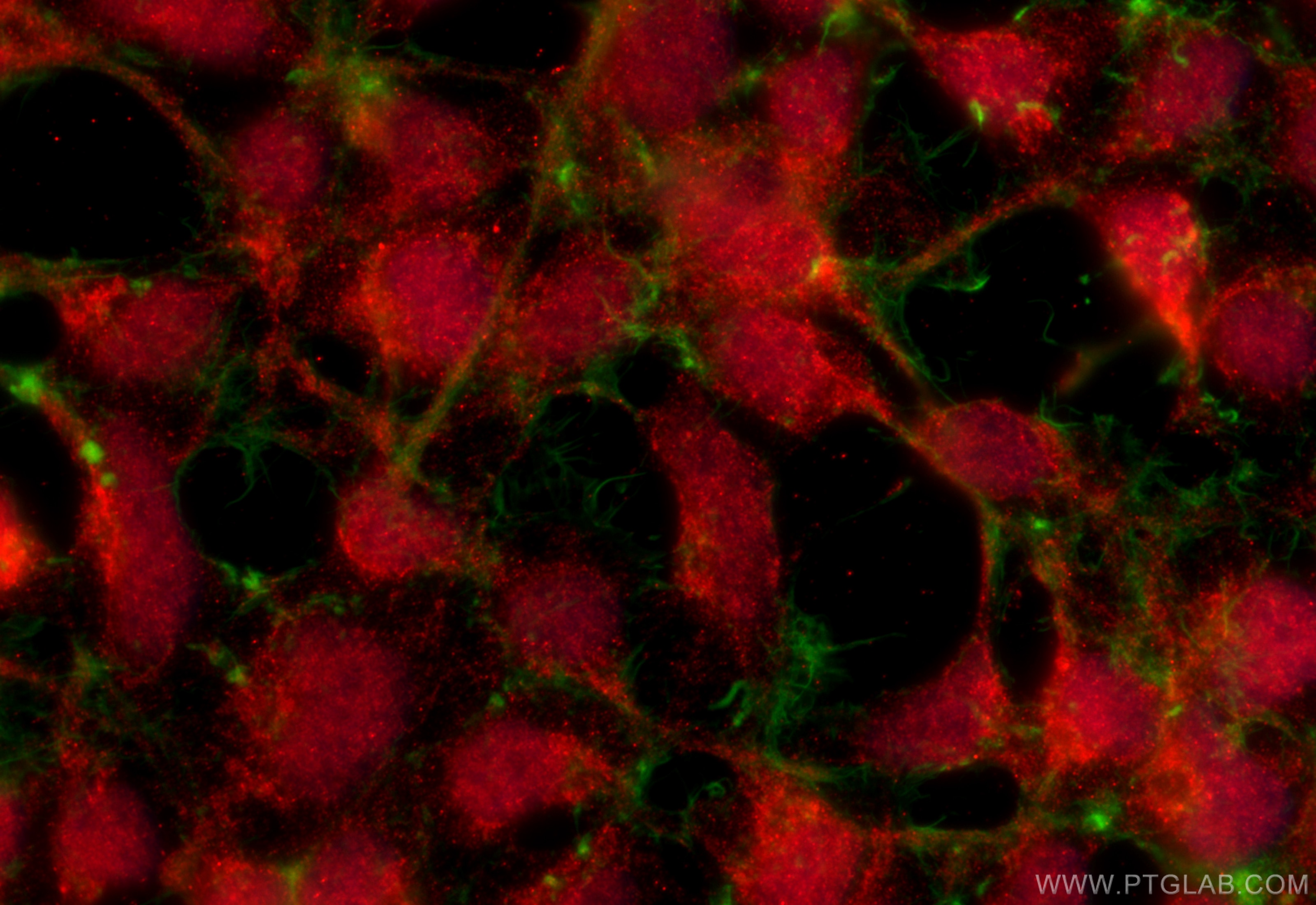
| Résultats positifs en IF/ICC | cellules HEK-293, |
Dilution recommandée
| Application | Dilution |
|---|---|
| Western Blot (WB) | WB : 1:1000-1:4000 |
| Immunoprécipitation (IP) | IP : 0.5-4.0 ug for 1.0-3.0 mg of total protein lysate |
| Immunohistochimie (IHC) | IHC : 1:50-1:500 |
| Immunofluorescence (IF)/ICC | IF/ICC : 1:200-1:800 |
| It is recommended that this reagent should be titrated in each testing system to obtain optimal results. | |
| Sample-dependent, check data in validation data gallery | |
Applications publiées
| KD/KO | See 3 publications below |
| WB | See 18 publications below |
| IHC | See 2 publications below |
| IF | See 3 publications below |
| IP | See 1 publications below |
Informations sur le produit
11107-1-AP cible CUL3 dans les applications de WB, IHC, IF/ICC, IP, ELISA et montre une réactivité avec des échantillons Humain, rat, souris
| Réactivité | Humain, rat, souris |
| Réactivité citée | rat, Humain, souris |
| Hôte / Isotype | Lapin / IgG |
| Clonalité | Polyclonal |
| Type | Anticorps |
| Immunogène | CUL3 Protéine recombinante Ag1555 |
| Nom complet | cullin 3 |
| Masse moléculaire calculée | 89 kDa |
| Poids moléculaire observé | 80-89 kDa |
| Numéro d’acquisition GenBank | BC039598 |
| Symbole du gène | CUL3 |
| Identification du gène (NCBI) | 8452 |
| Conjugaison | Non conjugué |
| Forme | Liquide |
| Méthode de purification | Purification par affinité contre l'antigène |
| Tampon de stockage | PBS with 0.02% sodium azide and 50% glycerol |
| Conditions de stockage | Stocker à -20°C. Stable pendant un an après l'expédition. L'aliquotage n'est pas nécessaire pour le stockage à -20oC Les 20ul contiennent 0,1% de BSA. |
Informations générales
Cullin-RING-based BCR (BTB-CUL3-RBX1) E3 ligase are the largest family of ubiquitin ligases which mediate the ubiquitination and subsequent proteasomal degradation of target proteins. One of seven cullin proteins serves as a scaffold protein for the assembly of the multisubunit ubiquitin ligase complex. CUL3 (Cullin-3) ubiquitin ligase complex targets multiple substrate for ubiquitination including cyclin E and of cyclin D1. Defects in CUL3 are the cause of Pseudohypoaldosteronism type 2E (PHA2E) characterized by severe hypertension, hyperkalemia, hyperchloremia, hyperchloremic metabolic acidosis, and correction of physiologic abnormalities by thiazide diuretics.
Protocole
| Product Specific Protocols | |
|---|---|
| WB protocol for CUL3 antibody 11107-1-AP | Download protocol |
| IHC protocol for CUL3 antibody 11107-1-AP | Download protocol |
| IF protocol for CUL3 antibody 11107-1-AP | Download protocol |
| IP protocol for CUL3 antibody 11107-1-AP | Download protocol |
| Standard Protocols | |
|---|---|
| Click here to view our Standard Protocols |
Publications
| Species | Application | Title |
|---|---|---|
Adv Sci (Weinh) DDRGK1 Enhances Osteosarcoma Chemoresistance via Inhibiting KEAP1-Mediated NRF2 Ubiquitination | ||
Free Radic Biol Med β-Sitosterol targets ASS1 for Nrf2 ubiquitin-dependent degradation, inducing ROS-mediated apoptosis via the PTEN/PI3K/AKT signaling pathway in ovarian cancer | ||
Sci Total Environ USP15 participates in DBP-induced testicular oxidative stress injury through regulating the Keap1/Nrf2 signaling pathway. | ||
Food Chem Tangeretin maintains antioxidant activity by reducing CUL3 mediated NRF2 ubiquitination. | ||
Aging (Albany NY) Tumor suppressor DCAF15 inhibits epithelial-mesenchymal transition by targeting ZEB1 for proteasomal degradation in hepatocellular carcinoma.
| ||
FEBS J CUL3 induces mitochondrial dysfunction via MRPL12 ubiquitination in renal tubular epithelial cells |
Avis
The reviews below have been submitted by verified Proteintech customers who received an incentive for providing their feedback.
FH Rajni (Verified Customer) (06-06-2025) | The Cul3 antibody performed well, producing clear and specific bands in Western blot. It demonstrated good sensitivity and target specificity under our experimental conditions.
|
FH Brittney (Verified Customer) (11-26-2018) | 10ug load of protein. Cells lysed in RIPA buffer then added to 2X Laemmli buffer for denaturing. Heated at 95deg for 10 minutes as well.Multiple bands, but all follow same pattern. Assumed the band at ~75 kD was the correct one.
|
