Anticorps Monoclonal anti-Calretinin
Calretinin Monoclonal Antibody for WB, IHC, IF/ICC, IF-P, IF-Fro, ELISA
Hôte / Isotype
Mouse / IgG1
Réactivité testée
Humain, porc, rat, souris
Applications
WB, IHC, IF/ICC, IF-P, IF-Fro, ELISA
Conjugaison
Non conjugué
CloneNo.
2D7A9
N° de cat : 66496-1-Ig
Synonymes
Galerie de données de validation
 at dilution of 1:20000 incubated at room temperature for 1.5 hours. The membrane was stripped and reblotted with HRP-conjugated Beta Actin Monoclonal antibody (HRP-66009) as loading control.")
 at dilution of 1:20000 incubated at room temperature for 1.5 hours.")
 at dilution of 1:20000 incubated at room temperature for 1.5 hours.")
 at dilution of 1:40000 incubated at room temperature for 1.5 hours.")
 at dilution of 1:40000 incubated at room temperature for 1.5 hours.")
 at dilution of 1:40000 incubated at room temperature for 1.5 hours.")
 at dilution of 1:40000 incubated at room temperature for 1.5 hours.")
 at dilution of 1:20000 incubated at room temperature for 1.5 hours.")
 at dilution of 1:5000 (under 10x lens). Heat mediated antigen retrieval with Tris-EDTA buffer (pH 9.0).")
 at dilution of 1:4000 (under 10x lens). Heat mediated antigen retrieval with Tris-EDTA buffer (pH 9.0).")
 at dilution of 1:5000 (under 40x lens). Heat mediated antigen retrieval with Tris-EDTA buffer (pH 9.0).")
 at dilution of 1:4000 (under 40x lens). Heat mediated antigen retrieval with Tris-EDTA buffer (pH 9.0).")
 at dilution of 1:16000 (under 10x lens). Heat mediated antigen retrieval with Tris-EDTA buffer (pH 9.0).")
 at dilution of 1:5000 (under 4x lens). Heat mediated antigen retrieval with Tris-EDTA buffer (pH 9.0).")
 at dilution of 1:5000 (under 40x lens). Heat mediated antigen retrieval with Tris-EDTA buffer (pH 9.0).")
 at dilution of 1:4000 (under 10x lens). Heat mediated antigen retrieval with Tris-EDTA buffer (pH 9.0).")
 at dilution of 1:200 (under 10x lens. Heat mediated antigen retrieval with Tris-EDTA buffer (pH 9.0).")
 at dilution of 1:200 (under 40x lens. Heat mediated antigen retrieval with Tris-EDTA buffer (pH 9.0).")
 at dilution of 1:200 (under 10x lens. Heat mediated antigen retrieval with Tris-EDTA buffer (pH 9.0).")
 at dilution of 1:200 (under 40x lens. Heat mediated antigen retrieval with Tris-EDTA buffer (pH 9.0).")
 fixed human appendicitis tissue using Calretinin antibody (66496-1-Ig, Clone: 2D7A9 ) at dilution of 1:400 and CoraLite®488-Conjugated AffiniPure Goat Anti-Mouse IgG(H+L).")
 fixed human appendicitis tissue using Calretinin antibody (66496-1-Ig, Clone: 2D7A9 ) at dilution of 1:400 and CoraLite®488-Conjugated AffiniPure Goat Anti-Mouse IgG(H+L).")
 fixed paraffin-embedded rat cerebellum tissue using Calretinin antibody (66496-1-Ig, Clone: 2D7A9 ) at dilution of 1:800 and CoraLite®488-Conjugated Goat Anti-Mouse IgG(H+L) (SA00013-1). Heat mediated antigen retrieval with Tris-EDTA buffer (pH 9.0).")
 fixed frozen OCT-embedded rat cerebellum tissue using Calretinin antibody (66496-1-Ig, Clone: 2D7A9 ) at dilution of 1:800 and CoraLite®488-Conjugated Goat Anti-Mouse IgG(H+L) (SA00013-1).")
 fixed frozen OCT-embedded mouse cerebellum tissue using Calretinin antibody (66496-1-Ig, Clone: 2D7A9 ) at dilution of 1:800 and CoraLite®488-Conjugated Goat Anti-Mouse IgG(H+L) (SA00013-1).")
 fixed SH-SY5Y cells using Calretinin antibody (66496-1-Ig, Clone: 2D7A9 ) at dilution of 1:100 and CoraLite®488-Conjugated AffiniPure Goat Anti-Mouse IgG(H+L).")
Applications testées
| Résultats positifs en WB | tissu cérébral de porc, cellules U-251, cellules U2OS, tissu cérébral de lapin, tissu cérébral de rat, tissu cérébral de souris, tissu de cervelet de rat, tissu de cervelet de souris |
| Résultats positifs en IHC | tissu d'appendicite humain, tissu cérébral de rat, tissu cérébral humain, tissu de cervelet humain, tissu de côlon humain il est suggéré de démasquer l'antigène avec un tampon de TE buffer pH 9.0; (*) À défaut, 'le démasquage de l'antigène peut être 'effectué avec un tampon citrate pH 6,0. |
| Résultats positifs en IF-P | tissu d'appendicite humain, tissu de cervelet de rat |
| Résultats positifs en IF-Fro | tissu de cervelet de souris, tissu de cervelet de rat |
| Résultats positifs en IF/ICC | cellules SH-SY5Y |
Dilution recommandée
| Application | Dilution |
|---|---|
| Western Blot (WB) | WB : 1:5000-1:50000 |
| Immunohistochimie (IHC) | IHC : 1:2000-1:8000 |
| Immunofluorescence (IF)-P | IF-P : 1:200-1:800 |
| Immunofluorescence (IF)-FRO | IF-FRO : 1:400-1:1600 |
| Immunofluorescence (IF)/ICC | IF/ICC : 1:200-1:800 |
| It is recommended that this reagent should be titrated in each testing system to obtain optimal results. | |
| Sample-dependent, check data in validation data gallery | |
Applications publiées
| WB | See 3 publications below |
| IHC | See 2 publications below |
| IF | See 8 publications below |
Informations sur le produit
66496-1-Ig cible Calretinin dans les applications de WB, IHC, IF/ICC, IF-P, IF-Fro, ELISA et montre une réactivité avec des échantillons Humain, porc, rat, souris
| Réactivité | Humain, porc, rat, souris |
| Réactivité citée | Humain, souris |
| Hôte / Isotype | Mouse / IgG1 |
| Clonalité | Monoclonal |
| Type | Anticorps |
| Immunogène | Calretinin Protéine recombinante Ag2924 |
| Nom complet | calbindin 2 |
| Masse moléculaire calculée | 29 kDa |
| Poids moléculaire observé | 29 kDa |
| Numéro d’acquisition GenBank | BC015484 |
| Symbole du gène | Calretinin |
| Identification du gène (NCBI) | 794 |
| Conjugaison | Non conjugué |
| Forme | Liquide |
| Méthode de purification | Purification par protéine G |
| Tampon de stockage | PBS with 0.02% sodium azide and 50% glycerol |
| Conditions de stockage | Stocker à -20°C. Stable pendant un an après l'expédition. L'aliquotage n'est pas nécessaire pour le stockage à -20oC Les 20ul contiennent 0,1% de BSA. |
Informations générales
Calbindin 2 (calretinin), is an intracellular calcium-binding protein belonging to the troponin C superfamily. Members of this protein family have six EF-hand domains which bind calcium. This protein plays a role in diverse cellular functions, including message targeting and intracellular calcium buffering. It also functions as a modulator of neuronal excitability, and is a diagnostic marker for some human diseases, including Hirschsprung disease and some cancers. Calretinin is a useful marker for differentiating malignant mesothelioma from carcinomas.
Protocole
| Product Specific Protocols | |
|---|---|
| WB protocol for Calretinin antibody 66496-1-Ig | Download protocol |
| IHC protocol for Calretinin antibody 66496-1-Ig | Download protocol |
| IF protocol for Calretinin antibody 66496-1-Ig | Download protocol |
| Standard Protocols | |
|---|---|
| Click here to view our Standard Protocols |
Publications
| Species | Application | Title |
|---|---|---|
Cell Glia-to-Neuron Conversion by CRISPR-CasRx Alleviates Symptoms of Neurological Disease in Mice. | ||
Acta Neuropathol Commun TMEM106B deficiency impairs cerebellar myelination and synaptic integrity with Purkinje cell loss. | ||
Neurobiol Dis Sex specific correlation between GABAergic disruption in the dorsal hippocampus and flurothyl seizure susceptibility after neonatal hypoxic-ischemic brain injury. | ||
Neurobiol Dis Cytohesin-2 mediates group I metabotropic glutamate receptor-dependent mechanical allodynia through the activation of ADP ribosylation factor 6 in the spinal cord. | ||
J Cancer A Prognostic Model of Angiogenesis and Neutrophil Extracellular Traps Related Genes Manipulating Tumor Microenvironment in Colon Cancer | ||
Avis
The reviews below have been submitted by verified Proteintech customers who received an incentive for providing their feedback.
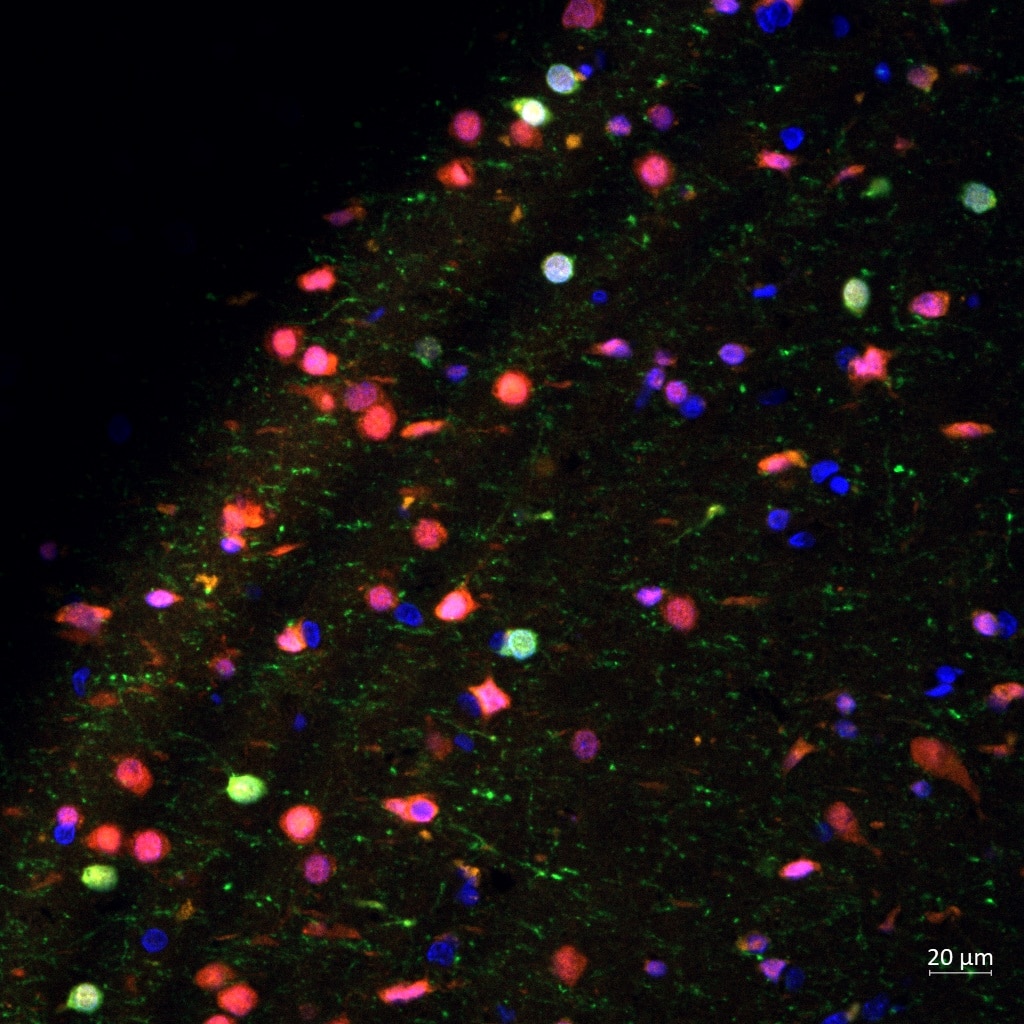
FH Reyes (Verified Customer) (09-05-2024) | Calretinin (green) worked magnificently on my human brain FFPE tissue marking some neurons (NeuN in red)
 |

