- Phare
- Validé par KD/KO
Anticorps Polyclonal de lapin anti-DDB1
DDB1 Polyclonal Antibody for WB, IHC, IP, ELISA
Hôte / Isotype
Lapin / IgG
Réactivité testée
Humain, rat, souris
Applications
WB, IHC, IF, IP, CoIP, ChIP, ELISA
Conjugaison
Non conjugué
N° de cat : 11380-1-AP
Synonymes
Galerie de données de validation
 at dilution of 1:8000 incubated at room temperature for 1.5 hours.")
 at dilution of 1:500 incubated at room temperature for 1.5 hours.")
 at dilution of 1:500 incubated at room temperature for 1.5 hours.")
 at dilution of 1:500 incubated at room temperature for 1.5 hours.")
 at dilution of 1:1000 incubated at room temperature for 1.5 hours.")
 at dilution of 1:8000 incubated at room temperature for 1.5 hours.")
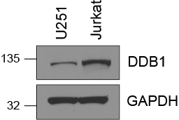
 with Jurkat cells lysate 3080ug.")
 at dilution of 1:200 (under 10x lens). Heat mediated antigen retrieval with Tris-EDTA buffer (pH 9.0).")
 at dilution of 1:200 (under 40x lens). Heat mediated antigen retrieval with Tris-EDTA buffer (pH 9.0).")
 at dilution of 1:1000 (under 20x lens). Heat mediated antigen retrieval with Tris-EDTA buffer (pH 9.0).")
 at dilution of 1:200 (under 10x lens). Heat mediated antigen retrieval with Tris-EDTA buffer (pH 9.0).")
 at dilution of 1:200 (under 40x lens). Heat mediated antigen retrieval with Tris-EDTA buffer (pH 9.0).")
Applications testées
| Résultats positifs en WB | cellules HCT 116, cellules C2C12, cellules HeLa, cellules HepG2, cellules Jurkat, cellules MCF-7, cellules MDA-MB-231, cellules NIH/3T3, tissu cérébral humain, tissu placentaire humain, tissu rénal humain, tissu testiculaire de rat, tissu testiculaire de souris |
| Résultats positifs en IP | cellules Jurkat |
| Résultats positifs en IHC | tissu de cancer du côlon humain, human colon cancer il est suggéré de démasquer l'antigène avec un tampon de TE buffer pH 9.0; (*) À défaut, 'le démasquage de l'antigène peut être 'effectué avec un tampon citrate pH 6,0. |
Dilution recommandée
| Application | Dilution |
|---|---|
| Western Blot (WB) | WB : 1:2000-1:16000 |
| Immunoprécipitation (IP) | IP : 0.5-4.0 ug for 1.0-3.0 mg of total protein lysate |
| Immunohistochimie (IHC) | IHC : 1:50-1:500 |
| It is recommended that this reagent should be titrated in each testing system to obtain optimal results. | |
| Sample-dependent, check data in validation data gallery | |
Applications publiées
| KD/KO | See 2 publications below |
| WB | See 10 publications below |
| IF | See 1 publications below |
| IP | See 1 publications below |
| CoIP | See 1 publications below |
| ChIP | See 1 publications below |
Informations sur le produit
11380-1-AP cible DDB1 dans les applications de WB, IHC, IF, IP, CoIP, ChIP, ELISA et montre une réactivité avec des échantillons Humain, rat, souris
| Réactivité | Humain, rat, souris |
| Réactivité citée | Humain, souris |
| Hôte / Isotype | Lapin / IgG |
| Clonalité | Polyclonal |
| Type | Anticorps |
| Immunogène | DDB1 Protéine recombinante Ag1901 |
| Nom complet | damage-specific DNA binding protein 1, 127kDa |
| Masse moléculaire calculée | 1140 aa, 127 kDa |
| Poids moléculaire observé | 127 kDa |
| Numéro d’acquisition GenBank | BC011686 |
| Symbole du gène | DDB1 |
| Identification du gène (NCBI) | 1642 |
| Conjugaison | Non conjugué |
| Forme | Liquide |
| Méthode de purification | Purification par affinité contre l'antigène |
| Tampon de stockage | PBS with 0.02% sodium azide and 50% glycerol |
| Conditions de stockage | Stocker à -20°C. Stable pendant un an après l'expédition. L'aliquotage n'est pas nécessaire pour le stockage à -20oC Les 20ul contiennent 0,1% de BSA. |
Informations générales
DDB1, also named as XAP1, XPCe, DDBa and XPE-BF, belongs to the DDB1 family. It is required for DNA repair. DDB1 binds to DDB2 to form the UV-damaged DNA-binding protein complex (the UV-DDB complex). The UV-DDB complex may recognize UV-induced DNA damage and recruit proteins of the nucleotide excision repair pathway (the NER pathway) to initiate DNA repair. The functional specificity of the DCX E3 ubiquitin-protein ligase complex is determined by the variable substrate recognition component recruited by DDB1. This antibody is specific to DDB1.
Protocole
| Product Specific Protocols | |
|---|---|
| WB protocol for DDB1 antibody 11380-1-AP | Download protocol |
| IHC protocol for DDB1 antibody 11380-1-AP | Download protocol |
| IP protocol for DDB1 antibody 11380-1-AP | Download protocol |
| Standard Protocols | |
|---|---|
| Click here to view our Standard Protocols |
Publications
| Species | Application | Title |
|---|---|---|
Front Immunol Ddb1 Is Essential for the Expansion of CD4+ Helper T Cells by Regulating Cell Cycle Progression and Cell Death. | ||
FASEB J DDB2 promotes melanoma cell growth by transcriptionally regulating the expression of KMT2A and predicts a poor prognosis | ||
iScience A Multidimensional Characterization of E3 Ubiquitin Ligase and Substrate Interaction Network. | ||
PLoS One 14-3-3ε mediates the cell fate decision-making pathways in response of hepatocellular carcinoma to Bleomycin-induced DNA damage. | ||
Virology DDB1 is a cellular substrate of NS3/4A protease and required for hepatitis C virus replication.
| ||
Cell Death Dis Cul4 E3 ubiquitin ligase regulates ovarian cancer drug resistance by targeting the antiapoptotic protein BIRC3.
|
Avis
The reviews below have been submitted by verified Proteintech customers who received an incentive for providing their feedback.
FH Bernadette (Verified Customer) (09-24-2025) | The antibody worked well at 1:2000 dilution in 5% milk shaken overnight in 4 degree fridge on HEK293 cells.
|
FH Sarah (Verified Customer) (07-03-2019) | Total cell lysate (15 ug) was resolved on a 4-12% Bis-Tris gel and transferred to nitrocellulose membrane. Membrane was incubated in blocking buffer (5% milk/0.1% Tween-20) for 1h. Membrane was incubated with anti-DDB1 in blocking buffer (1:1000) at 4C overnight. After washing, membrane was incubated in anti-rabbit-HRP in blocking bufffer (1:3000) for 1h at room temperature. Protein was detected using ECL reagent and imaged on a chemiluminescence detection system.
 |

