- Phare
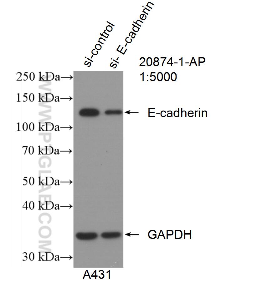
- Validé par KD/KO
Anticorps Polyclonal de lapin anti-E-cadherin
E-cadherin Polyclonal Antibody for WB, IHC, IF/ICC, IF-P, IF-Fro, IP, ELISA
Hôte / Isotype
Lapin / IgG
Réactivité testée
Humain, rat, souris et plus (4)
Applications
WB, IHC, IF/ICC, IF-P, IF-Fro, IP, CoIP, ELISA
Conjugaison
Non conjugué
N° de cat : 20874-1-AP
Synonymes
Galerie de données de validation
Applications testées
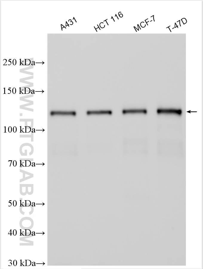
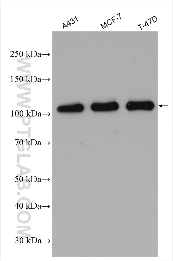
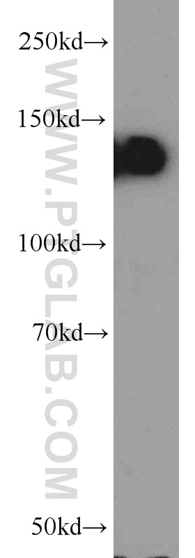
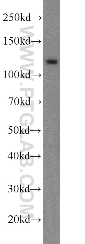
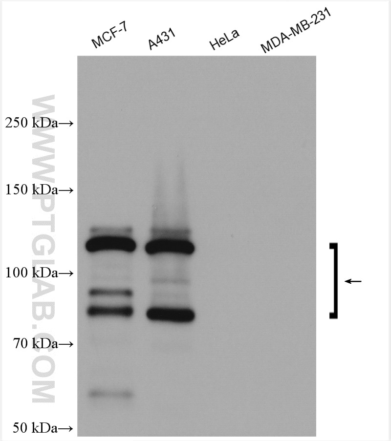
| Résultats positifs en WB | cellules A431, cellules DU 145, cellules HCT 116, cellules MCF-7, cellules T-47D, tissu testiculaire de souris |
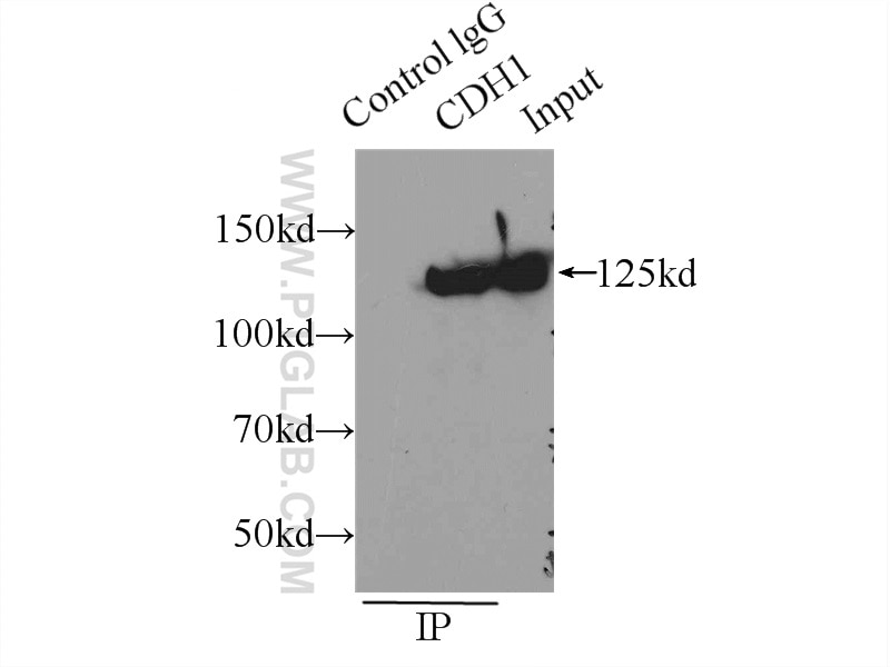
| Résultats positifs en IP | cellules A431 |
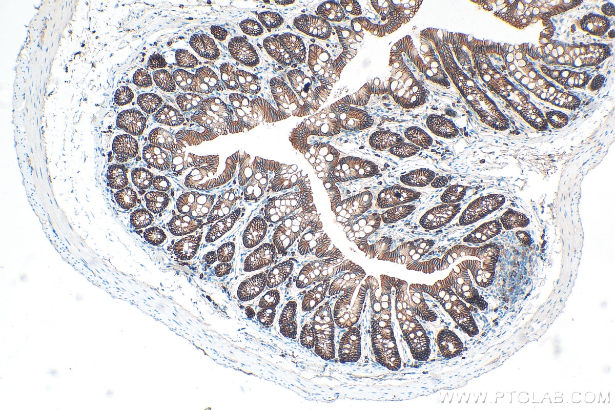
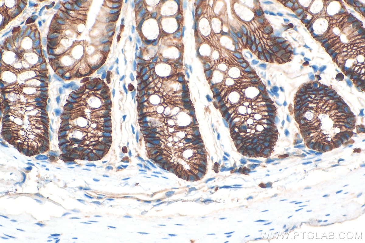
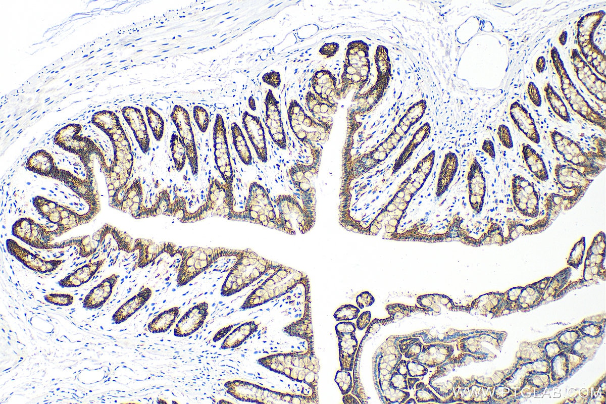
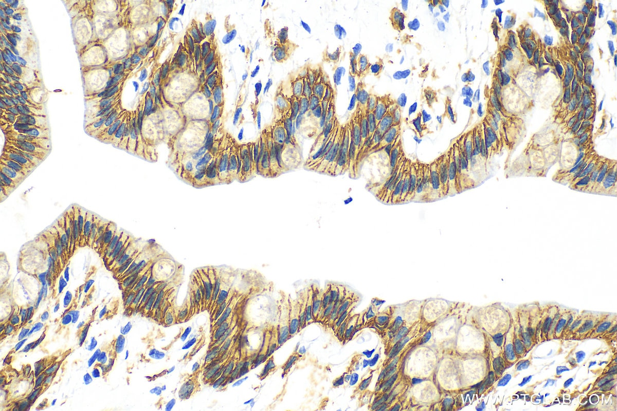
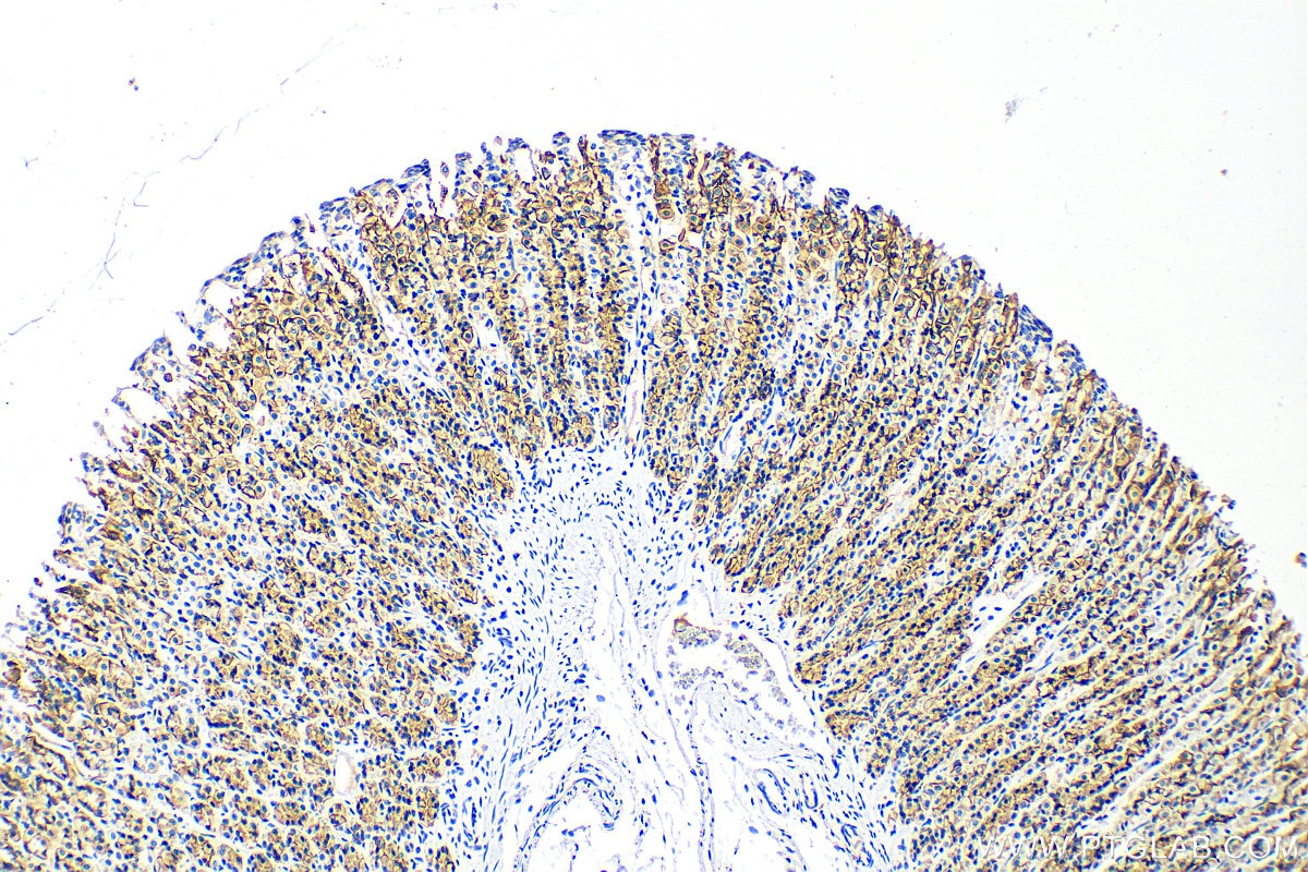
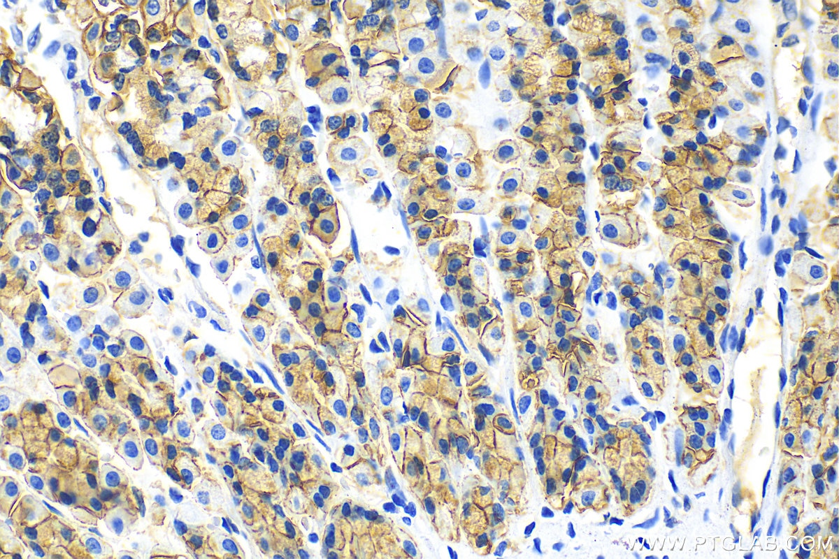
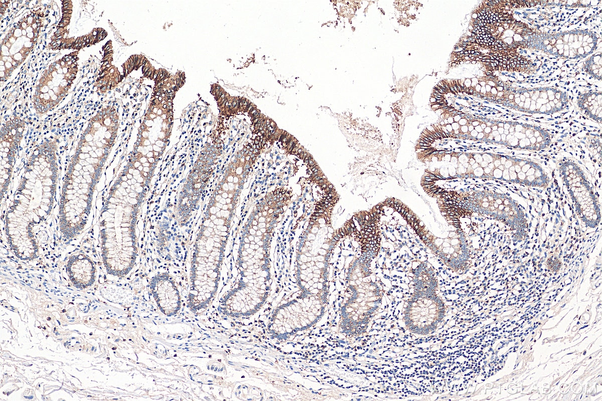
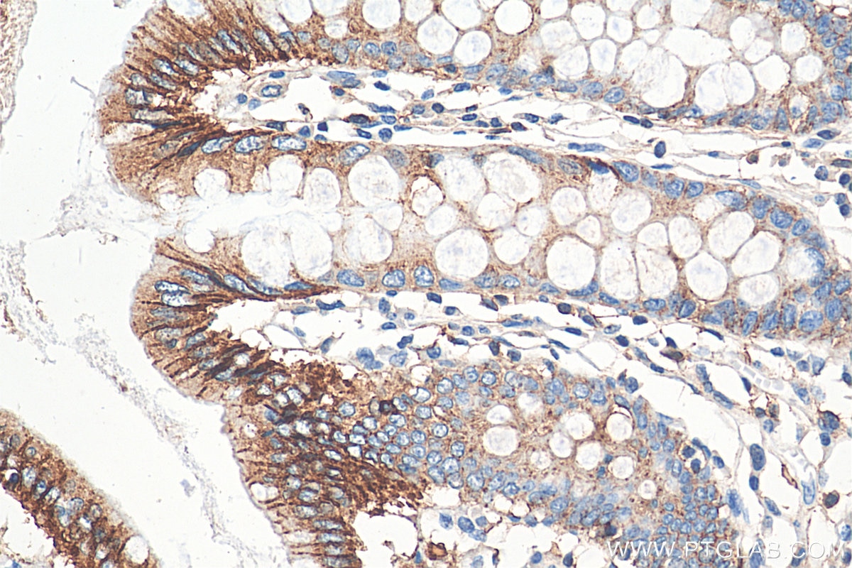
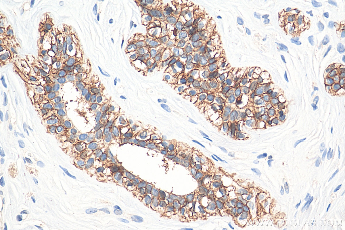
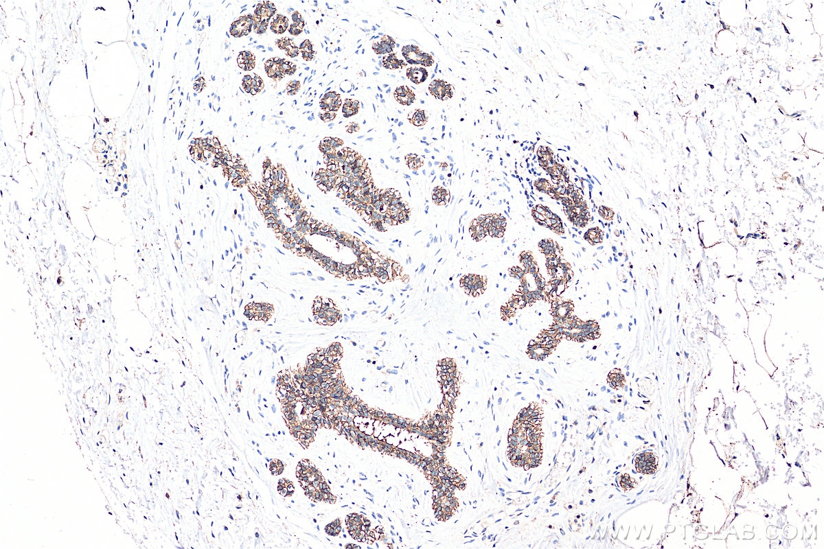
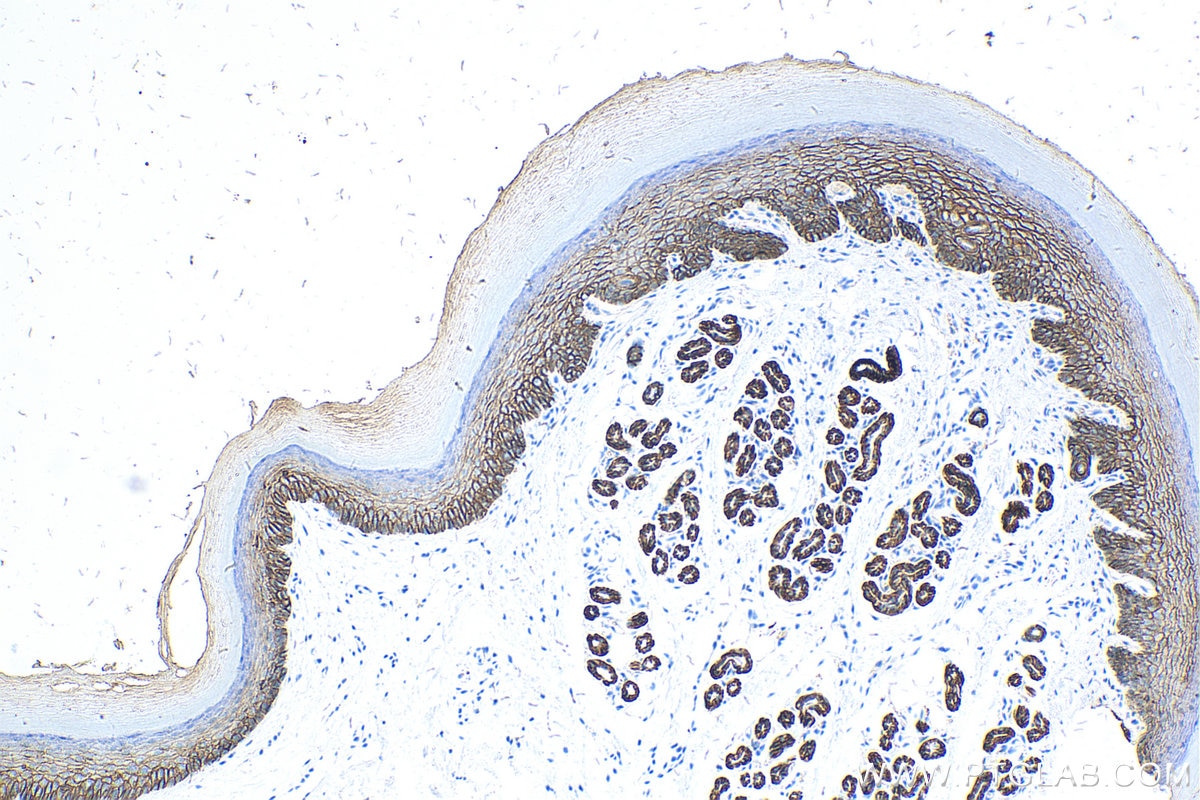
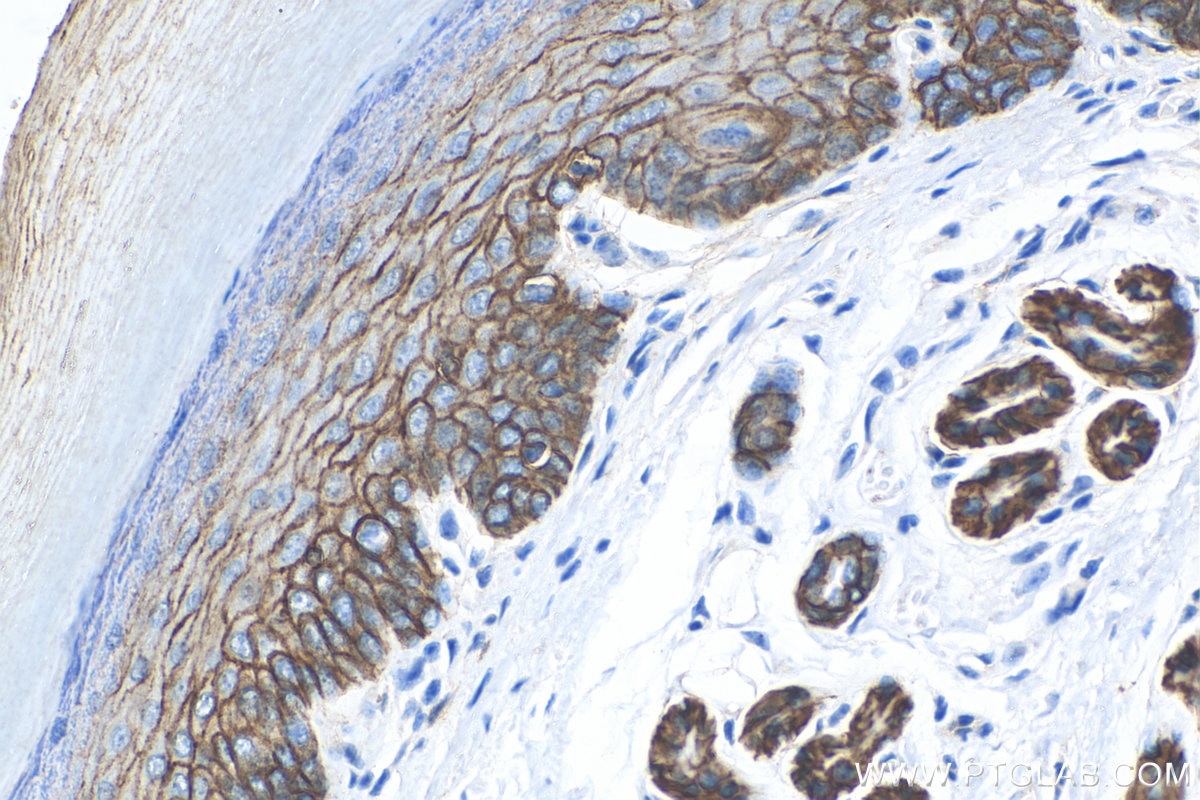
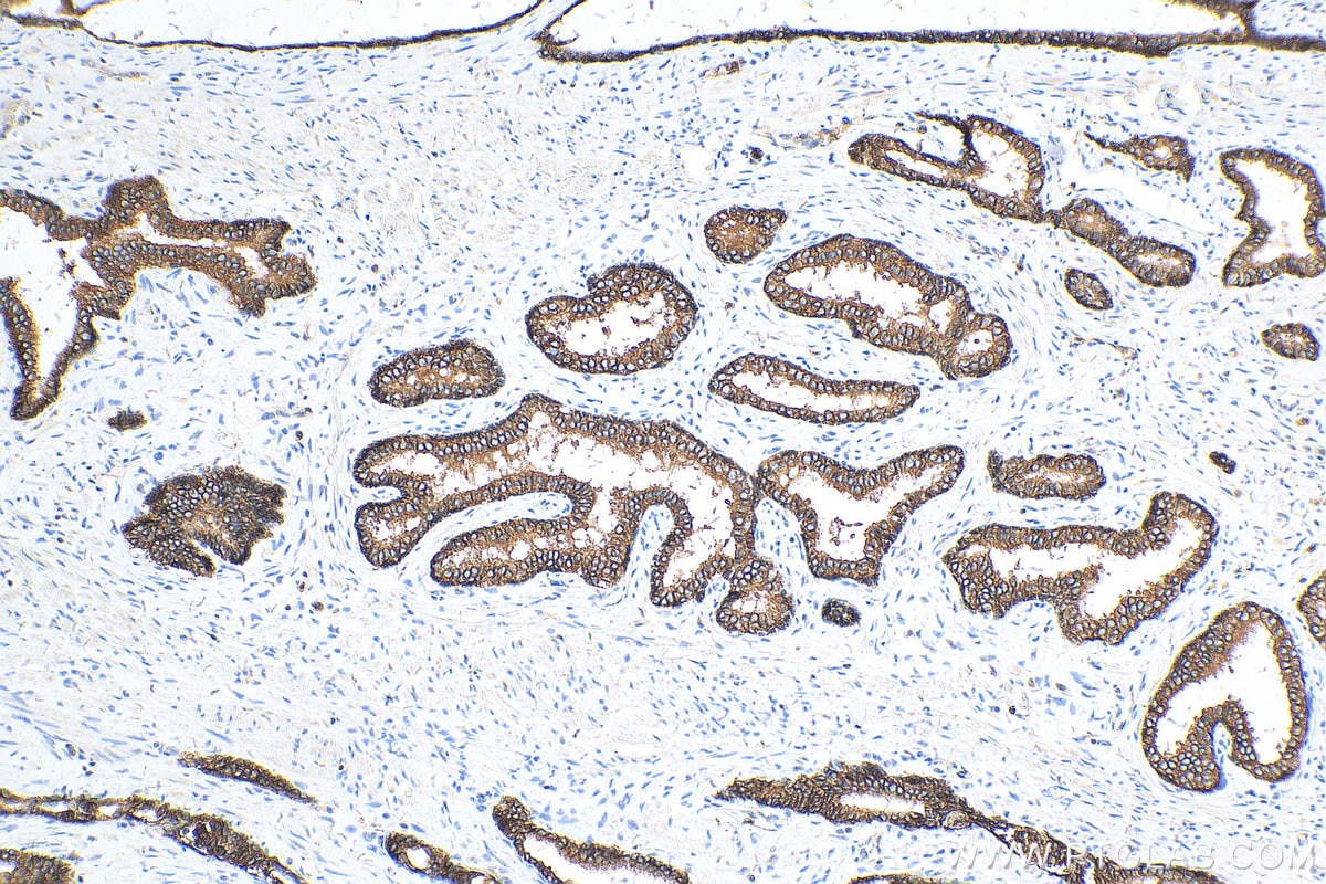
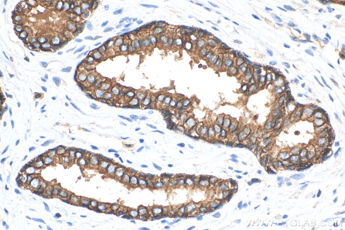
| Résultats positifs en IHC | tissu de côlon de souris, tissu cutané de souris, tissu de cancer de la prostate humain, tissu de cancer du sein humain, tissu de côlon de rat, tissu de côlon humain, tissu d'estomac de rat il est suggéré de démasquer l'antigène avec un tampon de TE buffer pH 9.0; (*) À défaut, 'le démasquage de l'antigène peut être 'effectué avec un tampon citrate pH 6,0. |
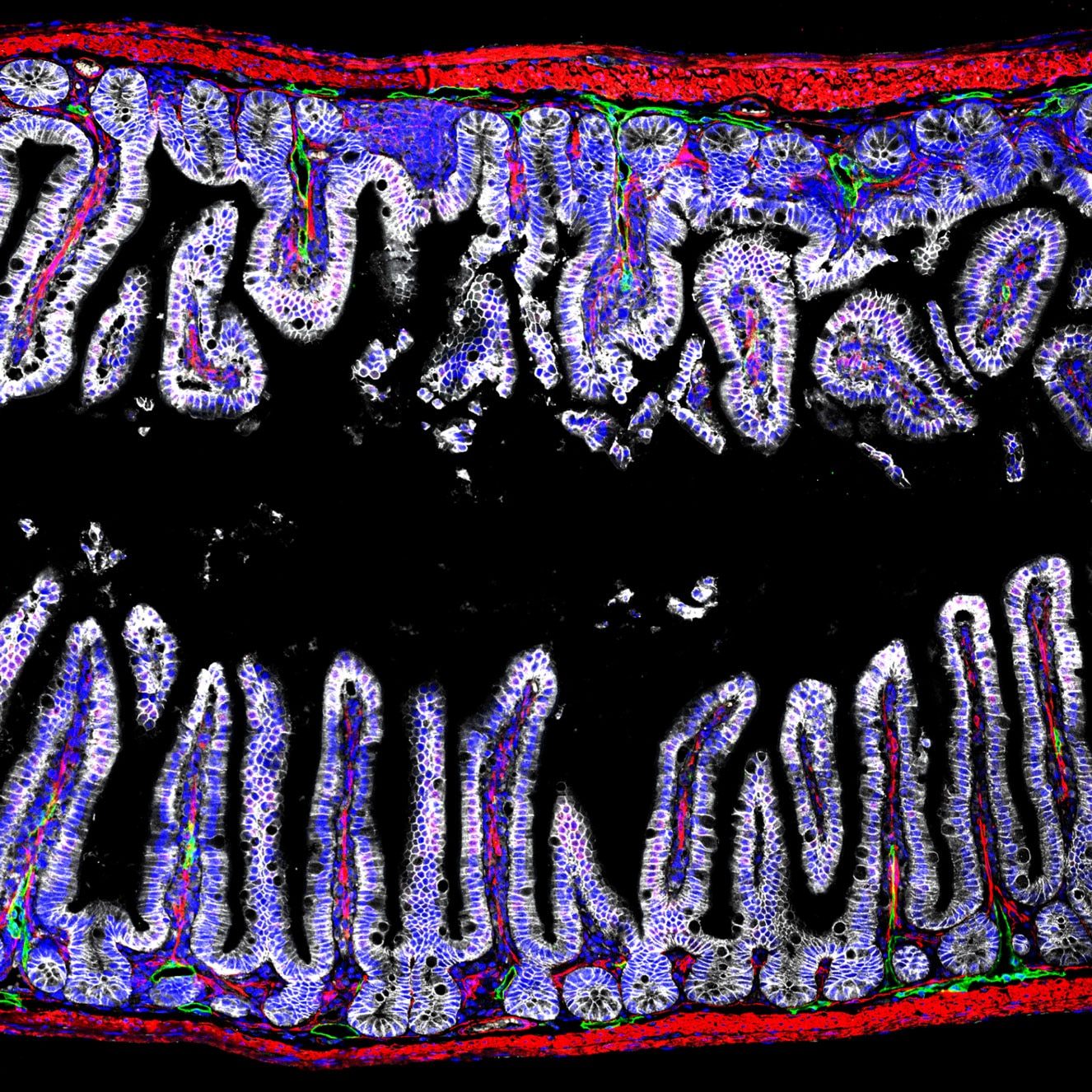
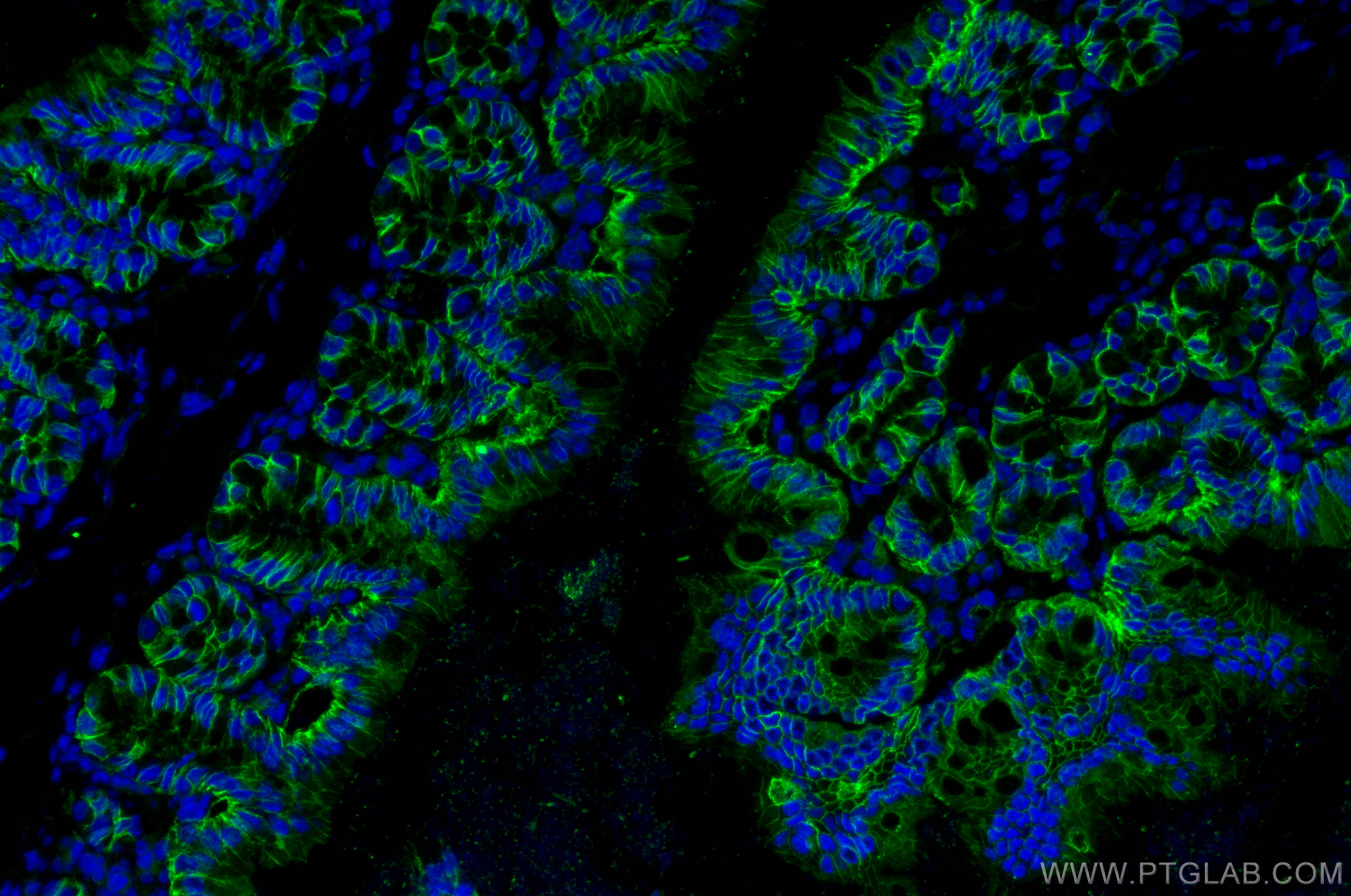
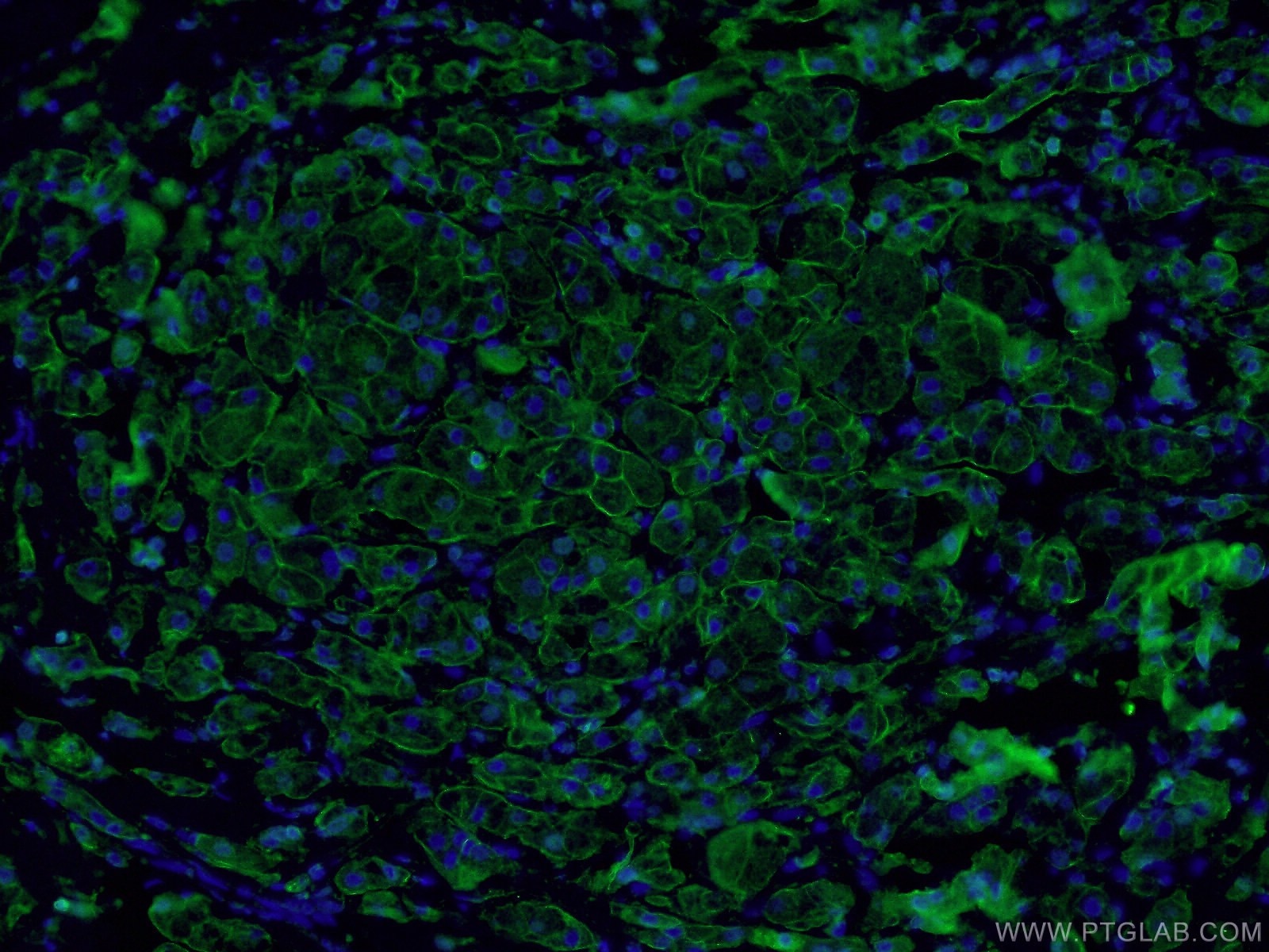
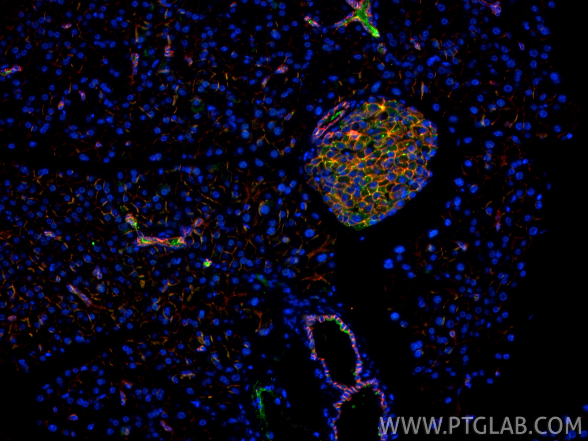
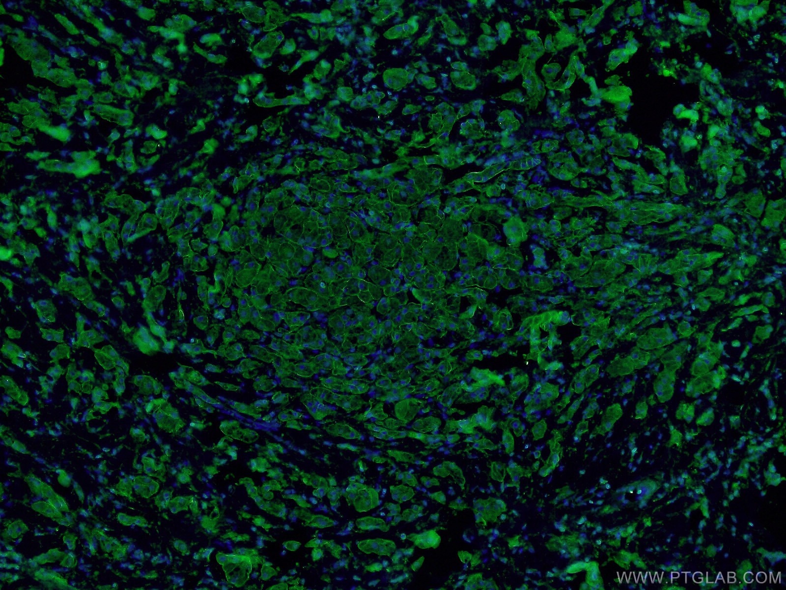
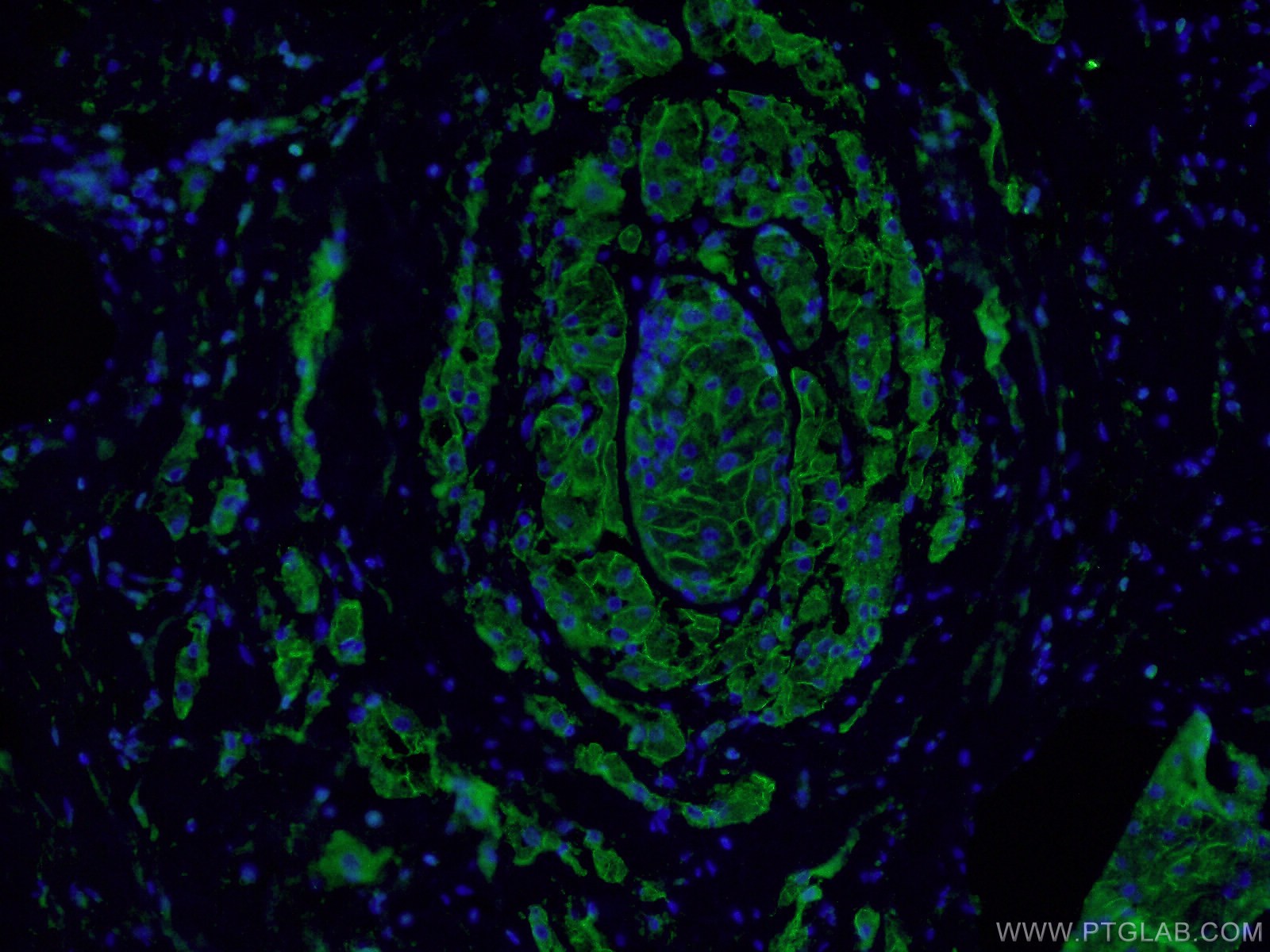
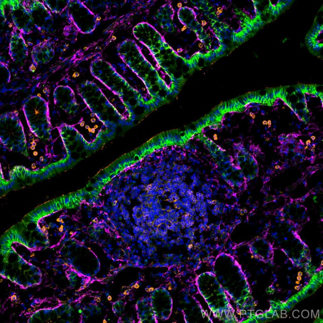
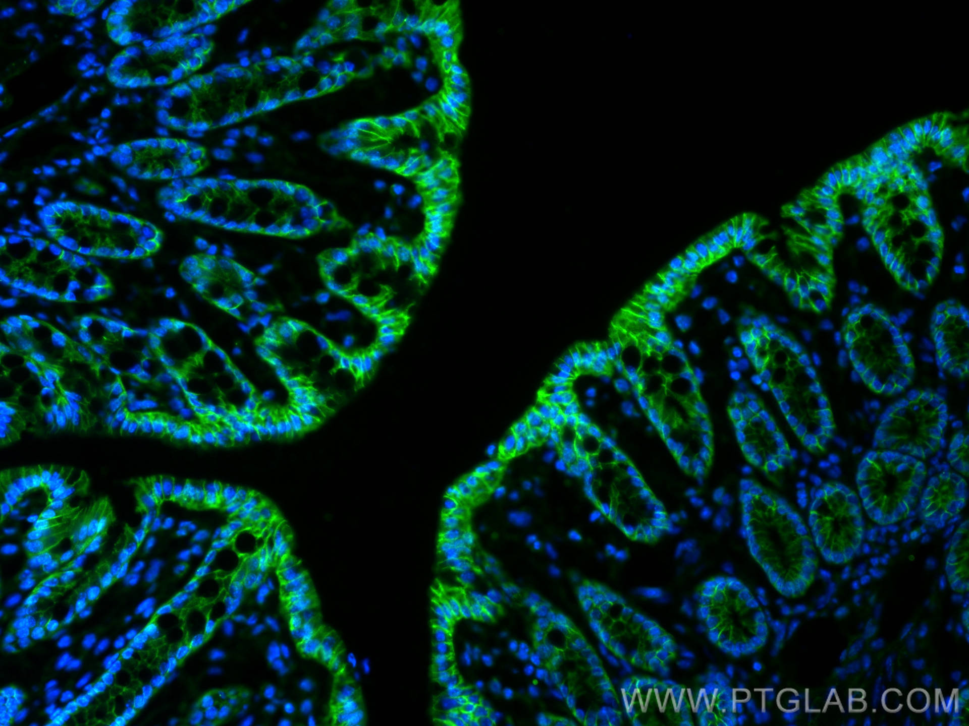
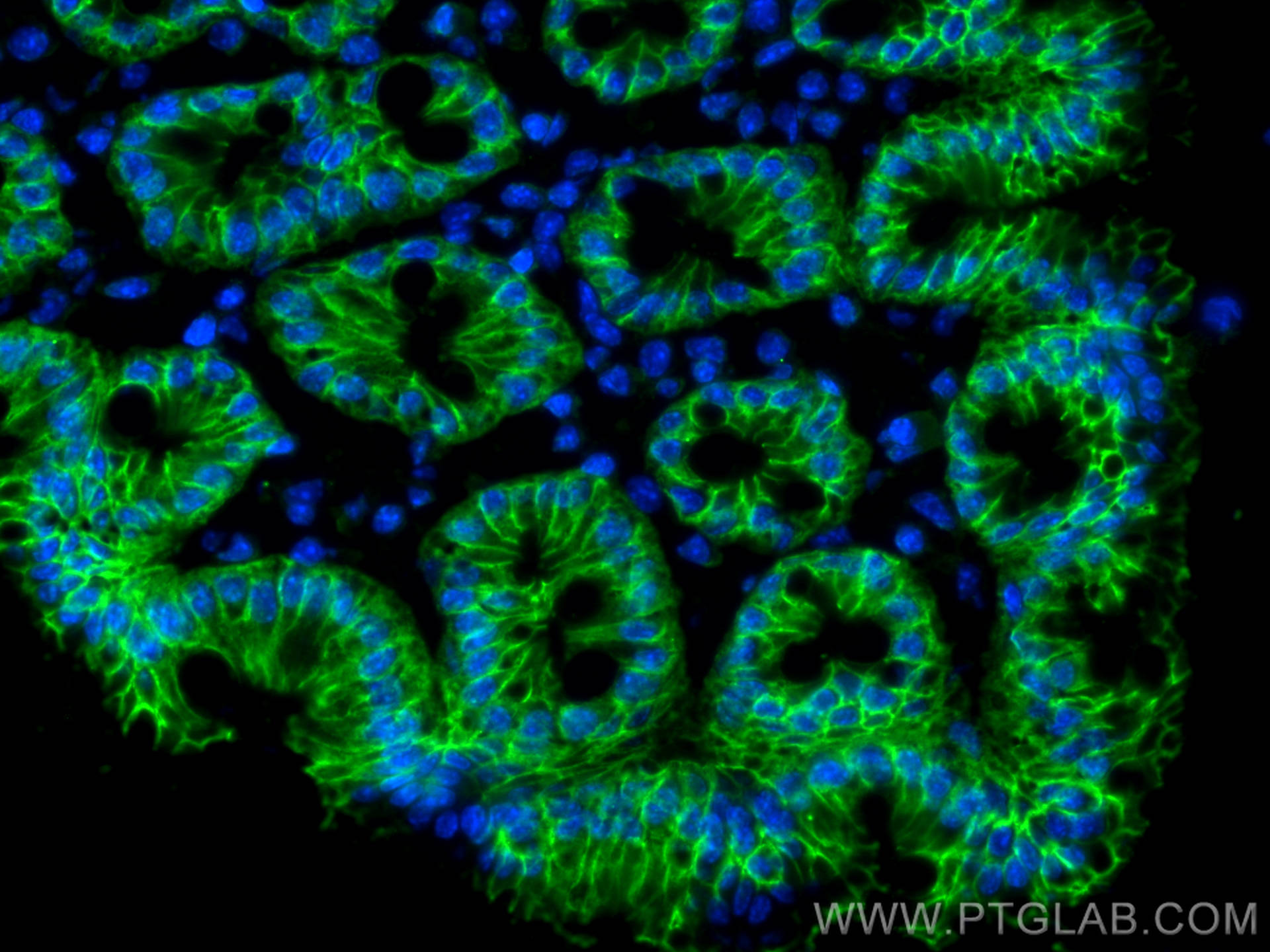
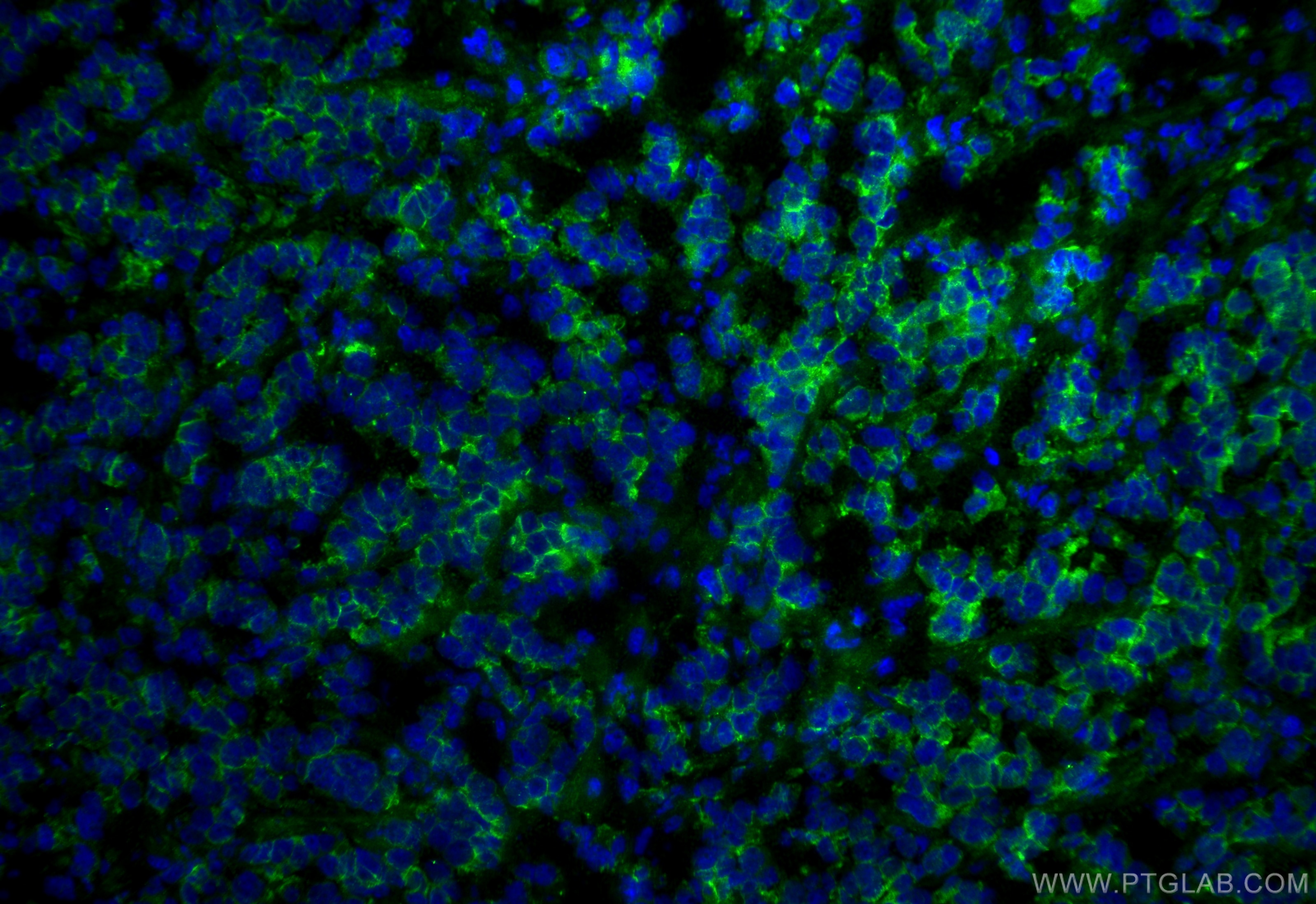
| Résultats positifs en IF-P | tissu de côlon de souris, tissu de cancer du sein humain, tissu d'intestin grêle de souris, tissu pancréatique de souris |
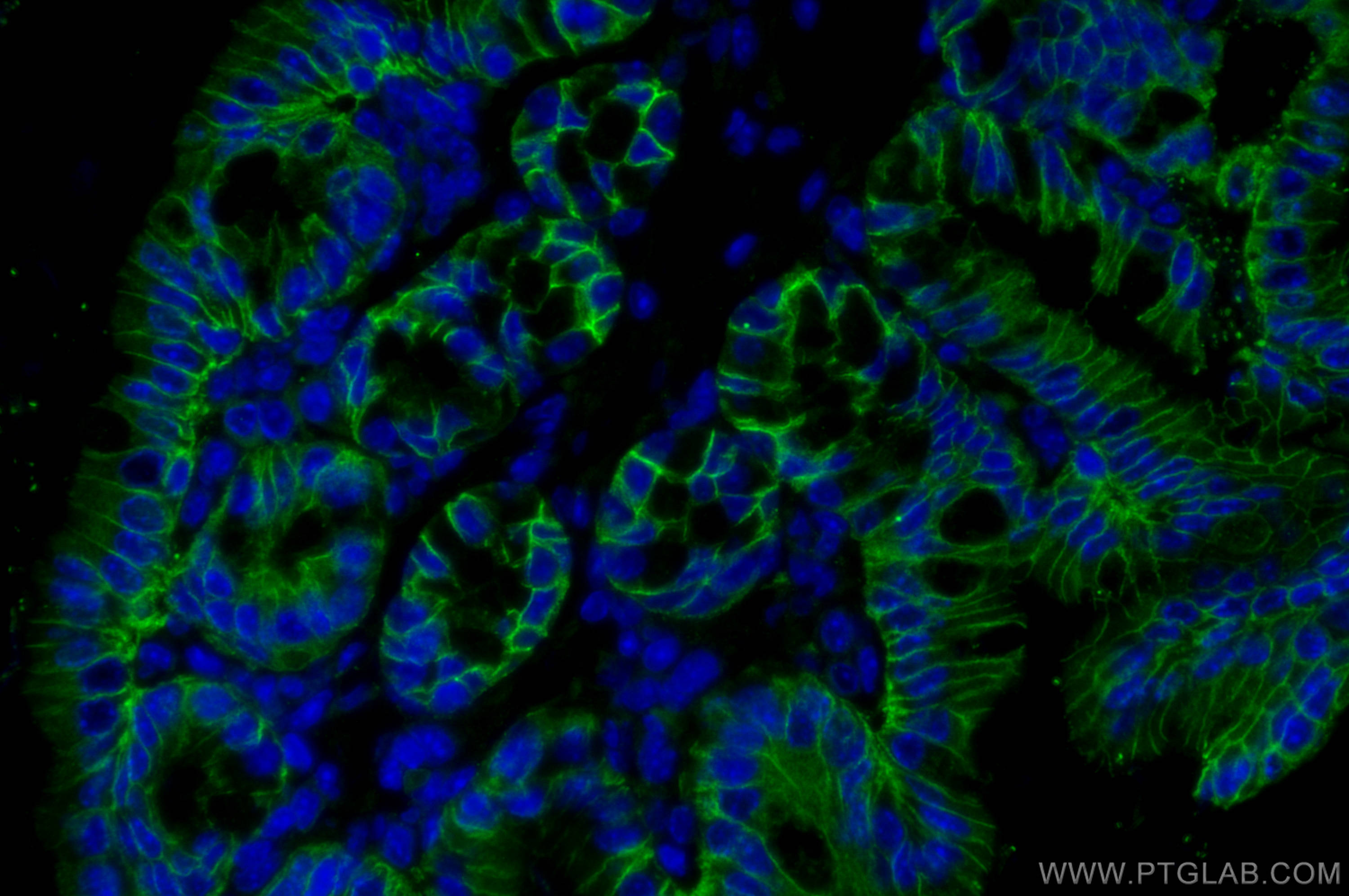
| Résultats positifs en IF-Fro | tissu de côlon de souris, mouse breast cancer |
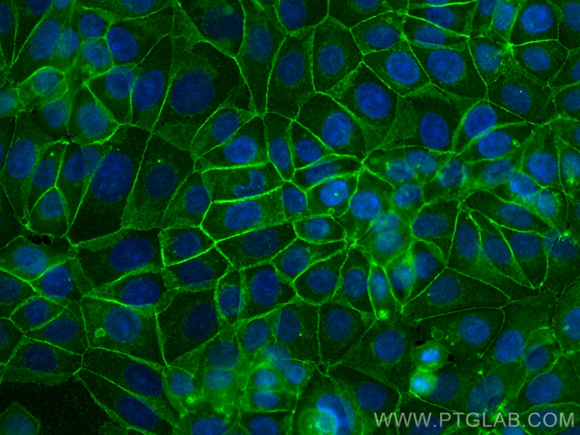
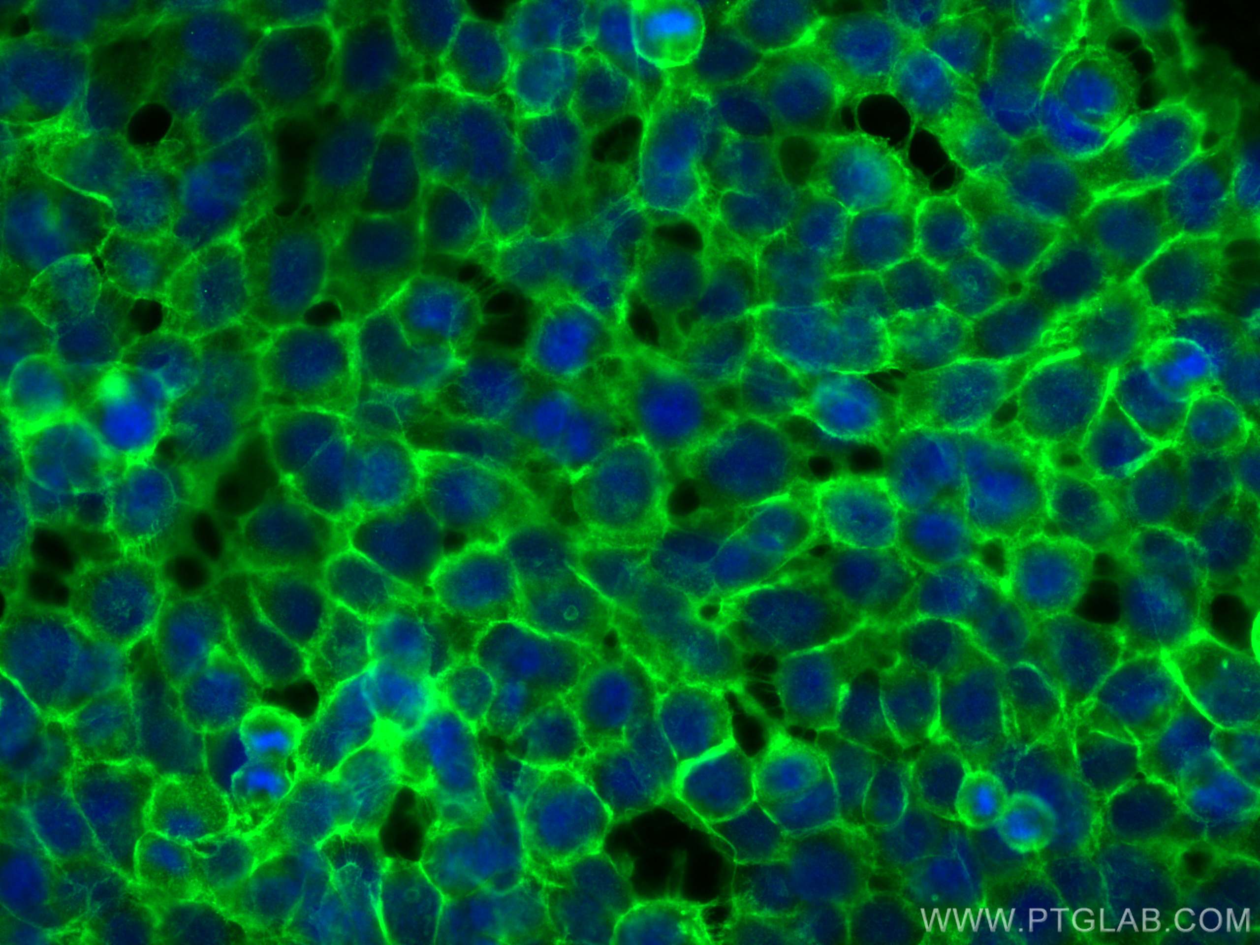
| Résultats positifs en IF/ICC | cellules MCF-7, cellules A431 |
Dilution recommandée
| Application | Dilution |
|---|---|
| Western Blot (WB) | WB : 1:20000-1:200000 |
| Immunoprécipitation (IP) | IP : 0.5-4.0 ug for 1.0-3.0 mg of total protein lysate |
| Immunohistochimie (IHC) | IHC : 1:5000-1:20000 |
| Immunofluorescence (IF)-P | IF-P : 1:200-1:800 |
| Immunofluorescence (IF)-FRO | IF-FRO : 1:50-1:500 |
| Immunofluorescence (IF)/ICC | IF/ICC : 1:200-1:800 |
| It is recommended that this reagent should be titrated in each testing system to obtain optimal results. | |
| Sample-dependent, check data in validation data gallery | |
Applications publiées
| KD/KO | See 3 publications below |
| WB | See 2126 publications below |
| IHC | See 400 publications below |
| IF | See 452 publications below |
| IP | See 1 publications below |
| CoIP | See 7 publications below |
Informations sur le produit
20874-1-AP cible E-cadherin dans les applications de WB, IHC, IF/ICC, IF-P, IF-Fro, IP, CoIP, ELISA et montre une réactivité avec des échantillons Humain, rat, souris
| Réactivité | Humain, rat, souris |
| Réactivité citée | rat, bovin, canin, Humain, poisson-zèbre, porc, souris |
| Hôte / Isotype | Lapin / IgG |
| Clonalité | Polyclonal |
| Type | Anticorps |
| Immunogène | E-cadherin Protéine recombinante Ag14973 |
| Nom complet | cadherin 1, type 1, E-cadherin (epithelial) |
| Masse moléculaire calculée | 882 aa, 97 kDa |
| Poids moléculaire observé | 120-125 kDa |
| Numéro d’acquisition GenBank | BC141838 |
| Symbole du gène | E-cadherin |
| Identification du gène (NCBI) | 999 |
| Conjugaison | Non conjugué |
| Forme | Liquide |
| Méthode de purification | Purification par affinité contre l'antigène |
| Tampon de stockage | PBS with 0.02% sodium azide and 50% glycerol |
| Conditions de stockage | Stocker à -20°C. Stable pendant un an après l'expédition. L'aliquotage n'est pas nécessaire pour le stockage à -20oC Les 20ul contiennent 0,1% de BSA. |
Informations générales
Cadherins are a family of transmembrane glycoproteins that mediate calcium-dependent cell-cell adhesion and play an important role in the maintenance of normal tissue architecture. E-cadherin (epithelial cadherin), also known as CDH1 (cadherin 1) or CAM 120/80, is a classical member of the cadherin superfamily which also include N-, P-, R-, and B-cadherins. E-cadherin is expressed on the cell surface in most epithelial tissues. The extracellular region of E-cadherin establishes calcium-dependent homophilic trans binding, providing specific interaction with adjacent cells, while the cytoplasmic domain is connected to the actin cytoskeleton through the interaction with p120-, α-, β-, and γ-catenin (plakoglobin). E-cadherin is important in the maintenance of the epithelial integrity, and is involved in mechanisms regulating proliferation, differentiation, and survival of epithelial cell. E-cadherin may also play a role in tumorigenesis. It is considered to be an invasion suppressor protein and its loss is an indicator of high tumor aggressiveness. E-cadherin is sensitive to trypsin digestion in the absence of Ca2+. This polyclonal antibody recognizes 120-125 kDa intact E-cadherin and its cleaved fragments of 80-120 kDa.
Protocole
| Product Specific Protocols | |
|---|---|
| WB protocol for E-cadherin antibody 20874-1-AP | Download protocol |
| IHC protocol for E-cadherin antibody 20874-1-AP | Download protocol |
| IF protocol for E-cadherin antibody 20874-1-AP | Download protocol |
| IP protocol for E-cadherin antibody 20874-1-AP | Download protocol |
| Standard Protocols | |
|---|---|
| Click here to view our Standard Protocols |
Publications
| Species | Application | Title |
|---|---|---|
Science Reversible reprogramming of cardiomyocytes to a fetal state drives heart regeneration in mice. | ||
Mol Cancer lncRNA ZNRD1-AS1 promotes malignant lung cell proliferation, migration, and angiogenesis via the miR-942/TNS1 axis and is positively regulated by the m6A reader YTHDC2 | ||
Adv Mater Supramolecular Hydrogel with Ultra-Rapid Cell-Mediated Network Adaptation for Enhancing Cellular Metabolic Energetics and Tissue Regeneration | ||
Nat Aging Single-cell and spatial RNA sequencing identify divergent microenvironments and progression signatures in early- versus late-onset prostate cancer | ||
ACS Nano Biomimetic Nanomedicine-Triggered in Situ Vaccination for Innate and Adaptive Immunity Activations for Bacterial Osteomyelitis Treatment. |
Avis
The reviews below have been submitted by verified Proteintech customers who received an incentive for providing their feedback.
FH Dhanwini (Verified Customer) (09-24-2025) | GOOD
|
FH Sneha (Verified Customer) (09-24-2025) | GOOD
|
FH Zeeshan (Verified Customer) (09-18-2025) | Work great
|
FH Michael (Verified Customer) (09-18-2025) | Works perfectly for western blots
|
FH Jimmy (Verified Customer) (05-20-2025) | Great results for western blot application
|
FH Davide (Verified Customer) (11-22-2022) | The antibody works optimally at 1:100. By the strenght of the signal it could probably also work at 1:50. Optimal product. E-CAD in red in the picture (abundant background signal due to non optimal blocking)
 |
FH Sophie (Verified Customer) (02-07-2022) | Highly efficient staining when used at 1/1000 overnight for IF on paraffin embedded tissues
|
FH Kishor (Verified Customer) (01-30-2019) | This antibody working very good for human cancer cells in western blotting (1:1000)
|
FH Martin (Verified Customer) (12-13-2017) |
|
