- Phare
- Validé par KD/KO
Anticorps Monoclonal anti-E-cadherin
E-cadherin Monoclonal Antibody for WB, IHC, IF/ICC, IF-P, IF-Fro, ELISA
Hôte / Isotype
Mouse / IgG2b
Réactivité testée
Humain, porc, rat, souris et plus (1)
Applications
WB, IHC, IF/ICC, IF-P, IF-Fro, ELISA
Conjugaison
Non conjugué
CloneNo.
6B11F11
N° de cat : 60335-1-Ig
Synonymes
Galerie de données de validation
 with sh-Control and sh-E-cadherin transfected A431 cells.")
 at dilution of 1:4000 incubated at room temperature for 1.5 hours.")
 at dilution of 1:1000 incubated at room temperature for 1.5 hours.")
 at dilution of 1:8000 incubated at room temperature for 1.5 hours.")
 at dilution of 1:8000 incubated at room temperature for 1.5 hours.")
 at dilution of 1:2000 (under 10x lens). Heat mediated antigen retrieval with Tris-EDTA buffer (pH 9.0).")
 at dilution of 1:2000 (under 40x lens). Heat mediated antigen retrieval with Tris-EDTA buffer (pH 9.0).")
 at dilution of 1:2000 (under 10x lens. Heat mediated antigen retrieval with Tris-EDTA buffer (pH 9.0).")
 at dilution of 1:2000 (under 40x lens. Heat mediated antigen retrieval with Tris-EDTA buffer (pH 9.0).")
 at dilution of 1:8000 (under 10x lens. Heat mediated antigen retrieval with Tris-EDTA buffer (pH 9.0).")
 at dilution of 1:10000 (under 10x lens). Heat mediated antigen retrieval with Tris-EDTA buffer (pH 9.0).")
 at dilution of 1:10000 (under 40x lens). Heat mediated antigen retrieval with Tris-EDTA buffer (pH 9.0).")
 at dilution of 1:300 (under 10x lens). Heat mediated antigen retrieval with Tris-EDTA buffer (pH 9.0).")
 at dilution of 1:300 (under 40x lens). Heat mediated antigen retrieval with Tris-EDTA buffer (pH 9.0).")
 at dilution of 1:4000 (under 10x lens. Heat mediated antigen retrieval with Tris-EDTA buffer (pH 9.0).")
 at dilution of 1:4000 (under 40x lens. Heat mediated antigen retrieval with Tris-EDTA buffer (pH 9.0).")
 at dilution of 1:2000 (under 10x lens). Heat mediated antigen retrieval with Tris-EDTA buffer (pH 9.0).")
 at dilution of 1:2000 (under 40x lens). Heat mediated antigen retrieval with Tris-EDTA buffer (pH 9.0).")
 fixed human kidney tissue using E-cadherin antibody (60335-1-Ig, Clone: 6B11F11 ) at dilution of 1:300 and CoraLite®594-Conjugated AffiniPure Goat Anti-Mouse IgG(H+L), (18150-1-AP, green). DNA was stained by DAPI (blue).")
 fixed human breast cancer tissue using E-cadherin antibody (60335-1-Ig, Clone: 6B11F11 ) at dilution of 1:400 and CoraLite®488-Conjugated AffiniPure Goat Anti-Mouse IgG(H+L).")
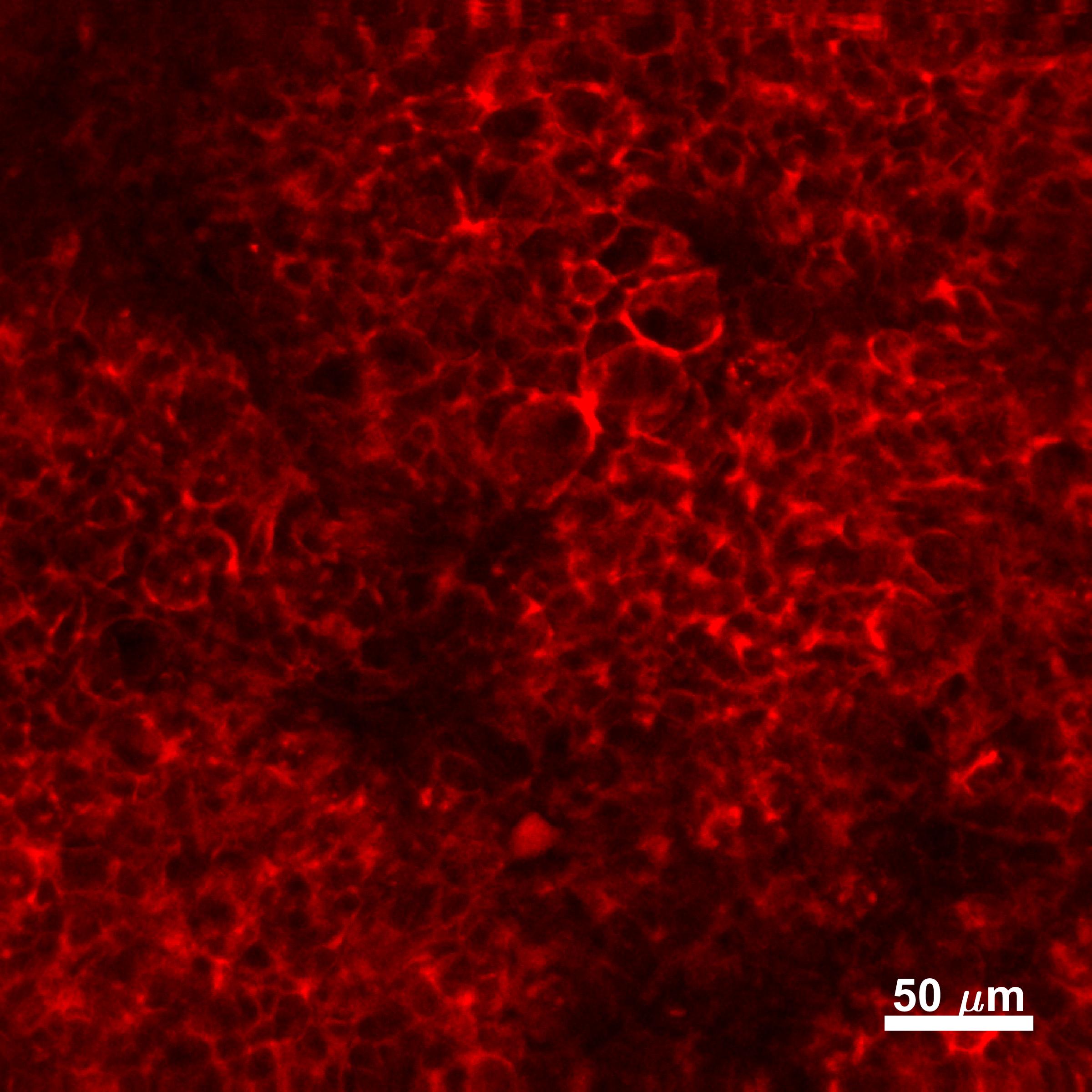
 fixed frozen OCT-embedded mouse brain tissue using E-cadherin antibody (60335-1-Ig, Clone: 6B11F11 ) at dilution of 1:400 and CoraLite®488-Conjugated Goat Anti-Mouse IgG(H+L) (SA00013-1).")
 fixed MCF-7 cells using E-cadherin antibody (60335-1-Ig, Clone: 6B11F11 ) at dilution of 1:400 and CoraLite®488-Conjugated AffiniPure Goat Anti-Mouse IgG(H+L).")
 fixed mouse breast cancer using E-cadherin antibody (60335-1-Ig, Clone: 6B11F11 ) at dilution of 1:800 and CoraLite®488-Conjugated Goat Anti-Mouse IgG(H+L) (SA00013-1).")
Applications testées
| Résultats positifs en WB | cellules PC-3, cellules A431, cellules MCF-7, cellules MKN-45, cellules SGC-7901, tissu cérébral de porc |
| Résultats positifs en IHC | tissu de cancer du sein humain, tissu de côlon de rat, tissu de côlon humain, tissu d'estomac de rat il est suggéré de démasquer l'antigène avec un tampon de TE buffer pH 9.0; (*) À défaut, 'le démasquage de l'antigène peut être 'effectué avec un tampon citrate pH 6,0. |
| Résultats positifs en IF-P | tissu de cancer du sein humain, cellules MCF-7, tissu rénal humain |
| Résultats positifs en IF-Fro | tissu cérébral de souris, |
| Résultats positifs en IF/ICC | cellules MCF-7, mouse breast cancer |
Dilution recommandée
| Application | Dilution |
|---|---|
| Western Blot (WB) | WB : 1:2000-1:8000 |
| Immunohistochimie (IHC) | IHC : 1:1000-1:4000 |
| Immunofluorescence (IF)-P | IF-P : 1:200-1:800 |
| Immunofluorescence (IF)-FRO | IF-FRO : 1:200-1:800 |
| Immunofluorescence (IF)/ICC | IF/ICC : 1:200-1:800 |
| It is recommended that this reagent should be titrated in each testing system to obtain optimal results. | |
| Sample-dependent, check data in validation data gallery | |
Applications publiées
| WB | See 190 publications below |
| IHC | See 27 publications below |
| IF | See 57 publications below |
Informations sur le produit
60335-1-Ig cible E-cadherin dans les applications de WB, IHC, IF/ICC, IF-P, IF-Fro, ELISA et montre une réactivité avec des échantillons Humain, porc, rat, souris
| Réactivité | Humain, porc, rat, souris |
| Réactivité citée | rat, Humain, porc, singe, souris |
| Hôte / Isotype | Mouse / IgG2b |
| Clonalité | Monoclonal |
| Type | Anticorps |
| Immunogène | E-cadherin Protéine recombinante Ag15085 |
| Nom complet | cadherin 1, type 1, E-cadherin (epithelial) |
| Masse moléculaire calculée | 882 aa, 97 kDa |
| Poids moléculaire observé | 120 kDa |
| Numéro d’acquisition GenBank | BC141838 |
| Symbole du gène | E-cadherin |
| Identification du gène (NCBI) | 999 |
| Conjugaison | Non conjugué |
| Forme | Liquide |
| Méthode de purification | Purification par protéine A |
| Tampon de stockage | PBS with 0.02% sodium azide and 50% glycerol |
| Conditions de stockage | Stocker à -20°C. Stable pendant un an après l'expédition. L'aliquotage n'est pas nécessaire pour le stockage à -20oC Les 20ul contiennent 0,1% de BSA. |
Informations générales
Cadherins are a family of transmembrane glycoproteins that mediate calcium-dependent cell-cell adhesion and play an important role in the maintenance of normal tissue architecture. E-cadherin (epithelial cadherin), also known as CDH1 (cadherin 1) or CAM 120/80, is a classical member of the cadherin superfamily which also include N-, P-, R-, and B-cadherins. E-cadherin is expressed on the cell surface in most epithelial tissues. The extracellular region of E-cadherin establishes calcium-dependent homophilic trans binding, providing specific interaction with adjacent cells, while the cytoplasmic domain is connected to the actin cytoskeleton through the interaction with p120-, α-, β-, and γ-catenin (plakoglobin). E-cadherin is important in the maintenance of the epithelial integrity, and is involved in mechanisms regulating proliferation, differentiation, and survival of epithelial cell. E-cadherin may also play a role in tumorigenesis. It is considered to be an invasion suppressor protein and its loss is an indicator of high tumor aggressiveness.
Protocole
| Product Specific Protocols | |
|---|---|
| WB protocol for E-cadherin antibody 60335-1-Ig | Download protocol |
| IHC protocol for E-cadherin antibody 60335-1-Ig | Download protocol |
| IF protocol for E-cadherin antibody 60335-1-Ig | Download protocol |
| Standard Protocols | |
|---|---|
| Click here to view our Standard Protocols |
Publications
| Species | Application | Title |
|---|---|---|
Theranostics Vitamin D binding protein (VDBP) hijacks twist1 to inhibit vasculogenic mimicry in hepatocellular carcinoma | ||
Theranostics FSH induces EMT in ovarian cancer via ALKBH5-regulated Snail m6A demethylation | ||
Biomaterials Urinary exosomes-based Engineered Nanovectors for Homologously Targeted Chemo-Chemodynamic Prostate Cancer Therapy via abrogating IGFR/AKT/NF-kB/IkB signaling. | ||
Redox Biol Riboflavin deficiency leads to irreversible cellular changes in the RPE and disrupts retinal function through alterations in cellular metabolic homeostasis. |
Avis
The reviews below have been submitted by verified Proteintech customers who received an incentive for providing their feedback.
FH Aqib (Verified Customer) (09-02-2024) | It works quite well. recommended
|
FH Saba (Verified Customer) (06-14-2022) | The IF staining was very good and satisfying.
|
FH Silvia (Verified Customer) (02-09-2022) | The antibody worked well on HT-29 cells at 1:800 dilution for IF.
|
FH Joshua (Verified Customer) (12-27-2019) | Caco-2 cells fixed in 4% paraformaldehyde. Stained overnight at 4C. Bright stain, minimal background
 |
FH Louisiane (Verified Customer) (02-06-2019) | Cells were fixed with 4% PFA for 10 min, permeabilized with 0.1% Triton-X100 for 5 min and blocked with 1% FBS/1% BSA in PBS for 3 h. Antibodies were diluted in 1% FBS/1% BSA in PBS. Primary antibody: 2 h. Alexa Fluor anti-mouse secondary antibody (1:250): 1 h.Cells were imaged by confocal microscopy - no labeling was observed.
|

