- Phare
- Validé par KD/KO
Anticorps Monoclonal anti-EGFR
EGFR Monoclonal Antibody for WB, IHC, ELISA
Hôte / Isotype
Mouse / IgG1
Réactivité testée
Humain
Applications
WB, IHC, IF, ELISA
Conjugaison
Non conjugué
CloneNo.
2A2H10
N° de cat : 66455-1-Ig
Synonymes
Galerie de données de validation
 at dilution of 1:20000 incubated at room temperature for 1.5 hours.")
 at dilution of 1:20000 incubated at room temperature for 1.5 hours.")
 with sh-Control and sh-EGFR transfected HepG2 cells.")
 at dilution of 1:20000 incubated at room temperature for 1.5 hours.")
 at dilution of 1:9700 incubated at room temperature for 1.5 hours.")
 at dilution of 1:9700 incubated at room temperature for 1.5 hours.")
 at dilution of 1:9700 incubated at room temperature for 1.5 hours.")
 at dilution of 1:9700 incubated at room temperature for 1.5 hours.")
 at dilution of 1:9700 incubated at room temperature for 1.5 hours.")
 at dilution of 1:9700 incubated at room temperature for 1.5 hours.")
 at dilution of 1:9700 incubated at room temperature for 1.5 hours.")
 at dilution of 1:2000 (under 10x lens). Heat mediated antigen retrieval with Tris-EDTA buffer (pH 9.0).")
 at dilution of 1:4000 (under 10x lens). Heat mediated antigen retrieval with Tris-EDTA buffer (pH 9.0).")
 at dilution of 1:4000 (under 40x lens). Heat mediated antigen retrieval with Tris-EDTA buffer (pH 9.0).")
 at dilution of 1:4000 (under 20x lens). Heat mediated antigen retrieval with Tris-EDTA buffer (pH 9.0).")
 at dilution of 1:4000 (under 20x lens). Heat mediated antigen retrieval with Tris-EDTA buffer (pH 9.0).")
 at dilution of 1:800 (under 10x lens. Heat mediated antigen retrieval with Tris-EDTA buffer (pH 9.0).")
 at dilution of 1:800 (under 40x lens. Heat mediated antigen retrieval with Tris-EDTA buffer (pH 9.0).")
 at dilution of 1:1000 (under 10x lens. Heat mediated antigen retrieval with Tris-EDTA buffer (pH 9.0).")
 at dilution of 1:1000 (under 40x lens. Heat mediated antigen retrieval with Tris-EDTA buffer (pH 9.0).")
 at dilution of 1:1000 (under 10x lens. Heat mediated antigen retrieval with Tris-EDTA buffer (pH 9.0).")
 at dilution of 1:1000 (under 40x lens. Heat mediated antigen retrieval with Tris-EDTA buffer (pH 9.0).")
 at dilution of 1:2000 (under 10x lens). Heat mediated antigen retrieval with Tris-EDTA buffer (pH 9.0).")
 at dilution of 1:2000 (under 40x lens). Heat mediated antigen retrieval with Tris-EDTA buffer (pH 9.0).")
 at dilution of 1:2000 (under 10x lens). Heat mediated antigen retrieval with Tris-EDTA buffer (pH 9.0).")
 at dilution of 1:2000 (under 40x lens). Heat mediated antigen retrieval with Tris-EDTA buffer (pH 9.0).")
 at dilution of 1:2000 (under 10x lens). Heat mediated antigen retrieval with Tris-EDTA buffer (pH 9.0).")
 at dilution of 1:2000 (under 40x lens). Heat mediated antigen retrieval with Tris-EDTA buffer (pH 9.0).")
Applications testées
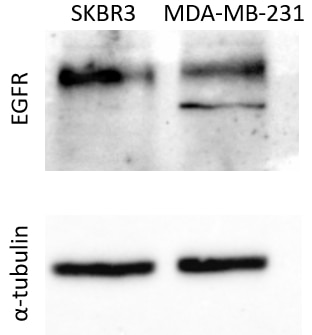
| Résultats positifs en WB | cellules A431, cellules A549, cellules EC109, cellules HeLa, cellules HepG2, cellules LNCaP, cellules MDA-MB-231, cellules MDA-MB-468, cellules PC-3, cellules PC-3, cellules SKOV-3 |
| Résultats positifs en IHC | tissu d'amygdalite humain, tissu de cancer de la peau humain, tissu de cancer du col de l'utérus humain, tissu de cancer du côlon humain, tissu de cancer du poumon humain, tissu de cancer du sein humain, tissu de gliome humain, tissu placentaire humain il est suggéré de démasquer l'antigène avec un tampon de TE buffer pH 9.0; (*) À défaut, 'le démasquage de l'antigène peut être 'effectué avec un tampon citrate pH 6,0. |
Dilution recommandée
| Application | Dilution |
|---|---|
| Western Blot (WB) | WB : 1:5000-1:50000 |
| Immunohistochimie (IHC) | IHC : 1:2000-1:8000 |
| It is recommended that this reagent should be titrated in each testing system to obtain optimal results. | |
| Sample-dependent, check data in validation data gallery | |
Applications publiées
| KD/KO | See 6 publications below |
| WB | See 85 publications below |
| IHC | See 14 publications below |
| IF | See 10 publications below |
Informations sur le produit
66455-1-Ig cible EGFR dans les applications de WB, IHC, IF, ELISA et montre une réactivité avec des échantillons Humain
| Réactivité | Humain |
| Réactivité citée | Humain |
| Hôte / Isotype | Mouse / IgG1 |
| Clonalité | Monoclonal |
| Type | Anticorps |
| Immunogène | EGFR Protéine recombinante Ag24947 |
| Nom complet | epidermal growth factor receptor (erythroblastic leukemia viral (v-erb-b) oncogene homolog, avian) |
| Masse moléculaire calculée | 1210 aa, 134 kDa |
| Poids moléculaire observé | 145-165 kDa |
| Numéro d’acquisition GenBank | BC094761 |
| Symbole du gène | EGFR |
| Identification du gène (NCBI) | 1956 |
| Conjugaison | Non conjugué |
| Forme | Liquide |
| Méthode de purification | Purification par protéine G |
| Tampon de stockage | PBS with 0.02% sodium azide and 50% glycerol |
| Conditions de stockage | Stocker à -20°C. Stable pendant un an après l'expédition. L'aliquotage n'est pas nécessaire pour le stockage à -20oC Les 20ul contiennent 0,1% de BSA. |
Informations générales
EGFR, also named as ERBB1, is a cell-surface receptor for members of the epidermal growth factor family (EGF-family) of extracellular protein ligands. Binding of the protein to a ligand induces receptor dimerization and tyrosine autophosphorylation and leads to cell proliferation. The gene resides on chromosome 7p12, encoding a 170 kDa membrane-associated glycoprotein. Recent studies have shown EGFR plays a critical role in cancer development and progression, including cell proliferation, apoptosis, angiogenesis, and metastatic spread. Mutations in this gene are associated with lung cancer.
Protocole
| Product Specific Protocols | |
|---|---|
| WB protocol for EGFR antibody 66455-1-Ig | Download protocol |
| IHC protocol for EGFR antibody 66455-1-Ig | Download protocol |
| Standard Protocols | |
|---|---|
| Click here to view our Standard Protocols |
Publications
| Species | Application | Title |
|---|---|---|
Nat Commun EGFR core fucosylation, induced by hepatitis C virus, promotes TRIM40-mediated-RIG-I ubiquitination and suppresses interferon-I antiviral defenses | ||
Mol Cell N7-Methylguanosine tRNA modification enhances oncogenic mRNA translation and promotes intrahepatic cholangiocarcinoma progression.
| ||
Cancer Res Inhibition of EGFR Overcomes Acquired Lenvatinib Resistance Driven by STAT3-ABCB1 Signaling in Hepatocellular Carcinoma | ||
Pharmacol Res Upregulation of CSNK1A1 induced by ITGB5 confers to hepatocellular carcinoma resistance to sorafenib in vivo by disrupting the EPS15/EGFR complex | ||
Cell Death Dis Neurokinin-1 receptor promotes non-small cell lung cancer progression through transactivation of EGFR. |
Avis
The reviews below have been submitted by verified Proteintech customers who received an incentive for providing their feedback.
FH k. (Verified Customer) (10-26-2023) | This antibody worked well for human cells and mouse liver cell proteins at 1:500 or 1:1000 concentrations at 4 °C over a night of incubation.
|
FH Christos (Verified Customer) (02-13-2023) | -35ug protein extract were loaded per well -Transfer was performed for 2hr at 400mA at 4oC, on a Nitrocellulose Blotting Membrane -Membrane blocking was performed in 5% non-fat milk in PBS-Tween20 at room temperature, under mild shacking -Antibodies dilutions were performed in 5% non-fat milk in PBS-Tween20. -Incubations with the primary antibodies were performed as followed: 1)EGFR: 1:1000 for 1.5hr at 4oC 2)tubulin (sc32293, Santa Cruz): 1:5000 for 1.5hr at 4oC -Incubations with the secondary antibodies were performed with Rb pAb to Ms IgG (HRP) (ab 6728, Abcam) at a 1:20000 dilution, for 1hr at 4oC.
 |
FH Guorong (Verified Customer) (03-31-2022) | A band of approximately 160 kDa was detected
 |
FH Carly (Verified Customer) (11-17-2020) | Tested using EDTA plasma on an antibody microarray
|

