- Phare
- Validé par KD/KO
Anticorps Polyclonal de lapin anti-EIF5B
EIF5B Polyclonal Antibody for WB, IHC, IF/ICC, ELISA
Hôte / Isotype
Lapin / IgG
Réactivité testée
Humain, rat, souris et plus (1)
Applications
WB, IHC, IF/ICC, ELISA
Conjugaison
Non conjugué
N° de cat : 13527-1-AP
Synonymes
Galerie de données de validation
Applications testées
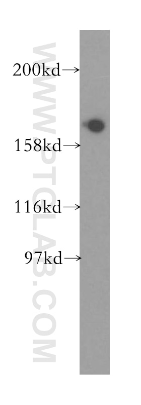
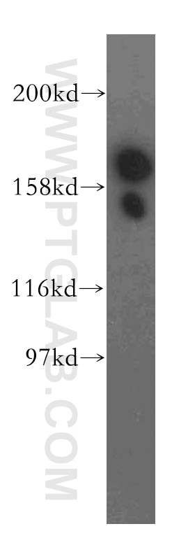
| Résultats positifs en WB | tissu cérébral de souris, cellules A549 |
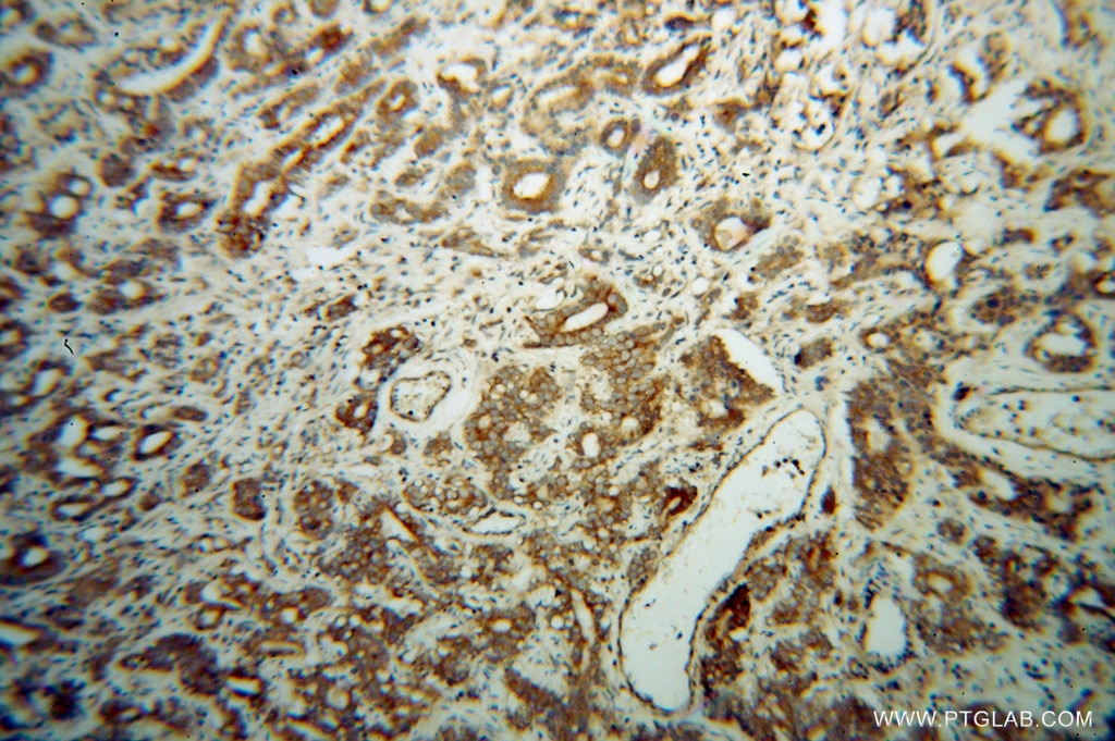
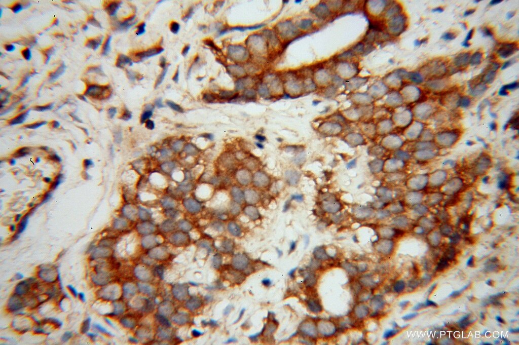
| Résultats positifs en IHC | tissu de gliome humain il est suggéré de démasquer l'antigène avec un tampon de TE buffer pH 9.0; (*) À défaut, 'le démasquage de l'antigène peut être 'effectué avec un tampon citrate pH 6,0. |
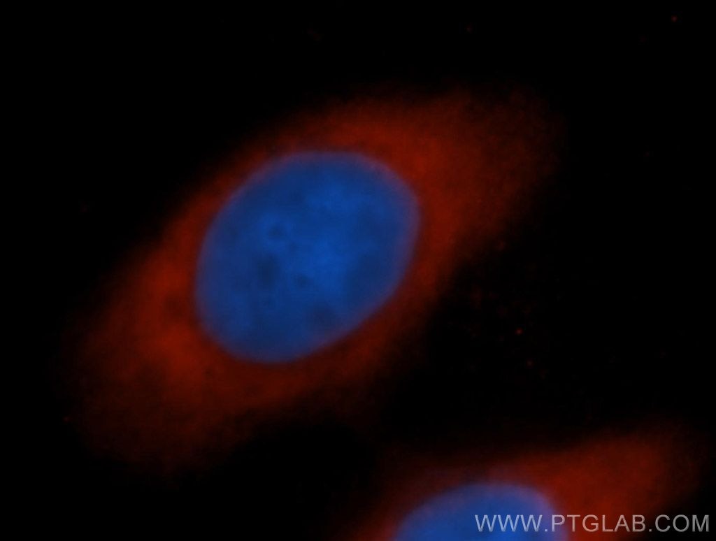
| Résultats positifs en IF/ICC | cellules MCF-7 |
Dilution recommandée
| Application | Dilution |
|---|---|
| Western Blot (WB) | WB : 1:500-1:2000 |
| Immunohistochimie (IHC) | IHC : 1:20-1:200 |
| Immunofluorescence (IF)/ICC | IF/ICC : 1:20-1:200 |
| It is recommended that this reagent should be titrated in each testing system to obtain optimal results. | |
| Sample-dependent, check data in validation data gallery | |
Applications publiées
| KD/KO | See 3 publications below |
| WB | See 7 publications below |
Informations sur le produit
13527-1-AP cible EIF5B dans les applications de WB, IHC, IF/ICC, ELISA et montre une réactivité avec des échantillons Humain, rat, souris
| Réactivité | Humain, rat, souris |
| Réactivité citée | Humain, porc |
| Hôte / Isotype | Lapin / IgG |
| Clonalité | Polyclonal |
| Type | Anticorps |
| Immunogène | EIF5B Protéine recombinante Ag4404 |
| Nom complet | eukaryotic translation initiation factor 5B |
| Masse moléculaire calculée | 1220 aa, 139 kDa |
| Poids moléculaire observé | 175 kDa |
| Numéro d’acquisition GenBank | BC032639 |
| Symbole du gène | EIF5B |
| Identification du gène (NCBI) | 9669 |
| Conjugaison | Non conjugué |
| Forme | Liquide |
| Méthode de purification | Purification par affinité contre l'antigène |
| Tampon de stockage | PBS with 0.02% sodium azide and 50% glycerol |
| Conditions de stockage | Stocker à -20°C. Stable pendant un an après l'expédition. L'aliquotage n'est pas nécessaire pour le stockage à -20oC Les 20ul contiennent 0,1% de BSA. |
Informations générales
Translation initiation requires the delivery of the initiator methionine tRNA to the 40S ribosomal subunit. The initiator methionine tRNA is delivered by the heterotrimeric complex EIF2 in a ternary complex with GTP that interacts with the 40S subunit. The resulting complex then binds to an mRNA and scans for the AUG start codon. Eukaryotic translation initiation factor 5B (EIF5B) plays a role in recognition of the AUG codon in conjunction with translation factor eIF2, which functions to general translation initiation by promoting the binding of the formylmethionine-tRNA to ribosomes, and promotes joining of the 60S ribosomal subunit. A single crossreactive polypeptide of 175 kDa was detected, whereas the predicted size of the protein was 139 kDa. This size discrepancy may be the result of posttranslational modifications of EIF5B or, perhaps more likely, of unusual behavior in SDS-PAGE caused by the highly charged N-terminal region of EIF5B (PMID: 10200264 ).
Protocole
| Product Specific Protocols | |
|---|---|
| WB protocol for EIF5B antibody 13527-1-AP | Download protocol |
| IHC protocol for EIF5B antibody 13527-1-AP | Download protocol |
| IF protocol for EIF5B antibody 13527-1-AP | Download protocol |
| Standard Protocols | |
|---|---|
| Click here to view our Standard Protocols |
Publications
| Species | Application | Title |
|---|---|---|
Cell Mol Life Sci eIF2A, an initiator tRNA carrier refractory to eIF2α kinases, functions synergistically with eIF5B. | ||
bioRxiv Start codon-associated ribosomal frameshifting mediates nutrient stress adaptation
| ||
bioRxiv LARP1 senses free ribosomes to coordinate supply and demand of ribosomal proteins
| ||
Sci Adv Hepatovirus translation requires PDGFA-associated protein 1, an eIF4E-binding protein regulating endoplasmic reticulum stress responses | ||
J Virol VP3 protein of Senecavirus A promotes viral IRES-driven translation and attenuates innate immunity by specifically relocalizing hnRNPA2B1 |
Avis
The reviews below have been submitted by verified Proteintech customers who received an incentive for providing their feedback.
FH Bill (Verified Customer) (12-23-2019) | This antibody give every strong signal on EIF5B.
|
