- Phare
- Validé par KD/KO
Anticorps Polyclonal de lapin anti-Phospho-ERK1/2 (Thr202/Tyr204)
Phospho-ERK1/2 (Thr202/Tyr204) Polyclonal Antibody for WB, IP, ELISA
Hôte / Isotype
Lapin / IgG
Réactivité testée
Humain, souris et plus (5)
Applications
WB, IHC, IF, IP, ELISA
Conjugaison
Non conjugué
N° de cat : 28733-1-AP
Synonymes
Galerie de données de validation
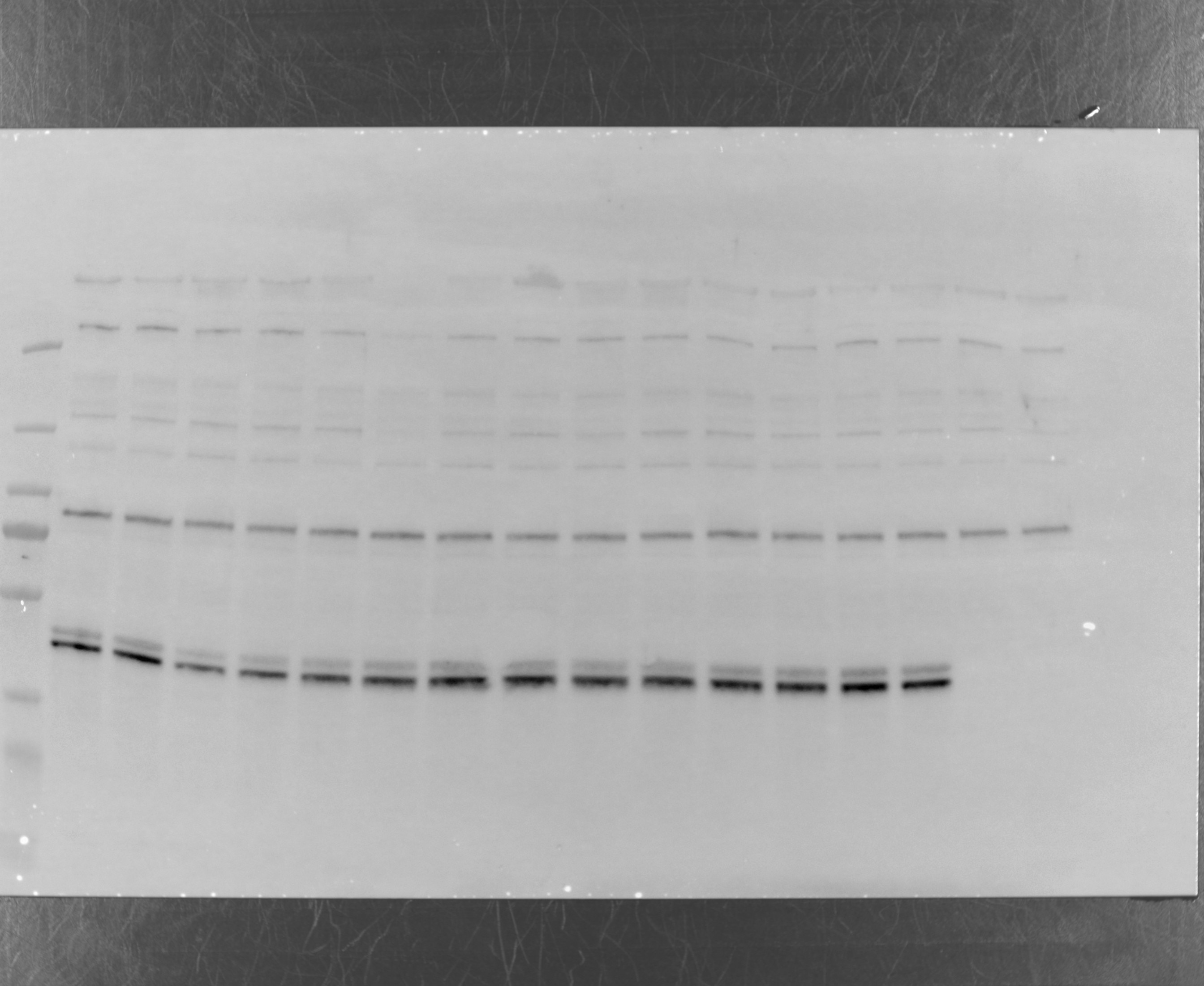
 at dilution of 1:3000 incubated at 4℃ overnight. The membrane was stripped and re-blotted with GAPDH antibody as loading control.")
 antibody) at dilution of 1:4500 incubated at 4℃ overnight.")
 antibody) at dilution of 1:2000 incubated at room temperature for 1.5 hours.")
 (IP:28733-1-AP, 2ug; Detection:28733-1-AP 1:1000) with Calyculin A treated PC-3 cells lysate 1552 ug.")
Applications testées
| Résultats positifs en WB | cellules NIH/3T3, cellules HEK-293T traitées à la calyculine A |
| Résultats positifs en IP | cellules PC-3 traitées à la calyculine A, |
Dilution recommandée
| Application | Dilution |
|---|---|
| Western Blot (WB) | WB : 1:1000-1:4000 |
| Immunoprécipitation (IP) | IP : 0.5-4.0 ug for 1.0-3.0 mg of total protein lysate |
| It is recommended that this reagent should be titrated in each testing system to obtain optimal results. | |
| Sample-dependent, check data in validation data gallery | |
Applications publiées
| KD/KO | See 1 publications below |
| WB | See 510 publications below |
| IHC | See 30 publications below |
| IF | See 19 publications below |
Informations sur le produit
28733-1-AP cible Phospho-ERK1/2 (Thr202/Tyr204) dans les applications de WB, IHC, IF, IP, ELISA et montre une réactivité avec des échantillons Humain, souris
| Réactivité | Humain, souris |
| Réactivité citée | rat, Chèvre, Humain, poisson-zèbre, porc, poulet, souris |
| Hôte / Isotype | Lapin / IgG |
| Clonalité | Polyclonal |
| Type | Anticorps |
| Immunogène | Peptide |
| Nom complet | mitogen-activated protein kinase 3 |
| Masse moléculaire calculée | 38-43 kDa |
| Poids moléculaire observé | 38-43 kDa |
| Numéro d’acquisition GenBank | NM_002746 |
| Symbole du gène | ERK1 |
| Identification du gène (NCBI) | 5595 |
| Conjugaison | Non conjugué |
| Forme | Liquide |
| Méthode de purification | Purification par affinité contre l'antigène |
| Tampon de stockage | PBS with 0.02% sodium azide and 50% glycerol |
| Conditions de stockage | Stocker à -20°C. Stable pendant un an après l'expédition. L'aliquotage n'est pas nécessaire pour le stockage à -20oC Les 20ul contiennent 0,1% de BSA. |
Informations générales
Serine/threonine kinase which acts as an essential component of the MAP kinase signal transduction pathway. MAPK1/ERK2 and MAPK3/ERK1 are the 2 MAPKs which play an important role in the MAPK/ERK cascade. They participate also in a signaling cascade initiated by activated KIT and KITLG/SCF. Depending on the cellular context, the MAPK/ERK cascade mediates diverse biological functions such as cell growth, adhesion, survival and differentiation through the regulation of transcription, translation, cytoskeletal rearrangements. The MAPK/ERK cascade plays also a role in initiation and regulation of meiosis, mitosis, and postmitotic functions in differentiated cells by phosphorylating a number of transcription factors. MEK1 and MEK2 activate p44 and p42 through phosphorylation of activation loop residues Thr202/Tyr204 and Thr185/Tyr187, respectively. Several downstream targets of p44/42 have been identified, including p90RSK and the transcription factor Elk-1. The antibody recognizes ERK2 phosphorylation sites Thr185 and Tyr187.
Protocole
| Product Specific Protocols | |
|---|---|
| WB protocol for Phospho-ERK1/2 (Thr202/Tyr204) antibody 28733-1-AP | Download protocol |
| IP protocol for Phospho-ERK1/2 (Thr202/Tyr204) antibody 28733-1-AP | Download protocol |
| Standard Protocols | |
|---|---|
| Click here to view our Standard Protocols |
Publications
| Species | Application | Title |
|---|---|---|
Signal Transduct Target Ther Circulating tumor cells shielded with extracellular vesicle-derived CD45 evade T cell attack to enable metastasis | ||
Mol Cancer Cholesterol promotes EGFR-TKIs resistance in NSCLC by inducing EGFR/Src/Erk/SP1 signaling-mediated ERRα re-expression. | ||
Biomaterials Myocardial delivery of miR30d with peptide-functionalized milk-derived extracellular vesicles for targeted treatment of hypertrophic heart failure | ||
Exp Mol Med Helicobacter pylori CagA-mediated ether lipid biosynthesis promotes ferroptosis susceptibility in gastric cancer | ||
Redox Biol Twin defect-rich Pt ultrathin nanowire nanozymes alleviate inflammatory skin diseases by scavenging reactive oxygen species | ||
EMBO Mol Med Luteolin detoxifies DEHP and prevents liver injury by degrading Uroc1 protein in mice |
Avis
The reviews below have been submitted by verified Proteintech customers who received an incentive for providing their feedback.
FH Charlotte (Verified Customer) (07-15-2024) | The last (most intense) band correspond to pERK. It worked nicely
 |
FH Alexandra (Verified Customer) (06-06-2023) | P19 cells were aggregated in bacteria petri dishes for 6h (point1) and 12 and 24 h (point 2 and 3) in the presence of 1uM RA (retinoic acid). For neuronal differentiation the RA treatment lasts for 4 days, the cells are trypsinized and cultured in serum free neurobasal medium supplemented with N2, glutamax and penstrep. Cell aggregates were collected by centrifugation, washed in 1X PBS and lysed in denaturing lysis buffer. For WB i used PVDF, blocking in 5%milk and the antibody incubation was O/N at 4C. Please do not include the Parp1 KO annotation if you will use this data. In the PDF you can find the ladder and the exposition as well. If the data did not get uploaded (i cannot tell) let me know and i will send it by email.
|
FH Macarena Lucia (Verified Customer) (10-17-2022) | nice band
|

