- Phare
- Validé par KD/KO
Anticorps Polyclonal de lapin anti-ERp57/ERp60
ERp57/ERp60 Polyclonal Antibody for FC (Intra)
Hôte / Isotype
Lapin / IgG
Réactivité testée
Humain, rat, souris
Applications
FC (Intra)
Conjugaison
CoraLite® Plus 647 Fluorescent Dye
N° de cat : CL647-15967
Synonymes
Galerie de données de validation
Applications testées
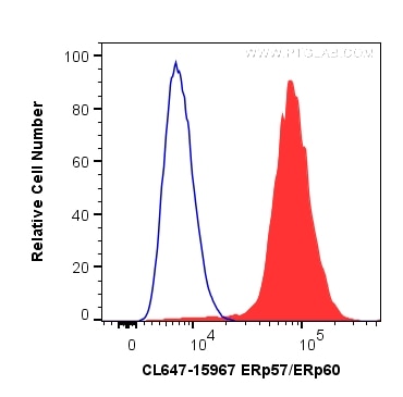
| Résultats positifs en FC (Intra) | cellules HepG2, |
Dilution recommandée
| Application | Dilution |
|---|---|
| Flow Cytometry (FC) (INTRA) | FC (INTRA) : 0.20 ug per 10^6 cells in a 100 µl suspension |
| It is recommended that this reagent should be titrated in each testing system to obtain optimal results. | |
| Sample-dependent, check data in validation data gallery | |
Informations sur le produit
CL647-15967 cible ERp57/ERp60 dans les applications de FC (Intra) et montre une réactivité avec des échantillons Humain, rat, souris
| Réactivité | Humain, rat, souris |
| Hôte / Isotype | Lapin / IgG |
| Clonalité | Polyclonal |
| Type | Anticorps |
| Immunogène | ERp57/ERp60 Protéine recombinante Ag8741 |
| Nom complet | protein disulfide isomerase family A, member 3 |
| Masse moléculaire calculée | 505 aa, 57 kDa |
| Poids moléculaire observé | 57 kDa |
| Numéro d’acquisition GenBank | BC014433 |
| Symbole du gène | PDIA3 |
| Identification du gène (NCBI) | 2923 |
| Conjugaison | CoraLite® Plus 647 Fluorescent Dye |
| Excitation/Emission maxima wavelengths | 654 nm / 674 nm |
| Forme | Liquide |
| Méthode de purification | Purification par affinité contre l'antigène |
| Tampon de stockage | PBS with 50% glycerol, 0.05% Proclin300, 0.5% BSA |
| Conditions de stockage | Stocker à -20 °C. Éviter toute exposition à la lumière. Stable pendant un an après l'expédition. L'aliquotage n'est pas nécessaire pour le stockage à -20oC Les 20ul contiennent 0,1% de BSA. |
Informations générales
PDIA3, also named as P58, ER60, ERp57, ERp60, ERp61, GRP57, GRP58 and PI-PLC, is a member of the PDI family, participates in the oxidation, reduction, and isomerization of disulfide bonds for correct folding of secretory proteins before modification and transport in the endoplasmic reticulum. It is associated with apoptosis or inhibition of cancer cell growth. PDIA3 was once thought to be a phospholipase; however, it has been demonstrated that this protein actually has protein disulfide isomerase activity. It is thought that complexes of lectins and PDIA3 mediate protein folding by promoting formation of disulfide bonds in their glycoprotein substrates.
Protocole
| Product Specific Protocols | |
|---|---|
| FC protocol for CL Plus 647 ERp57/ERp60 antibody CL647-15967 | Download protocol |
| Standard Protocols | |
|---|---|
| Click here to view our Standard Protocols |


