- Phare
- Validé par KD/KO
Anticorps Monoclonal anti-Fas/CD95
Fas/CD95 Monoclonal Antibody for WB, IHC, ELISA
Hôte / Isotype
Mouse / IgG1
Réactivité testée
Humain et plus (1)
Applications
WB, IHC, IF, ELISA
Conjugaison
Non conjugué
CloneNo.
1G7F7
N° de cat : 60196-1-Ig
Synonymes
Galerie de données de validation
Applications testées
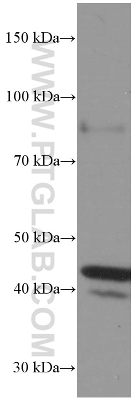
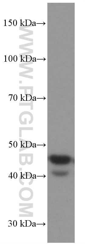
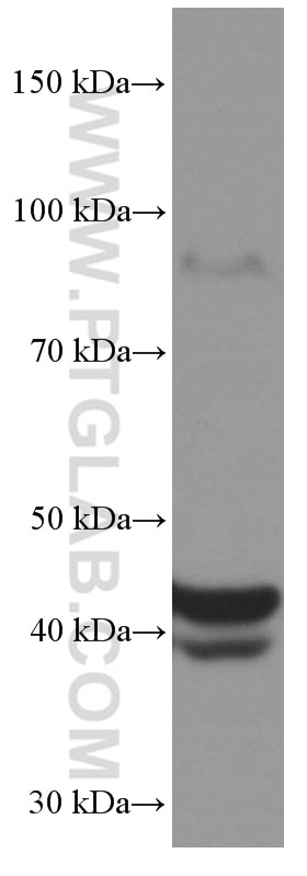
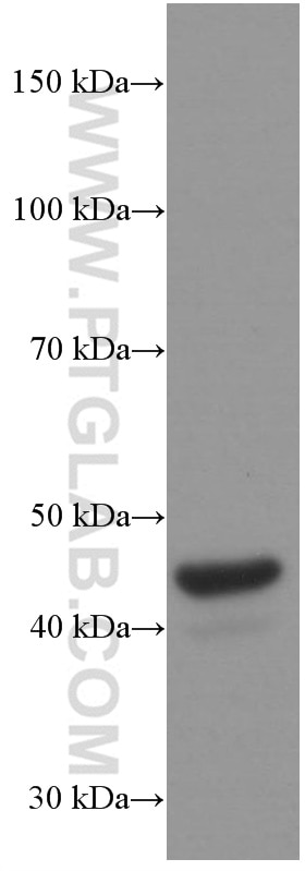
| Résultats positifs en WB | cellules Jurkat, cellules HeLa, cellules K-562, cellules U-937 |
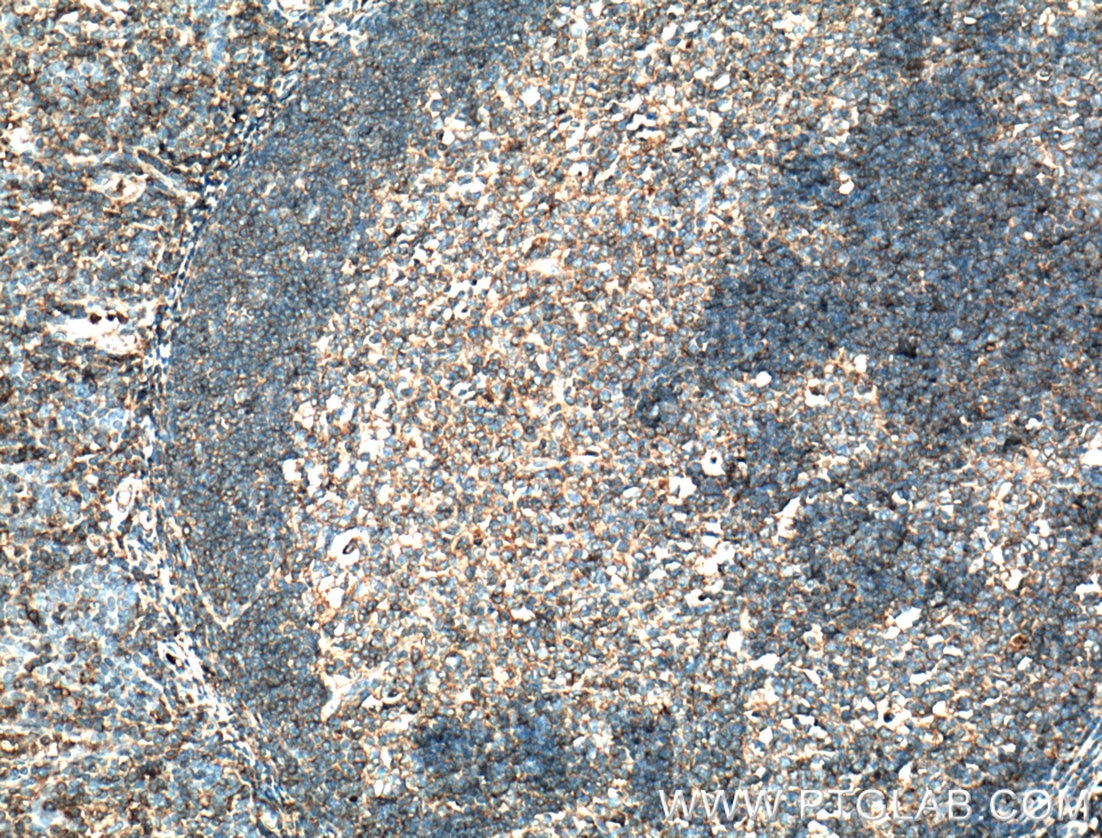
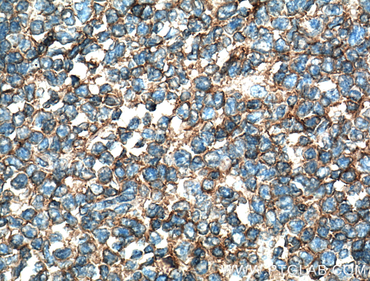
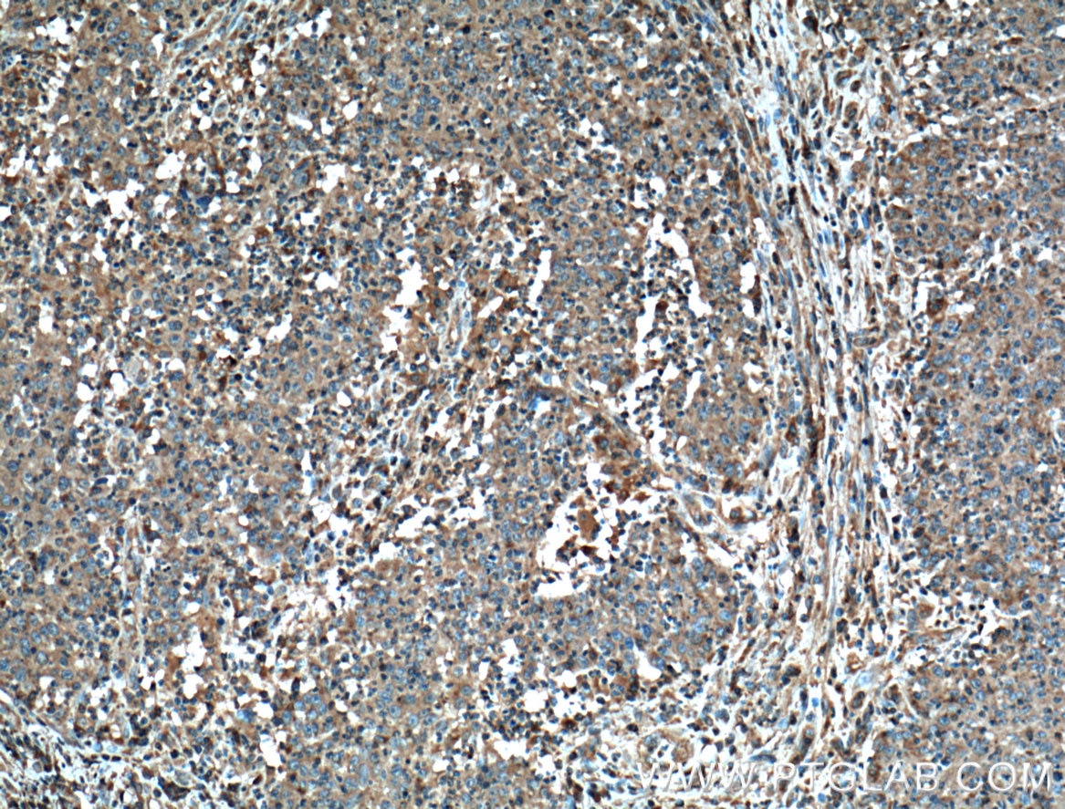
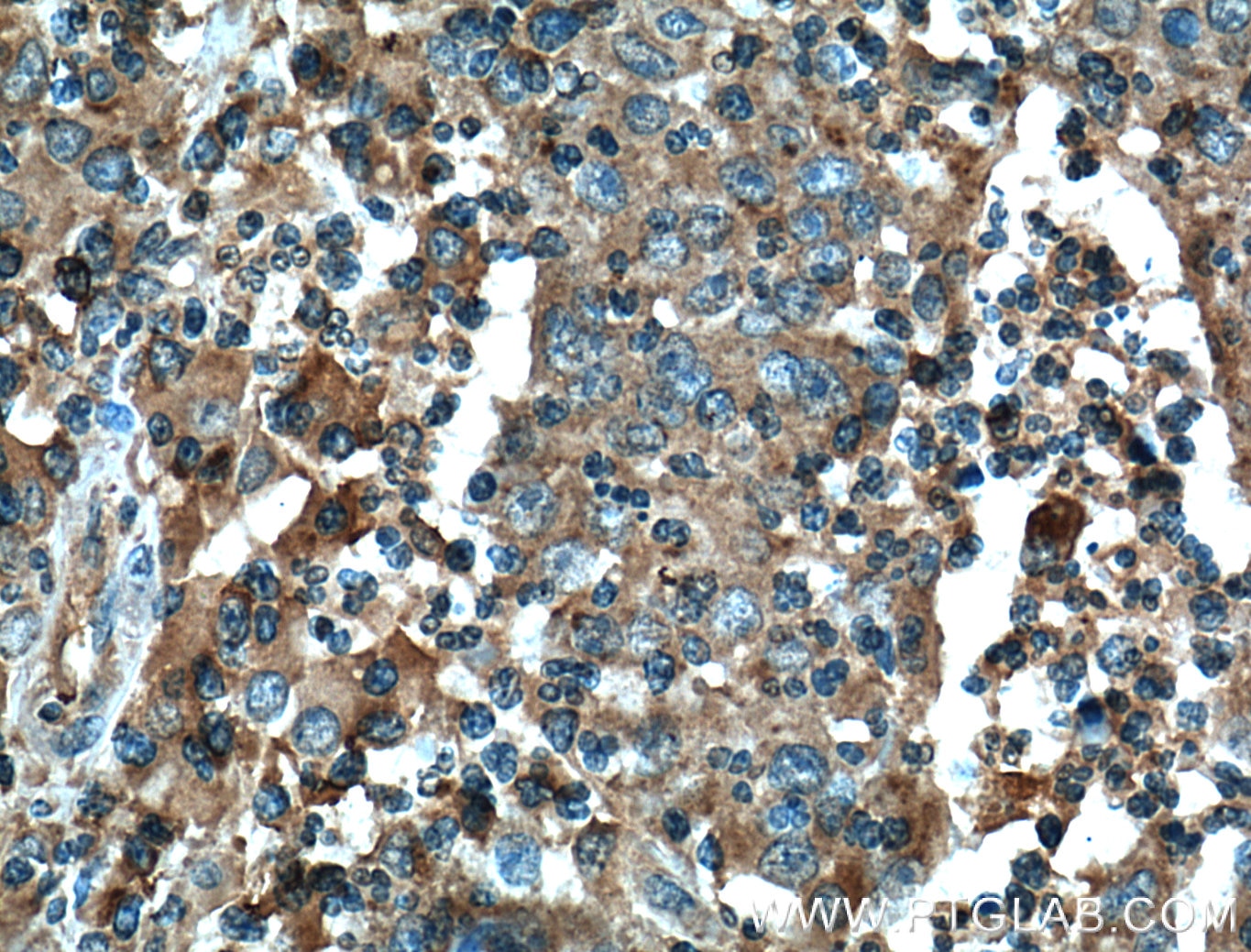
| Résultats positifs en IHC | tissu d'amygdalite humain, tissu de cancer du côlon humain il est suggéré de démasquer l'antigène avec un tampon de TE buffer pH 9.0; (*) À défaut, 'le démasquage de l'antigène peut être 'effectué avec un tampon citrate pH 6,0. |
Dilution recommandée
| Application | Dilution |
|---|---|
| Western Blot (WB) | WB : 1:1000-1:8000 |
| Immunohistochimie (IHC) | IHC : 1:50-1:500 |
| It is recommended that this reagent should be titrated in each testing system to obtain optimal results. | |
| Sample-dependent, check data in validation data gallery | |
Applications publiées
| KD/KO | See 2 publications below |
| WB | See 13 publications below |
| IHC | See 4 publications below |
| IF | See 2 publications below |
Informations sur le produit
60196-1-Ig cible Fas/CD95 dans les applications de WB, IHC, IF, ELISA et montre une réactivité avec des échantillons Humain
| Réactivité | Humain |
| Réactivité citée | Humain, poisson-zèbre |
| Hôte / Isotype | Mouse / IgG1 |
| Clonalité | Monoclonal |
| Type | Anticorps |
| Immunogène | Fas/CD95 Protéine recombinante Ag3718 |
| Nom complet | Fas (TNF receptor superfamily, member 6) |
| Masse moléculaire calculée | 35-38 kDa |
| Poids moléculaire observé | 38-45 kDa |
| Numéro d’acquisition GenBank | BC012479 |
| Symbole du gène | Fas/CD95 |
| Identification du gène (NCBI) | 355 |
| Conjugaison | Non conjugué |
| Forme | Liquide |
| Méthode de purification | Purification par protéine G |
| Tampon de stockage | PBS with 0.02% sodium azide and 50% glycerol |
| Conditions de stockage | Stocker à -20°C. Stable pendant un an après l'expédition. L'aliquotage n'est pas nécessaire pour le stockage à -20oC Les 20ul contiennent 0,1% de BSA. |
Informations générales
FAS, also named as CD95, APO-1, APT1, FAS1 and TNFRSF6, is a receptor for TNFSF6/FASLG. It is a cell surface receptor belonging to the TNF receptor superfamily, can mediate apoptosis by ligation with an agonistic anti-Fas antibody or Fas ligand. Stimulation of Fas results in the aggregation of its intracellular death domains, leading to the formation of the death-inducing signaling complex (DISC). FAS-mediated apoptosis may have a role in the induction of peripheral tolerance, in the antigen-stimulated suicide of mature T-cells, or both. The secreted isoforms 2 to 6 block apoptosis (in vitro). This anti-Fas monoclonal antibody can be used to induce apoptosis in cell cultures through Fas by imitating the Fas-ligand.
Protocole
| Product Specific Protocols | |
|---|---|
| WB protocol for Fas/CD95 antibody 60196-1-Ig | Download protocol |
| IHC protocol for Fas/CD95 antibody 60196-1-Ig | Download protocol |
| Standard Protocols | |
|---|---|
| Click here to view our Standard Protocols |
Publications
| Species | Application | Title |
|---|---|---|
Cell Neutrophil elastase selectively kills cancer cells and attenuates tumorigenesis.
| ||
Biochim Biophys Acta Down-regulation of islet amyloid polypeptide expression induces death of human annulus fibrosus cells via mitochondrial and death receptor pathways. | ||
Mol Neurobiol Ox-LDL Induces Neuron Apoptosis and Worsens Neurological Outcomes in aSAH via Fas/FADD Pathway | ||
J Physiol Biochem miR-128-3p regulates 3T3-L1 adipogenesis and lipolysis by targeting Pparg and Sertad2. | ||
iScience Loss of Adipose Growth Hormone Receptor in Mice Enhances Local Fatty Acid Trapping and Impairs Brown Adipose Tissue Thermogenesis. | ||
Chemosphere Possible involvement of Fas/FasL-dependent apoptotic pathway in α-bisabolol induced cardiotoxicity in zebrafish embryos. |
