- Phare
- Validé par KD/KO
Anticorps Monoclonal anti-c-Fos
c-Fos Monoclonal Antibody for WB, ELISA
Hôte / Isotype
Mouse / IgG1
Réactivité testée
Humain, rat, souris et plus (1)
Applications
WB, IHC, IP, CoIP, ELISA
Conjugaison
Non conjugué
CloneNo.
1G2C5
N° de cat : 66590-1-Ig
Synonymes
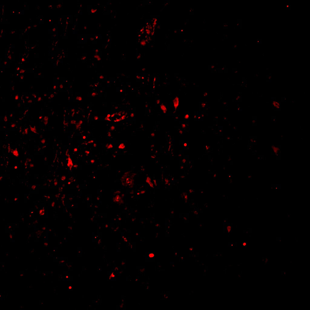
Galerie de données de validation
 at dilution of 1:10000 incubated at room temperature for 1.5 hours.")
 with sh-Control and sh-c-Fos transfected HepG2 cells.")
 at dilution of 1:10000 incubated at room temperature for 1.5 hours.")
 at dilution of 1:5000 incubated at room temperature for 1.5 hours.")
 at dilution of 1:5000 incubated at room temperature for 1.5 hours.")
Applications testées
| Résultats positifs en WB | cellules HeLa, cellules HepG2, cellules HSC-T6, cellules Jurkat, cellules K-562, cellules NIH/3T3, cellules RAW 264.7, cellules THP-1, cellules U-937 |
Dilution recommandée
| Application | Dilution |
|---|---|
| Western Blot (WB) | WB : 1:5000-1:50000 |
| It is recommended that this reagent should be titrated in each testing system to obtain optimal results. | |
| Sample-dependent, check data in validation data gallery | |
Applications publiées
| KD/KO | See 3 publications below |
| WB | See 87 publications below |
| IHC | See 14 publications below |
| IP | See 3 publications below |
| CoIP | See 1 publications below |
Informations sur le produit
66590-1-Ig cible c-Fos dans les applications de WB, IHC, IP, CoIP, ELISA et montre une réactivité avec des échantillons Humain, rat, souris
| Réactivité | Humain, rat, souris |
| Réactivité citée | rat, Humain, Lapin, souris |
| Hôte / Isotype | Mouse / IgG1 |
| Clonalité | Monoclonal |
| Type | Anticorps |
| Immunogène | c-Fos Protéine recombinante Ag24340 |
| Nom complet | FOS |
| Masse moléculaire calculée | 41 kDa |
| Poids moléculaire observé | 55-60 kDa |
| Numéro d’acquisition GenBank | BC004490 |
| Symbole du gène | c-Fos |
| Identification du gène (NCBI) | 2353 |
| Conjugaison | Non conjugué |
| Forme | Liquide |
| Méthode de purification | Purification par protéine G |
| Tampon de stockage | PBS with 0.02% sodium azide and 50% glycerol |
| Conditions de stockage | Stocker à -20°C. Stable pendant un an après l'expédition. L'aliquotage n'est pas nécessaire pour le stockage à -20oC Les 20ul contiennent 0,1% de BSA. |
Informations générales
c-Fos, also named as FOS and G0/G1 switch regulatory protein 7, is a 380 amino acid protein, which contains 1 bZIP (basic-leucine zipper) domain and belongs to the bZIP family. c-Fos is expressed at very low levels in quiescent cells. When cells are stimulated to reenter growth, c-Fos undergo 2 waves of expression, the first one peaks 7.5 minutes following FBS induction. At this stage, the c-Fos protein is localized endoplasmic reticulum. The second wave of expression occurs at about 20 minutes after induction and peaks at 1 hour. At this stage, the c-FOS protein becomes nuclear. c-Fos is a very short-lived intracellular protein, which is very easy to degrade. The calculated molecular weight of c-Fos is 40 kDa, but Phosphorylated c-Fos protein is about 60-65 kDa. It is involved in important cellular events, including cell proliferation, differentiation and survival; genes associated with hypoxia; and angiogenesis; which makes its dysregulation an important factor for cancer development. It can also induce a loss of cell polarity and epithelial-mesenchymal transition, leading to invasive and metastatic growth in mammary epithelial cells. Expression of c-Fos is an indirect marker of neuronal activity because c-Fos is often expressed when neurons fire action potentials. Upregulation of c-Fos mRNA in a neuron indicates recent activity.
Protocole
| Product Specific Protocols | |
|---|---|
| WB protocol for c-Fos antibody 66590-1-Ig | Download protocol |
| Standard Protocols | |
|---|---|
| Click here to view our Standard Protocols |
Publications
| Species | Application | Title |
|---|---|---|
Adv Mater Noninvasive Optogenetics Realized by iPSC-Derived Tentacled Carrier in Alzheimer's Disease Treatment | ||
Theranostics KDM6A promotes imatinib resistance through YY1-mediated transcriptional upregulation of TRKA independently of its demethylase activity in chronic myelogenous leukemia.
| ||
Phytomedicine Withaferin A protects against epilepsy by promoting LCN2-mediated astrocyte polarization to stopping neuronal ferroptosis | ||
Phytomedicine Gastrodin alleviates NTG-induced migraine-like pain via inhibiting succinate/HIF-1α/TRPM2 signaling pathway in trigeminal ganglion | ||
J Headache Pain SS-31 alleviated nociceptive responses and restored mitochondrial function in a headache mouse model via Sirt3/Pgc-1α positive feedback loop |
Avis
The reviews below have been submitted by verified Proteintech customers who received an incentive for providing their feedback.
FH Reyes (Verified Customer) (02-14-2025) | cFOS (a nuclear marker) did not perform as expected in my epileptic human FFPE tissue. It seems to appear in the nucleus in a few cells, but also a strong marking in the cyoplasm of others.
 |
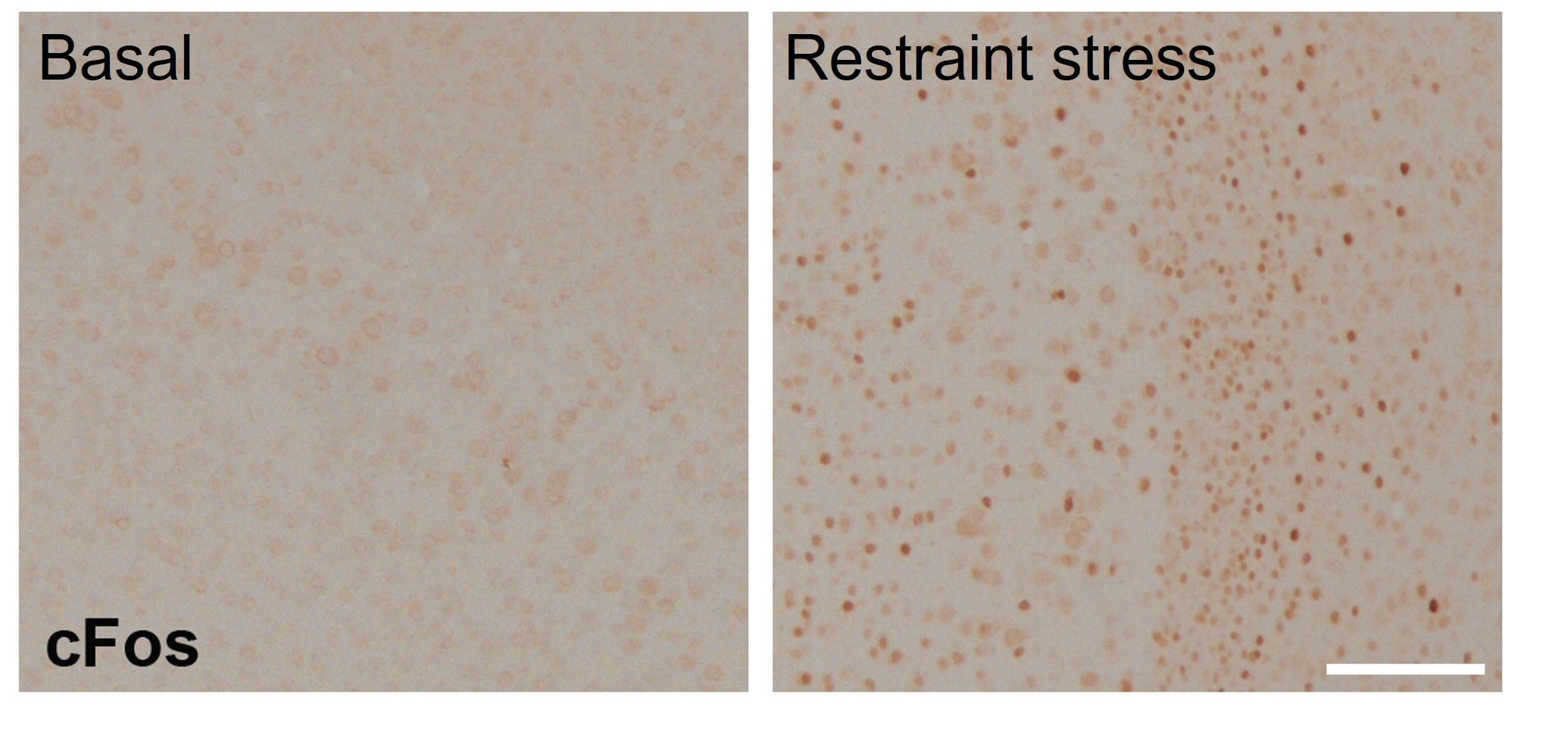
FH Tatyana (Verified Customer) (01-21-2023) | Suitable for IHC in the paraffinised brain sections of mice (cortex). Samples were fixed in 4% and standard IHC procedure with antigen retrieval and DAB detection was performed. Antibody was incubated at 1:1000 dilution overnight at 4C. Provided a good specific signal in neurons that increased after restraint stress.
 |

