- Phare
- Validé par KD/KO
Anticorps Polyclonal de lapin anti-FUS/TLS
FUS/TLS Polyclonal Antibody for WB, IHC, IF/ICC, IF-P, IF-Fro, IP, ELISA
Hôte / Isotype
Lapin / IgG
Réactivité testée
Humain, rat, souris et plus (1)
Applications
WB, IHC, IF/ICC, IF-P, IF-Fro, IP, CoIP, ChIP, RIP, ELISA
Conjugaison
Non conjugué
N° de cat : 11570-1-AP
Synonymes
Galerie de données de validation
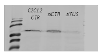
 with si-Control and si-FUS transfected HEK 293 cells.")
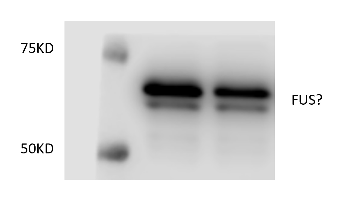
 lysates prepared with RIPA buffer, 30 μg protein loaded. 11570-1-AP incubated at 1:5000 at 4°C overnight in 5% milk in TBST. Ponceau stained transfers shown on right. Data provided by YCharOS, an open science company with a mission to validate commercial antibodies to improve scientific reproducibility and transparency.")
 at dilution of 1:30000 incubated at room temperature for 1.5 hours.")
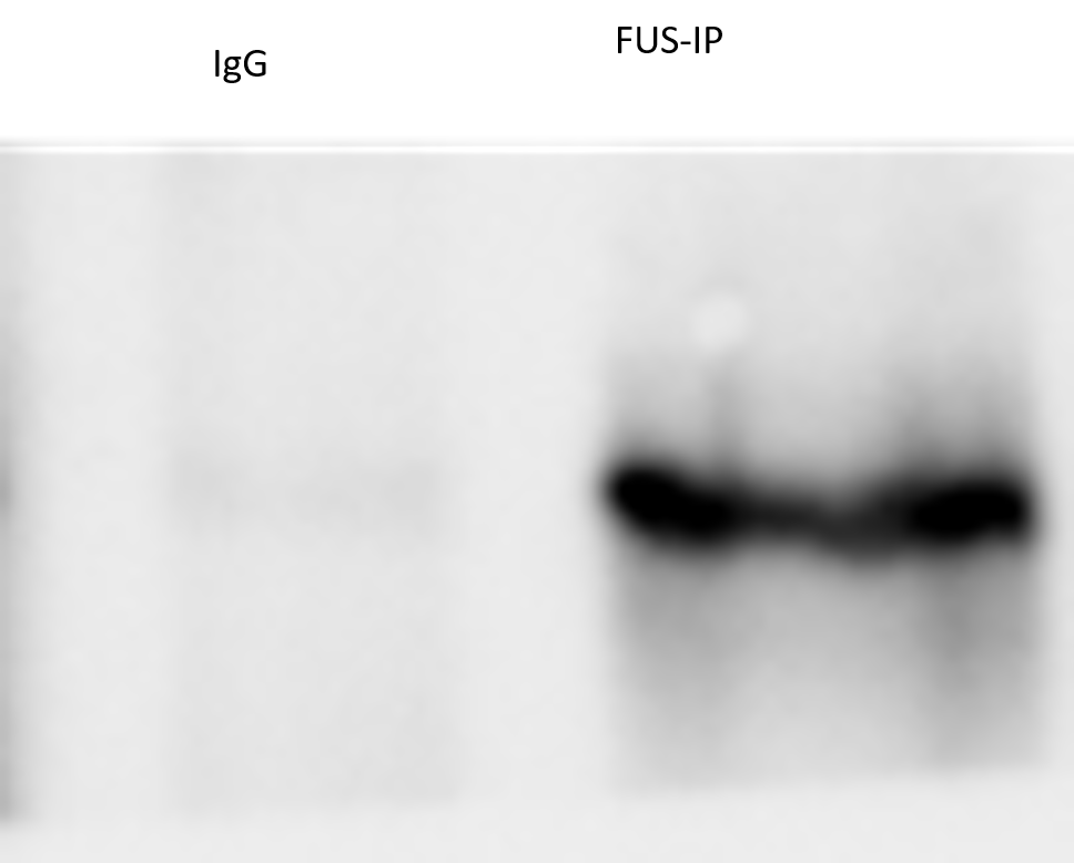
 with K-562 cells lysate 1760 ug.")
 at dilution of 1:200 (under 10x lens). Heat mediated antigen retrieval with Tris-EDTA buffer (pH 9.0).")
 at dilution of 1:200 (under 40x lens). Heat mediated antigen retrieval with Tris-EDTA buffer (pH 9.0).")
 at dilution of 1:200 (under 10x lens).")
 at dilution of 1:200 (under 40x lens).")
 at dilution of 1:200 (under 10x lens).")
 at dilution of 1:200 (under 40x lens).")
 at dilution of 1:2000 (under 10x lens). Heat mediated antigen retrieval with Tris-EDTA buffer (pH 9.0).")
 at dilution of 1:2000 (under 40x lens). Heat mediated antigen retrieval with Tris-EDTA buffer (pH 9.0).")
 fixed paraffin-embedded mouse liver tissue using FUS/TLS antibody (11570-1-AP) at dilution of 1:400 and CoraLite®488-Conjugated Goat Anti-Rabbit IgG(H+L) (SA00013-2). Heat mediated antigen retrieval with Tris-EDTA buffer (pH 9.0).")
 fixed mouse colon tissue using FUS/TLS antibody (11570-1-AP) at dilution of 1:200, CoraLite®594 smooth muscle actin antibody (CL594-14395, red), CD45 antibody (80297-1-RR, Clone: 6O19, orange), E-cadherin antibody (20874-1-AP, Magenta).")
 fixed frozen OCT-embedded rat brain tissue using FUS/TLS antibody (11570-1-AP) at dilution of 1:200 and CoraLite®488-Conjugated Goat Anti-Rabbit IgG(H+L) (SA00013-2).")
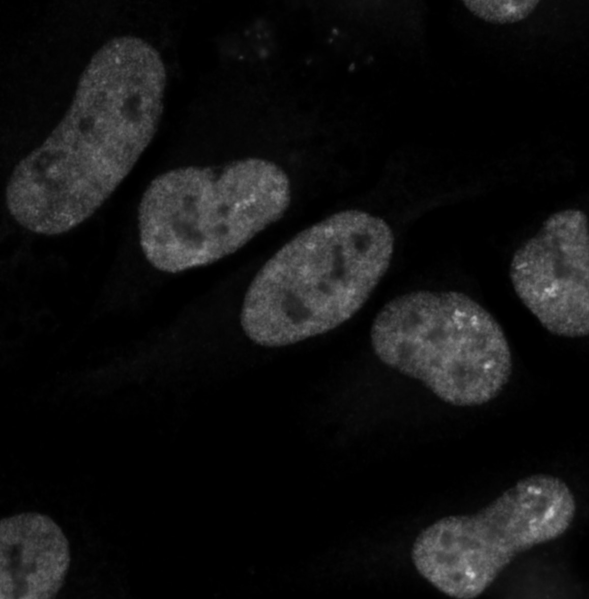
 and FUS KO cells (red outline) labelled with a green or a far-red fluorescence dye, respectively. Cells fixed with 4% PFA and stained with 11570-1-AP at 1:2000 plus DAPI. Bars = 10 μm. Data provided by YCharOS, an open science company with a mission to validate commercial antibodies to improve scientific reproducibility and transparency.")
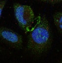
 fixed HepG2 cells using FUS/TLS antibody (11570-1-AP) at dilution of 1:200 and Multi-rAb CoraLite ® Plus 488-Goat Anti-Rabbit Recombinant Secondary Antibody (H+L) (RGAR002), CL594-phalloidin (red).")
 fixed HeLa cells using FUS/TLS antibody (11570-1-AP) at dilution of 1:400 and CoraLite®488-Conjugated AffiniPure Goat Anti-Rabbit IgG(H+L).")
Applications testées
| Résultats positifs en WB | cellules HEK-293, cellules HeLa, cellules HepG2, cellules Jurkat, cellules K-562, cellules SH-SY5Y, tissu cérébral de rat, tissu cérébral de souris |
| Résultats positifs en IP | cellules K-562, |
| Résultats positifs en IHC | tissu cérébral de souris, tissu cérébral de rat, tissu de cancer du sein humain, tissu de tumeur ovarienne humain il est suggéré de démasquer l'antigène avec un tampon de TE buffer pH 9.0; (*) À défaut, 'le démasquage de l'antigène peut être 'effectué avec un tampon citrate pH 6,0. |
| Résultats positifs en IF-P | tissu de côlon de souris, tissu hépatique de souris |
| Résultats positifs en IF-Fro | tissu cérébral de rat, |
| Résultats positifs en IF/ICC | cellules HepG2, cellules HeLa |
Dilution recommandée
| Application | Dilution |
|---|---|
| Western Blot (WB) | WB : 1:5000-1:50000 |
| Immunoprécipitation (IP) | IP : 0.5-4.0 ug for 1.0-3.0 mg of total protein lysate |
| Immunohistochimie (IHC) | IHC : 1:50-1:500 |
| Immunofluorescence (IF)-P | IF-P : 1:50-1:500 |
| Immunofluorescence (IF)-FRO | IF-FRO : 1:50-1:500 |
| Immunofluorescence (IF)/ICC | IF/ICC : 1:50-1:500 |
| It is recommended that this reagent should be titrated in each testing system to obtain optimal results. | |
| Sample-dependent, check data in validation data gallery | |
Informations sur le produit
11570-1-AP cible FUS/TLS dans les applications de WB, IHC, IF/ICC, IF-P, IF-Fro, IP, CoIP, ChIP, RIP, ELISA et montre une réactivité avec des échantillons Humain, rat, souris
| Réactivité | Humain, rat, souris |
| Réactivité citée | rat, Humain, poulet, souris |
| Hôte / Isotype | Lapin / IgG |
| Clonalité | Polyclonal |
| Type | Anticorps |
| Immunogène | FUS/TLS Protéine recombinante Ag2150 |
| Nom complet | fusion (involved in t(12;16) in malignant liposarcoma) |
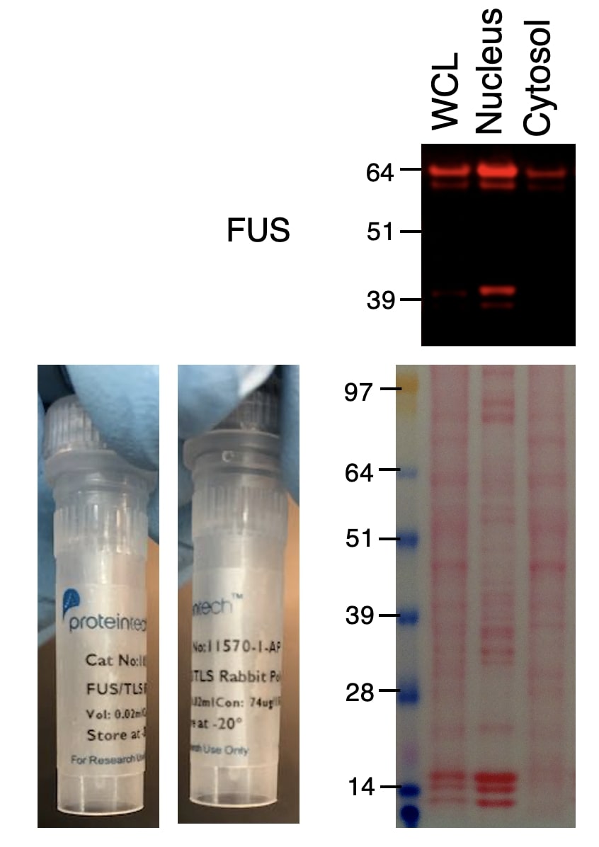
| Masse moléculaire calculée | 75 kDa |
| Poids moléculaire observé | 68-75 kDa |
| Numéro d’acquisition GenBank | BC026062 |
| Symbole du gène | FUS/TLS |
| Identification du gène (NCBI) | 2521 |
| Conjugaison | Non conjugué |
| Forme | Liquide |
| Méthode de purification | Purification par affinité contre l'antigène |
| Tampon de stockage | PBS with 0.02% sodium azide and 50% glycerol |
| Conditions de stockage | Stocker à -20°C. Stable pendant un an après l'expédition. L'aliquotage n'est pas nécessaire pour le stockage à -20oC Les 20ul contiennent 0,1% de BSA. |
Informations générales
FUS (also named TLS and POMp75) belongs to the RRM TET family. FUS may play a role in the maintenance of genomic integrity; it binds both single-stranded and double-stranded DNA and promotes ATP-independent annealing of complementary single-stranded DNAs and D-loop formation in superhelical double-stranded DNA. FUS is also an RNA-binding protein, and its links to neurodegenerative disease proffer the intriguing possibility that altered RNA metabolism or RNA processing may underlie or contribute to neuron degeneration[PMID: 22640227]. FUS may be a cause of angiomatoid fibrous histiocytoma (AFH) and is implicated in certain forms of amyotrophic lateral sclerosis (ALS) and frontotemporal dementias (FTDs) such as frontotemporal lobar dementia with ubiquitin inclusions (FTLD-U)[PMID: 22640227]. This antibody is a rabbit polyclonal antibody raised against an internal region of human FUS. FUS was detected double bands of 68-74 kDa (PMID:31519807).
Protocole
| Product Specific Protocols | |
|---|---|
| WB protocol for FUS/TLS antibody 11570-1-AP | Download protocol |
| IHC protocol for FUS/TLS antibody 11570-1-AP | Download protocol |
| IF protocol for FUS/TLS antibody 11570-1-AP | Download protocol |
| IP protocol for FUS/TLS antibody 11570-1-AP | Download protocol |
| Standard Protocols | |
|---|---|
| Click here to view our Standard Protocols |
Publications
| Species | Application | Title |
|---|---|---|
Nature Mutations in UBQLN2 cause dominant X-linked juvenile and adult-onset ALS and ALS/dementia. | ||
Nat Med Antisense oligonucleotide silencing of FUS expression as a therapeutic approach in amyotrophic lateral sclerosis. | ||
Cell Nuclear-Import Receptors Reverse Aberrant Phase Transitions of RNA-Binding Proteins with Prion-like Domains. | ||
Cell Metab NEAT1 is essential for metabolic changes that promote breast cancer growth and metastasis. | ||
Nat Neurosci FUS-mediated regulation of acetylcholine receptor transcription at neuromuscular junctions is compromised in amyotrophic lateral sclerosis.
|
Avis
The reviews below have been submitted by verified Proteintech customers who received an incentive for providing their feedback.
FH Xiaochen (Verified Customer) (07-08-2024) | Sensitivie for IF and show image with good quelity.
 |
FH Xhuljana (Verified Customer) (03-01-2024) | Used in siRNA transfected C2C12 cells
 |
FH Zhongwen (Verified Customer) (09-25-2023) | I can find two bands in the target region. I am not sure which one is the target band.
 |
FH manohar (Verified Customer) (07-10-2023) | Nitrocellulose membrane is used with 5% milk as blocking and antibody diluted in 1% milk and incubated overnight.
|
FH Tatyana (Verified Customer) (05-14-2023) | ICC using 5% goat serum/PBST buffer, 2 hours at RT. Good specific nuclear signal.
 |
FH shashirekha (Verified Customer) (12-23-2020) | Used for immunopreciptation at 1:1000 dilution. Works very well
 |
FH H (Verified Customer) (04-06-2020) | The antibody worked well for HCT116 cell line. Nuclear cytosolic fractionation clearly showed that FUS is dominantly in nucleus, and sub-fraction is present in cytosol.
 |
FH Karthik (Verified Customer) (04-24-2019) | Magenta- FUSBlue- MAP2FUS staining consistent obtained with this antibody is consistent with literature
|
FH Yen-Chen (Verified Customer) (12-03-2018) |
|

