- Phare
- Validé par KD/KO
Anticorps Monoclonal anti-G3BP1
G3BP1 Monoclonal Antibody for WB, IHC, IF/ICC, ELISA
Hôte / Isotype
Mouse / IgG1
Réactivité testée
Humain, porc, rat, souris
Applications
WB, IHC, IF/ICC, IP, CoIP, RIP, ELISA
Conjugaison
Non conjugué
CloneNo.
1E4A2
N° de cat : 66486-1-Ig
Synonymes
Galerie de données de validation
 at dilution of 1:20000 incubated at room temperature for 1.5 hours.")
 at dilution of 1:40000 incubated at room temperature for 1.5 hours. The membrane was stripped and reblotted with HRP-conjugated GAPDH Monoclonal antibody (HRP-60004) as loading control.")
 at dilution of 1:10000 incubated at room temperature for 1.5 hours.")
 at dilution of 1:10000 incubated at room temperature for 1.5 hours.")
 at dilution of 1:20000 incubated at room temperature for 1.5 hours.")
 at dilution of 1:200 (under 10x lens).")
 at dilution of 1:200 (under 40x lens).")
 at dilution of 1:5000 (under 10x lens). Heat mediated antigen retrieval with Tris-EDTA buffer (pH 9.0).")
 at dilution of 1:5000 (under 40x lens). Heat mediated antigen retrieval with Tris-EDTA buffer (pH 9.0).")
 at dilution of 1:200 (under 10x lens. Heat mediated antigen retrieval with Tris-EDTA buffer (pH 9.0).")
 at dilution of 1:200 (under 40x lens. Heat mediated antigen retrieval with Tris-EDTA buffer (pH 9.0).")
 at dilution of 1:200 (under 10x lens).")
 at dilution of 1:200 (under 40x lens).")
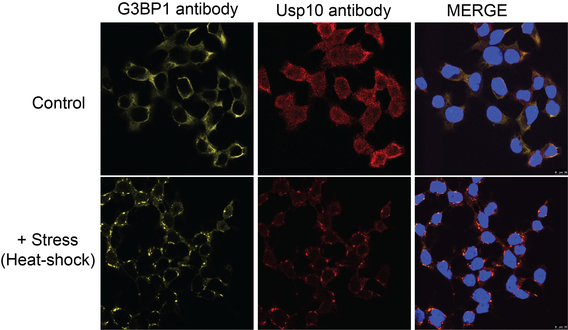
 fixed sodium arsenite treated HeLa cells using G3BP1 antibody (66486-1-Ig, Clone: 1E4A2 ) at dilution of 1:1000 and CoraLite®488-Conjugated AffiniPure Goat Anti-Mouse IgG(H+L), CL594-Phalloidin (red).")
Applications testées
| Résultats positifs en WB | cellules LNCaP, cellules 4T1, cellules HEK-293, cellules HeLa, cellules HepG2, cellules HSC-T6, cellules Jurkat, cellules K-562, cellules NIH/3T3, cellules PC-12, cellules RAW 264.7, tissu cérébral de porc, tissu cérébral de souris |
| Résultats positifs en IHC | tissu testiculaire humain, tissu cérébral de rat, tissu de côlon humain, tissu de lymphome humain il est suggéré de démasquer l'antigène avec un tampon de TE buffer pH 9.0; (*) À défaut, 'le démasquage de l'antigène peut être 'effectué avec un tampon citrate pH 6,0. |
| Résultats positifs en IF/ICC | sodium arsenite treated HeLa cells, |
Dilution recommandée
| Application | Dilution |
|---|---|
| Western Blot (WB) | WB : 1:5000-1:50000 |
| Immunohistochimie (IHC) | IHC : 1:50-1:500 |
| Immunofluorescence (IF)/ICC | IF/ICC : 1:500-1:2000 |
| It is recommended that this reagent should be titrated in each testing system to obtain optimal results. | |
| Sample-dependent, check data in validation data gallery | |
Applications publiées
| KD/KO | See 5 publications below |
| WB | See 32 publications below |
| IF | See 50 publications below |
| IP | See 2 publications below |
| CoIP | See 4 publications below |
| RIP | See 1 publications below |
Informations sur le produit
66486-1-Ig cible G3BP1 dans les applications de WB, IHC, IF/ICC, IP, CoIP, RIP, ELISA et montre une réactivité avec des échantillons Humain, porc, rat, souris
| Réactivité | Humain, porc, rat, souris |
| Réactivité citée | rat, Humain, porc, souris |
| Hôte / Isotype | Mouse / IgG1 |
| Clonalité | Monoclonal |
| Type | Anticorps |
| Immunogène | G3BP1 Protéine recombinante Ag3728 |
| Nom complet | GTPase activating protein (SH3 domain) binding protein 1 |
| Masse moléculaire calculée | 466 aa, 52 kDa |
| Poids moléculaire observé | 68 kDa |
| Numéro d’acquisition GenBank | BC006997 |
| Symbole du gène | G3BP1 |
| Identification du gène (NCBI) | 10146 |
| Conjugaison | Non conjugué |
| Forme | Liquide |
| Méthode de purification | Purification par protéine G |
| Tampon de stockage | PBS with 0.02% sodium azide and 50% glycerol |
| Conditions de stockage | Stocker à -20°C. Stable pendant un an après l'expédition. L'aliquotage n'est pas nécessaire pour le stockage à -20oC Les 20ul contiennent 0,1% de BSA. |
Informations générales
GAP SH3 Binding Protein 1 (G3BP1), also named as G3BP, is an effector of stress granule (SG) assembly. SG biology plays an important role in the pathophysiology of TDP-43 in ALS and FTLD-U. G3BP1 can be used as a marker of SG. It has been shown to function downstream of Ras and play a role in RNA metabolism, signal transduction, and proliferation. G3BP1 is a ubiquitously expressed protein that localizes to the cytoplasm in proliferating cells and to the nucleus in non-proliferating cells. G3BP1 has recently been implicated in cancer biology.
Protocole
| Product Specific Protocols | |
|---|---|
| WB protocol for G3BP1 antibody 66486-1-Ig | Download protocol |
| IHC protocol for G3BP1 antibody 66486-1-Ig | Download protocol |
| IF protocol for G3BP1 antibody 66486-1-Ig | Download protocol |
| Standard Protocols | |
|---|---|
| Click here to view our Standard Protocols |
Publications
| Species | Application | Title |
|---|---|---|
Nature DDX3X acts as a live-or-die checkpoint in stressed cells by regulating NLRP3 inflammasome. | ||
Sci Transl Med Precise genomic editing of pathogenic mutations in RBM20 rescues dilated cardiomyopathy | ||
Nat Struct Mol Biol TDP-43 aggregation induced by oxidative stress causes global mitochondrial imbalance in ALS. | ||
Brain Behav Immun Transcriptomic and proteomic profiling of bi-partite and tri-partite murine iPSC-derived neurospheroids under steady-state and inflammatory condition | ||
Autophagy Stress granule homeostasis is modulated by TRIM21-mediated ubiquitination of G3BP1 and autophagy-dependent elimination of stress granules |
Avis
The reviews below have been submitted by verified Proteintech customers who received an incentive for providing their feedback.
FH Xiaochen (Verified Customer) (11-11-2024) | wonderful image
 |
FH Haibo (Verified Customer) (10-26-2020) | This is a excellent antibody to visualize stress granules tested in HEK293 and human fibroblasts.
|
FH Manohar (Verified Customer) (09-23-2020) | 1% milk is used
|
FH David (Verified Customer) (01-13-2020) | Good signal in control cells and immunopositive puncta seen in response to stress (e.g. sodium arsenite).
|
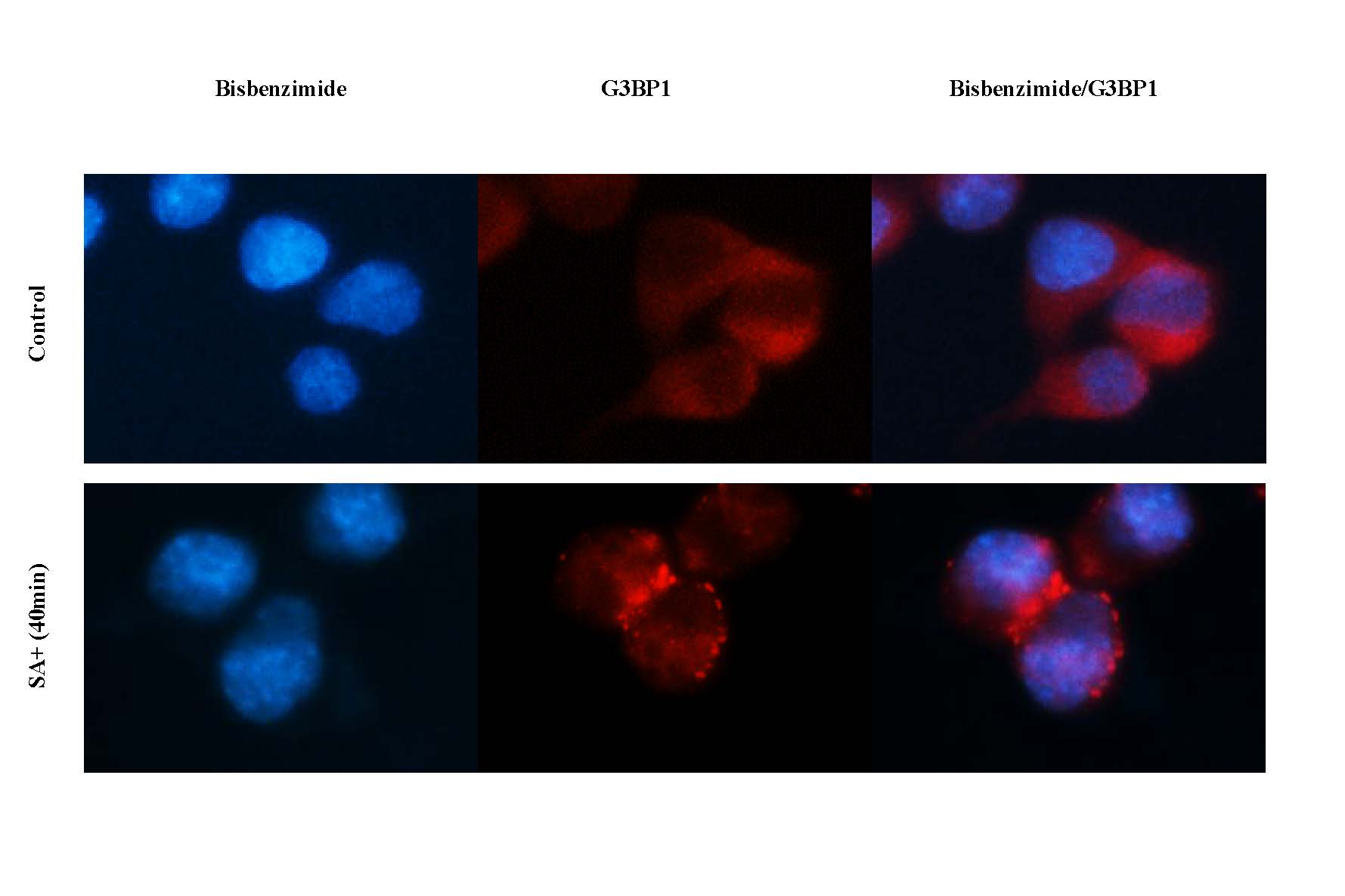
FH Azita (Verified Customer) (10-04-2019) | Assembly of stress granula upon treatment with sodium arsenite for 40 min. (It works great)
 |
FH Kyosuke (Verified Customer) (06-12-2019) | It works well on HEK293T for Western blot.
|
FH Natalia (Verified Customer) (06-06-2019) | PFA fixated cells
 |

