- Phare
- Validé par KD/KO
Anticorps Monoclonal anti-GLUT1
GLUT1 Monoclonal Antibody for WB, IHC, IF/ICC, IF-P, FC (Intra), ELISA
Hôte / Isotype
Mouse / IgG1
Réactivité testée
Humain, souris et plus (1)
Applications
WB, IHC, IF/ICC, IF-P, FC (Intra), ELISA
Conjugaison
Non conjugué
CloneNo.
2A5A2
N° de cat : 66290-1-Ig
Synonymes
Galerie de données de validation
 at dilution of 1:1500 incubated at room temperature for 1.5 hours.")
 at dilution of 1:1500 incubated at room temperature for 1.5 hours.")
 at dilution of 1:3000 incubated at room temperature for 1.5 hours.")
 at dilution of 1:2000 (under 40x lens). Heat mediated antigen retrieval with Tris-EDTA buffer (pH 9.0).")
 at dilution of 1:300 (under 10x lens).")
 at dilution of 1:300 (under 40x lens).")
 at dilution of 1:10000 (under 10x lens). Heat mediated antigen retrieval with Tris-EDTA buffer (pH 9.0).")
 at dilution of 1:2000 (under 10x lens). Heat mediated antigen retrieval with Tris-EDTA buffer (pH 9.0).")
 fixed human lung cancer tissue using GLUT1 antibody (66290-1-Ig, Clone: 2A5A2 ) at dilution of 1:400 and CoraLite®488-Conjugated AffiniPure Goat Anti-Mouse IgG(H+L).")
 fixed HeLa cells using GLUT1 antibody (66290-1-Ig, Clone: 2A5A2 ) at dilution of 1:400 and Multi-rAb CoraLite ® Plus 488-Goat Anti-Mouse Recombinant Secondary Antibody (H+L) (RGAM002).")
 and CoraLite®488-Conjugated AffiniPure Goat Anti-Mouse IgG(H+L) at dilution 1:1000 (red), or 0.4 ug Mouse IgG1 Isotype Control (MOPC-21) (65124-1-Ig, Clone: MOPC-21) (blue). Cells were fixed with 4% PFA and permeabilized with Flow Cytometry Perm Buffer (PF00011-C).")
Applications testées
| Résultats positifs en WB | cellules HEK-293, cellules NIH/3T3 |
| Résultats positifs en IHC | tissu de cancer du poumon humain, human rectal cancer tissue il est suggéré de démasquer l'antigène avec un tampon de TE buffer pH 9.0; (*) À défaut, 'le démasquage de l'antigène peut être 'effectué avec un tampon citrate pH 6,0. |
| Résultats positifs en IF-P | tissu de cancer du poumon humain, |
| Résultats positifs en IF/ICC | cellules HeLa, |
| Résultats positifs en FC (Intra) | cellules Jurkat, |
Dilution recommandée
| Application | Dilution |
|---|---|
| Western Blot (WB) | WB : 1:500-1:3000 |
| Immunohistochimie (IHC) | IHC : 1:1000-1:4000 |
| Immunofluorescence (IF)-P | IF-P : 1:200-1:800 |
| Immunofluorescence (IF)/ICC | IF/ICC : 1:200-1:800 |
| Flow Cytometry (FC) (INTRA) | FC (INTRA) : 0.40 ug per 10^6 cells in a 100 µl suspension |
| It is recommended that this reagent should be titrated in each testing system to obtain optimal results. | |
| Sample-dependent, check data in validation data gallery | |
Applications publiées
| KD/KO | See 3 publications below |
| WB | See 56 publications below |
| IHC | See 16 publications below |
| IF | See 10 publications below |
| FC | See 1 publications below |
Informations sur le produit
66290-1-Ig cible GLUT1 dans les applications de WB, IHC, IF/ICC, IF-P, FC (Intra), ELISA et montre une réactivité avec des échantillons Humain, souris
| Réactivité | Humain, souris |
| Réactivité citée | rat, Humain, souris |
| Hôte / Isotype | Mouse / IgG1 |
| Clonalité | Monoclonal |
| Type | Anticorps |
| Immunogène | GLUT1 Protéine recombinante Ag17108 |
| Nom complet | solute carrier family 2 (facilitated glucose transporter), member 1 |
| Masse moléculaire calculée | 492 aa, 54 kDa |
| Poids moléculaire observé | 45-55 kDa |
| Numéro d’acquisition GenBank | BC121804 |
| Symbole du gène | GLUT1 |
| Identification du gène (NCBI) | 6513 |
| Conjugaison | Non conjugué |
| Forme | Liquide |
| Méthode de purification | Purification par protéine G |
| Tampon de stockage | PBS with 0.02% sodium azide and 50% glycerol |
| Conditions de stockage | Stocker à -20°C. Stable pendant un an après l'expédition. L'aliquotage n'est pas nécessaire pour le stockage à -20oC Les 20ul contiennent 0,1% de BSA. |
Informations générales
Glucose transporter 1 (GLUT1), also known as solute carrier family 2, facilitated glucose transporter member 1 (SLC2A1), is a uniporter protein responsible for the transport of glucose in many cell types and across the blood-brain barrier.
What is the molecular weight of GLUT1? Is GLUT1 post-translationally modified?
There are two forms of GLUT1 transporter that differ in their molecular weight. The 45-kDa form is found in glial cells, while the 55-kDa form is present in the endothelial cells regulating glucose transport over the blood-brain and blood-tissue barriers (PMID: 9630522). N-glycosylation of asparagine at position 42 is the only known post-translation modification of GLUT1 (PMID: 3839598).
What is the subcellular localization of GLUT1?
Glucose transporters, including GLUT1, are multiple-pass integral membrane proteins. GLUT1 is present at the plasma membrane but is also a subject of recycling between plasma membrane and endosomes.
What molecules can be transported by GLUT1?
The main substrate of GLUT1 transport is glucose, but it can also transport galactose, mannose, glucosamine, and reduced ascorbate.
What is the tissue expression pattern of GLUT1?
GLUT1 is expressed by many cell types but the highest levels are observed in erythrocytes and in the central nervous system (astrocytes). GLUT1 is responsible for glucose transfer across the blood-brain and blood-tissue barriers, including placental transport.
Protocole
| Product Specific Protocols | |
|---|---|
| WB protocol for GLUT1 antibody 66290-1-Ig | Download protocol |
| IHC protocol for GLUT1 antibody 66290-1-Ig | Download protocol |
| IF protocol for GLUT1 antibody 66290-1-Ig | Download protocol |
| Standard Protocols | |
|---|---|
| Click here to view our Standard Protocols |
Publications
| Species | Application | Title |
|---|---|---|
Cell Metab Acetate enables metabolic fitness and cognitive performance during sleep disruption | ||
Cell Metab Di-methylation of CD147-K234 Promotes the Progression of NSCLC by Enhancing Lactate Export. | ||
ACS Nano Dual Inhibition of Endoplasmic Reticulum Stress and Oxidation Stress Manipulates the Polarization of Macrophages under Hypoxia to Sensitize Immunotherapy. | ||
ACS Nano Choline Phosphate-Grafted Nanozymes as Universal Extracellular Vesicle Probes for Bladder Cancer Detection | ||
Mol Cell TCR activation directly stimulates PYGB-dependent glycogenolysis to fuel the early recall response in CD8+ memory T cells. | ||
Nat Commun Fasting inhibits aerobic glycolysis and proliferation in colorectal cancer via the Fdft1-mediated AKT/mTOR/HIF1α pathway suppression. |
Avis
The reviews below have been submitted by verified Proteintech customers who received an incentive for providing their feedback.
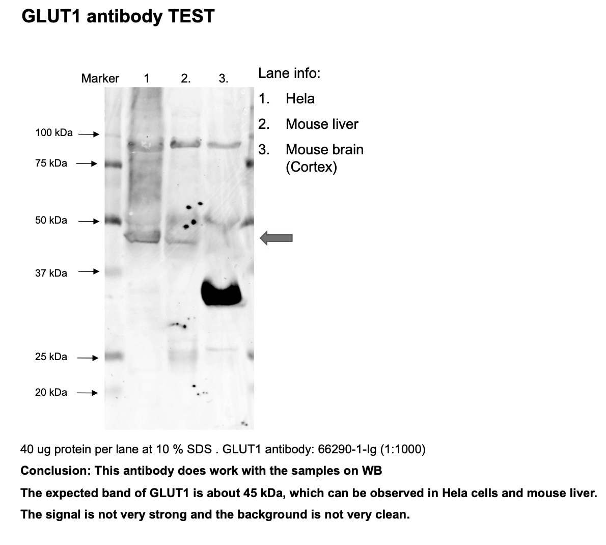
FH Hua (Verified Customer) (09-06-2023) | Works ok in WB. Signal is weak and background is not very clean.
 |
FH Iru (Verified Customer) (04-23-2019) | This antibody works ok-ish, even though the protocol says use 1:1500 to 1:3000, I have to use 1:500 to see the bands.
|

